 Movie
Movie Controller

[English] 日本語
 Yorodumi
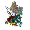
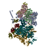
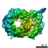
- EMDB-10259: Sub-tomogram average of the Nap adhesion complex from the human p... -
+ Open data

- Basic information
| Entry | Database: EMDB / ID: EMD-10259 | |||||||||
|---|---|---|---|---|---|---|---|---|---|---|
| Title | Sub-tomogram average of the Nap adhesion complex from the human pathogen Mycoplasma genitalium. | |||||||||
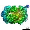
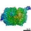
 Map data Map data | Sub-tomogram average of the surface adhesin (NAP) complex from Mycoplasma genitalium cells by cryo-electron tomography. | |||||||||
 Sample Sample |
| |||||||||
| Biological species |  Mycoplasma genitalium G37 (bacteria) Mycoplasma genitalium G37 (bacteria) | |||||||||
| Method | subtomogram averaging / cryo EM / Resolution: 15.0 Å | |||||||||
 Authors Authors | Scheffer MP / Aparicio D | |||||||||
| Funding support |  Germany, 2 items Germany, 2 items
| |||||||||
 Citation Citation | Journal: Nat Commun / Year: 2020 Title: Structure and mechanism of the Nap adhesion complex from the human pathogen Mycoplasma genitalium. Authors: David Aparicio / Margot P Scheffer / Marina Marcos-Silva / David Vizarraga / Lasse Sprankel / Mercè Ratera / Miriam S Weber / Anja Seybert / Sergi Torres-Puig / Luis Gonzalez-Gonzalez / ...Authors: David Aparicio / Margot P Scheffer / Marina Marcos-Silva / David Vizarraga / Lasse Sprankel / Mercè Ratera / Miriam S Weber / Anja Seybert / Sergi Torres-Puig / Luis Gonzalez-Gonzalez / Julian Reitz / Enrique Querol / Jaume Piñol / Oscar Q Pich / Ignacio Fita / Achilleas S Frangakis /  Abstract: Mycoplasma genitalium is a human pathogen adhering to host target epithelial cells and causing urethritis, cervicitis and pelvic inflammatory disease. Essential for infectivity is a transmembrane ...Mycoplasma genitalium is a human pathogen adhering to host target epithelial cells and causing urethritis, cervicitis and pelvic inflammatory disease. Essential for infectivity is a transmembrane adhesion complex called Nap comprising proteins P110 and P140. Here we report the crystal structure of P140 both alone and in complex with the N-terminal domain of P110. By cryo-electron microscopy (cryo-EM) and tomography (cryo-ET) we find closed and open Nap conformations, determined at 9.8 and 15 Å, respectively. Both crystal structures and the cryo-EM structure are found in a closed conformation, where the sialic acid binding site in P110 is occluded. By contrast, the cryo-ET structure shows an open conformation, where the binding site is accessible. Structural information, in combination with functional studies, suggests a mechanism for attachment and release of M. genitalium to and from the host cell receptor, in which Nap conformations alternate to sustain motility and guarantee infectivity. | |||||||||
| History |
|
- Structure visualization
| Movie |
 Movie viewer Movie viewer |
|---|---|
| Structure viewer | EM map: SurfViewMolmilJmol/JSmol |
| Supplemental images |

- Downloads & links
-EMDB archive
| Map data | emd_10259.map.gz | 2.4 MB | EMDB map data format | |
|---|---|---|---|---|
| Header (meta data) | emd-10259-v30.xmlemd-10259.xml | 10.1 KB 10.1 KB | Display Display | EMDB header |
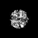
| Images | 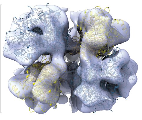 emd_10259.png emd_10259.png | 274.2 KB | ||
| Archive directory |  http://ftp.pdbj.org/pub/emdb/structures/EMD-10259ftp://ftp.pdbj.org/pub/emdb/structures/EMD-10259 http://ftp.pdbj.org/pub/emdb/structures/EMD-10259ftp://ftp.pdbj.org/pub/emdb/structures/EMD-10259 | HTTPS FTP |
-Related structure data


-Links
| EMDB pages | EMDB (EBI/PDBe) / EMDataResource |
|---|
-Map
| File | Download / File: emd_10259.map.gz / Format: CCP4 / Size: 8 MB / Type: IMAGE STORED AS FLOATING POINT NUMBER (4 BYTES) | ||||||||||||||||||||||||||||||||||||||||||||||||||||||||||||
|---|---|---|---|---|---|---|---|---|---|---|---|---|---|---|---|---|---|---|---|---|---|---|---|---|---|---|---|---|---|---|---|---|---|---|---|---|---|---|---|---|---|---|---|---|---|---|---|---|---|---|---|---|---|---|---|---|---|---|---|---|---|
| Annotation | Sub-tomogram average of the surface adhesin (NAP) complex from Mycoplasma genitalium cells by cryo-electron tomography. | ||||||||||||||||||||||||||||||||||||||||||||||||||||||||||||
| Projections & slices | Image control
Images are generated by Spider. | ||||||||||||||||||||||||||||||||||||||||||||||||||||||||||||
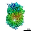
| Voxel size | X=Y=Z: 2.2 Å | ||||||||||||||||||||||||||||||||||||||||||||||||||||||||||||
| Density |
| ||||||||||||||||||||||||||||||||||||||||||||||||||||||||||||
| Symmetry | Space group: 1 | ||||||||||||||||||||||||||||||||||||||||||||||||||||||||||||
| Details | EMDB XML:
CCP4 map header:
| ||||||||||||||||||||||||||||||||||||||||||||||||||||||||||||
 Z (Sec.)
 Y (Row.)
 X (Col.)


-Supplemental data
- Sample components
-Entire : Sub-tomogram average of the Nap adhesin from Mycoplasma genitaliu...
| Entire | Name: Sub-tomogram average of the Nap adhesin from Mycoplasma genitalium cells. |
|---|---|
| Components |
|
-Supramolecule #1: Sub-tomogram average of the Nap adhesin from Mycoplasma genitaliu...
| Supramolecule | Name: Sub-tomogram average of the Nap adhesin from Mycoplasma genitalium cells. type: complex / ID: 1 / Parent: 0 |
|---|---|
| Source (natural) | Organism: Mycoplasma genitalium G37 (bacteria) |
-Experimental details
-Structure determination
| Method | cryo EM |
|---|---|
 Processing Processing | subtomogram averaging |
| Aggregation state | cell |
-Sample preparation
| Buffer | pH: 7.5 |
|---|---|
| Vitrification | Cryogen name: ETHANE |
| Details | Mildly detergent-lysed cells |
- Electron microscopy
| Microscope | FEI TITAN KRIOS |
|---|---|
| Image recording | Film or detector model: GATAN K2 SUMMIT (4k x 4k) / Detector mode: SUPER-RESOLUTION / Average electron dose: 50.0 e/Å2 |
| Electron beam | Acceleration voltage: 300 kV / Electron source:  FIELD EMISSION GUN FIELD EMISSION GUN |
| Electron optics | Illumination mode: FLOOD BEAM / Imaging mode: BRIGHT FIELD |
| Sample stage | Specimen holder model: FEI TITAN KRIOS AUTOGRID HOLDER / Cooling holder cryogen: NITROGEN |
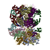
| Experimental equipment |  Model: Titan Krios / Image courtesy: FEI Company |
-Image processing
| Final reconstruction | Applied symmetry - Point group: C1 (asymmetric) / Resolution.type: BY AUTHOR / Resolution: 15.0 Å / Resolution method: FSC 0.5 CUT-OFF / Number subtomograms used: 8800 |
|---|---|
| Extraction | Number tomograms: 54 / Number images used: 11000 |
| CTF correction | Software - Name: Super-sampling SART (ver. 2) Software - details: Kunz, M. & Frangakis, A. S. Three-dimensional CTF correction improves the resolution of electron tomograms. Journal of structural biology 197, 114-122, doi: 10.1016 Details: Tomograms were reconstructed by super-sampling SART with 3D contrast transfer function (CTF) correction. |
| Final angle assignment | Type: NOT APPLICABLE |

