 Movie
Movie Controller


+ Open data

- Basic information
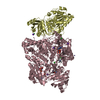
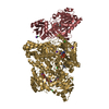
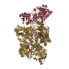
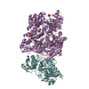
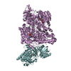
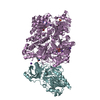
| Entry | Database: PDB / ID: 6s3u | ||||||
|---|---|---|---|---|---|---|---|
| Title | Adhesin P140 from Mycoplasma Genitalium | ||||||
 Components Components | Adhesin P1 | ||||||
 Keywords Keywords | PROTEIN BINDING / Adhesion complex | ||||||
| Function / homology | Adhesin P1, C-terminal domain / Adhesin P1, N-terminal / Adhesin P1 / Mycoplasma adhesin P1, C-terminal / Mycoplasma adhesin P1, N-terminal / adhesion of symbiont to microvasculature / plasma membrane / Adhesin P140 Function and homology information Function and homology information | ||||||
| Biological species |  Mycoplasma genitalium G37 (bacteria) Mycoplasma genitalium G37 (bacteria) | ||||||
| Method |  X-RAY DIFFRACTION / SYNCHROTRON / MOLECULAR REPLACEMENT / Resolution: 3.24 Å X-RAY DIFFRACTION / SYNCHROTRON / MOLECULAR REPLACEMENT / Resolution: 3.24 Å | ||||||
 Authors Authors | Fita, I. / Aparicio, D. | ||||||
| Funding support |  Spain, 1items Spain, 1items
| ||||||
 Citation Citation | Journal: Nat Commun / Year: 2020 Title: Structure and mechanism of the Nap adhesion complex from the human pathogen Mycoplasma genitalium. Authors: David Aparicio / Margot P Scheffer / Marina Marcos-Silva / David Vizarraga / Lasse Sprankel / Mercè Ratera / Miriam S Weber / Anja Seybert / Sergi Torres-Puig / Luis Gonzalez-Gonzalez / ...Authors: David Aparicio / Margot P Scheffer / Marina Marcos-Silva / David Vizarraga / Lasse Sprankel / Mercè Ratera / Miriam S Weber / Anja Seybert / Sergi Torres-Puig / Luis Gonzalez-Gonzalez / Julian Reitz / Enrique Querol / Jaume Piñol / Oscar Q Pich / Ignacio Fita / Achilleas S Frangakis /  Abstract: Mycoplasma genitalium is a human pathogen adhering to host target epithelial cells and causing urethritis, cervicitis and pelvic inflammatory disease. Essential for infectivity is a transmembrane ...Mycoplasma genitalium is a human pathogen adhering to host target epithelial cells and causing urethritis, cervicitis and pelvic inflammatory disease. Essential for infectivity is a transmembrane adhesion complex called Nap comprising proteins P110 and P140. Here we report the crystal structure of P140 both alone and in complex with the N-terminal domain of P110. By cryo-electron microscopy (cryo-EM) and tomography (cryo-ET) we find closed and open Nap conformations, determined at 9.8 and 15 Å, respectively. Both crystal structures and the cryo-EM structure are found in a closed conformation, where the sialic acid binding site in P110 is occluded. By contrast, the cryo-ET structure shows an open conformation, where the binding site is accessible. Structural information, in combination with functional studies, suggests a mechanism for attachment and release of M. genitalium to and from the host cell receptor, in which Nap conformations alternate to sustain motility and guarantee infectivity. | ||||||
| History |
|
- Structure visualization
| Structure viewer | Molecule: MolmilJmol/JSmol |
|---|
- Downloads & links
-Download
| PDBx/mmCIF format | 6s3u.cif.gz | 2.9 MB | Display | PDBx/mmCIF format |
|---|---|---|---|---|
| PDB format | pdb6s3u.ent.gz | 2.4 MB | Display | PDB format |
| PDBx/mmJSON format | 6s3u.json.gz | Tree view | PDBx/mmJSON format | |
| Others |  Other downloads Other downloads |
-Validation report
| Arichive directory | https://data.pdbj.org/pub/pdb/validation_reports/s3/6s3uftp://data.pdbj.org/pub/pdb/validation_reports/s3/6s3u | HTTPS FTP |
|---|
-Related structure data
| Related structure data |  6rutSC  6yrkC S: Starting model for refinement C: citing same article ( |
|---|---|
| Similar structure data |


-Links
 PDBj
PDBj- Assembly
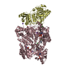
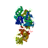
| Deposited unit | 
| ||||||||
|---|---|---|---|---|---|---|---|---|---|
| 1 | 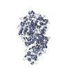
| ||||||||
| 2 | 
| ||||||||
| 3 | 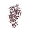
| ||||||||
| 4 | 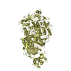
| ||||||||
| 5 | 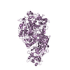
| ||||||||
| 6 | 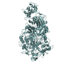
| ||||||||
| Unit cell |
|
-Components
| #1: Protein | Mass: 144661.219 Da / Num. of mol.: 6 Source method: isolated from a genetically manipulated source Source: (gene. exp.) Mycoplasma genitalium G37 (bacteria) / Gene: mgpA, MG191 / Variant: ATCC 33530 / G-37 / NCTC 10195 / Production host: |
|---|
-Experimental details
-Experiment
| Experiment | Method: X-RAY DIFFRACTION / Number of used crystals: 1 |
|---|
- Sample preparation
| Crystal | Density Matthews: 3.67 Å3/Da / Density % sol: 66.45 % |
|---|---|
| Crystal grow | Temperature: 293 K / Method: vapor diffusion, hanging drop Details: 20% PEG3350 0.1M BisTris Propane pH7.5 0.2M Sodium Sulphate Hydrate |
-Data collection
| Diffraction | Mean temperature: 80 K / Serial crystal experiment: N |
|---|---|
| Diffraction source | Source: SYNCHROTRON / Site: ALBA / Beamline: XALOC / Wavelength: 0.9791 Å |
| Detector | Type: DECTRIS PILATUS 2M / Detector: PIXEL / Date: May 21, 2012 |
| Radiation | Protocol: SINGLE WAVELENGTH / Monochromatic (M) / Laue (L): M / Scattering type: x-ray |
| Radiation wavelength | Wavelength: 0.9791 Å / Relative weight: 1 |
| Reflection | Resolution: 3.24→189.88 Å / Num. obs: 123883 / % possible obs: 63.5 % / Redundancy: 3.1 % / Biso Wilson estimate: 80.85 Å2 / CC1/2: 0.986 / Net I/σ(I): 5.8 |
| Reflection shell | Resolution: 3.24→3.58 Å / Num. unique obs: 6159 / CC1/2: 0.74 |
- Processing
| Software |
| |||||||||||||||||||||||||||||||||||||||||||||||||||||||||||||||||||||||||||||||||||||||||||||||||||||||||||||||||||||||||||||||||||||||||||||||||||||||||||||||||||||||||||||||
|---|---|---|---|---|---|---|---|---|---|---|---|---|---|---|---|---|---|---|---|---|---|---|---|---|---|---|---|---|---|---|---|---|---|---|---|---|---|---|---|---|---|---|---|---|---|---|---|---|---|---|---|---|---|---|---|---|---|---|---|---|---|---|---|---|---|---|---|---|---|---|---|---|---|---|---|---|---|---|---|---|---|---|---|---|---|---|---|---|---|---|---|---|---|---|---|---|---|---|---|---|---|---|---|---|---|---|---|---|---|---|---|---|---|---|---|---|---|---|---|---|---|---|---|---|---|---|---|---|---|---|---|---|---|---|---|---|---|---|---|---|---|---|---|---|---|---|---|---|---|---|---|---|---|---|---|---|---|---|---|---|---|---|---|---|---|---|---|---|---|---|---|---|---|---|---|---|
| Refinement | Method to determine structure: MOLECULAR REPLACEMENT Starting model: 6RUT Resolution: 3.24→37.36 Å / Cor.coef. Fo:Fc: 0.882 / Cor.coef. Fo:Fc free: 0.86 / Cross valid method: THROUGHOUT / σ(F): 0 / SU Rfree Blow DPI: 0.52
| |||||||||||||||||||||||||||||||||||||||||||||||||||||||||||||||||||||||||||||||||||||||||||||||||||||||||||||||||||||||||||||||||||||||||||||||||||||||||||||||||||||||||||||||
| Displacement parameters | Biso mean: 91.11 Å2
| |||||||||||||||||||||||||||||||||||||||||||||||||||||||||||||||||||||||||||||||||||||||||||||||||||||||||||||||||||||||||||||||||||||||||||||||||||||||||||||||||||||||||||||||
| Refine analyze | Luzzati coordinate error obs: 0.42 Å | |||||||||||||||||||||||||||||||||||||||||||||||||||||||||||||||||||||||||||||||||||||||||||||||||||||||||||||||||||||||||||||||||||||||||||||||||||||||||||||||||||||||||||||||
| Refinement step | Cycle: LAST / Resolution: 3.24→37.36 Å
| |||||||||||||||||||||||||||||||||||||||||||||||||||||||||||||||||||||||||||||||||||||||||||||||||||||||||||||||||||||||||||||||||||||||||||||||||||||||||||||||||||||||||||||||
| Refine LS restraints |
| |||||||||||||||||||||||||||||||||||||||||||||||||||||||||||||||||||||||||||||||||||||||||||||||||||||||||||||||||||||||||||||||||||||||||||||||||||||||||||||||||||||||||||||||
| LS refinement shell | Resolution: 3.24→3.45 Å / Total num. of bins used: 50
| |||||||||||||||||||||||||||||||||||||||||||||||||||||||||||||||||||||||||||||||||||||||||||||||||||||||||||||||||||||||||||||||||||||||||||||||||||||||||||||||||||||||||||||||
| Refinement TLS params. | Method: refined / Refine-ID: X-RAY DIFFRACTION
| |||||||||||||||||||||||||||||||||||||||||||||||||||||||||||||||||||||||||||||||||||||||||||||||||||||||||||||||||||||||||||||||||||||||||||||||||||||||||||||||||||||||||||||||
| Refinement TLS group |
|
