[English] 日本語
Yorodumi
- PDB-4ogc: Crystal structure of the Type II-C Cas9 enzyme from Actinomyces n... -

+
Open data


ID or keywords:

Loading...

-
Basic information

Entry
Database: PDB / ID: 4ogc
TitleCrystal structure of the Type II-C Cas9 enzyme from Actinomyces naeslundii
ComponentsHNH endonuclease domain protein
KeywordsHYDROLASE / CRISPR-Cas / Cas9 / HNH / RuvC / RNA-guided DNA endonuclease / cytoplasmic
Function / homology
Function and homology information


defense response to virus / endonuclease activity / Hydrolases; Acting on ester bonds / DNA binding / RNA binding / zinc ion binding
Similarity search - Function
Alpha-Beta Plaits - #3520 / CRISPR-associated endonuclease Cas9, beta-hairpin domain / Cas9 C-terminal domain / Cas9, topo homolgy domain / CRISPR-associated endonuclease Cas9 beta-hairpin domain / Topo homolgy domain in CRISPR-associated endonuclease Cas9 / Cas9 C-terminal domain / Cas9, alpha-helical lobe domain / Cas9 alpha-helical lobe domain / HNH endonuclease ...Alpha-Beta Plaits - #3520 / CRISPR-associated endonuclease Cas9, beta-hairpin domain / Cas9 C-terminal domain / Cas9, topo homolgy domain / CRISPR-associated endonuclease Cas9 beta-hairpin domain / Topo homolgy domain in CRISPR-associated endonuclease Cas9 / Cas9 C-terminal domain / Cas9, alpha-helical lobe domain / Cas9 alpha-helical lobe domain / HNH endonuclease / HNH endonuclease / RuvC endonuclease subdomain 3 / RuvC endonuclease subdomain 3 / HNH nucleases / Cas9-type HNH domain / Cas9-type HNH domain profile. / HNH nuclease / Ribonuclease H-like superfamily/Ribonuclease H / Nucleotidyltransferase; domain 5 / Ribonuclease H superfamily / Alpha-Beta Plaits / 2-Layer Sandwich / Alpha Beta
Similarity search - Domain/homology
ACETATE ION / : / SPERMIDINE / CRISPR-associated endonuclease Cas9
Similarity search - Component
Biological speciesActinomyces naeslundii (bacteria)
MethodX-RAY DIFFRACTION / SYNCHROTRON / MOLECULAR REPLACEMENT / Resolution: 2.8 Å
AuthorsJiang, F. / Ma, E. / Lin, S. / Doudna, J.A.
Citation
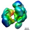
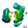
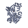
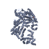
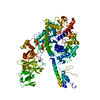
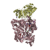
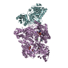
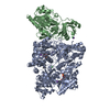
Journal: Science / Year: 2014
Title: Structures of Cas9 endonucleases reveal RNA-mediated conformational activation.
Authors: Martin Jinek / Fuguo Jiang / David W Taylor / Samuel H Sternberg / Emine Kaya / Enbo Ma / Carolin Anders / Michael Hauer / Kaihong Zhou / Steven Lin / Matias Kaplan / Anthony T Iavarone / ...Authors: Martin Jinek / Fuguo Jiang / David W Taylor / Samuel H Sternberg / Emine Kaya / Enbo Ma / Carolin Anders / Michael Hauer / Kaihong Zhou / Steven Lin / Matias Kaplan / Anthony T Iavarone / Emmanuelle Charpentier / Eva Nogales / Jennifer A Doudna /
Abstract: Type II CRISPR (clustered regularly interspaced short palindromic repeats)-Cas (CRISPR-associated) systems use an RNA-guided DNA endonuclease, Cas9, to generate double-strand breaks in invasive DNA ...Type II CRISPR (clustered regularly interspaced short palindromic repeats)-Cas (CRISPR-associated) systems use an RNA-guided DNA endonuclease, Cas9, to generate double-strand breaks in invasive DNA during an adaptive bacterial immune response. Cas9 has been harnessed as a powerful tool for genome editing and gene regulation in many eukaryotic organisms. We report 2.6 and 2.2 angstrom resolution crystal structures of two major Cas9 enzyme subtypes, revealing the structural core shared by all Cas9 family members. The architectures of Cas9 enzymes define nucleic acid binding clefts, and single-particle electron microscopy reconstructions show that the two structural lobes harboring these clefts undergo guide RNA-induced reorientation to form a central channel where DNA substrates are bound. The observation that extensive structural rearrangements occur before target DNA duplex binding implicates guide RNA loading as a key step in Cas9 activation.
History
DepositionJan 15, 2014Deposition site: RCSB / Processing site: RCSB
Revision 1.0Feb 12, 2014Provider: repository / Type: Initial release
Revision 1.1Mar 12, 2014Group: Database references
Revision 1.2Mar 26, 2014Group: Database references

-
Structure visualization

Structure viewerMolecule:
MolmilJmol/JSmol

Downloads & links

-
Assembly

Deposited unit
A: HNH endonuclease domain protein
hetero molecules


Theoretical massNumber of molelcules
Total (without water)125,3258
Polymers124,8971
Non-polymers4287
Water1629
1


  • Idetical with deposited unit
  • defined by author&software
TypeNameSymmetry operationNumber
identity operation1_555x,y,z1
MethodPISA
Unit cell
Length a, b, c (Å)74.610, 132.560, 80.040
Angle α, β, γ (deg.)90.00, 95.38, 90.00
Int Tables number4
Space group name H-MP1211

-
Components

-
Protein , 1 types, 1 molecules A

#1: Protein HNH endonuclease domain protein


Mass: 124896.641 Da / Num. of mol.: 1
Source method: isolated from a genetically manipulated source
Source: (gene. exp.) Actinomyces naeslundii (bacteria) / Strain: Howell 279 / Gene: HMPREF1129_2620 / Production host: Escherichia coli (E. coli) / Strain (production host): Rosetta (DE3) / References: UniProt: J3F2B0

-
Non-polymers , 6 types, 16 molecules

#2: Chemical ChemComp-ZN / ZINC ION


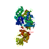
Mass: 65.409 Da / Num. of mol.: 1 / Source method: obtained synthetically / Formula: Zn
#3: Chemical ChemComp-MN / MANGANESE (II) ION


Mass: 54.938 Da / Num. of mol.: 2 / Source method: obtained synthetically / Formula: Mn
#4: Chemical ChemComp-MG / MAGNESIUM ION


Mass: 24.305 Da / Num. of mol.: 2 / Source method: obtained synthetically / Formula: Mg
#5: Chemical ChemComp-SPD / SPERMIDINE / N-(2-AMINO-PROPYL)-1,4-DIAMINOBUTANE / PA(34) / Spermidine


Mass: 145.246 Da / Num. of mol.: 1 / Source method: obtained synthetically / Formula: C7H19N3
#6: Chemical ChemComp-ACT / ACETATE ION / Acetate


Mass: 59.044 Da / Num. of mol.: 1 / Source method: obtained synthetically / Formula: C2H3O2
#7: Water ChemComp-HOH / water / Water


Mass: 18.015 Da / Num. of mol.: 9 / Source method: isolated from a natural source / Formula: H2O

-
Experimental details

-
Experiment

ExperimentMethod: X-RAY DIFFRACTION / Number of used crystals: 1

-
Sample preparation

CrystalDensity Matthews: 3.16 Å3/Da / Density % sol: 61.02 %
Crystal growTemperature: 293 K / Method: vapor diffusion, hanging drop / pH: 7
Details: 10% (w/v) PEG 8000, 0.25 M calcium acetate, 50 mM magnesium acetate and 5 mM spermidine., pH 7, VAPOR DIFFUSION, HANGING DROP, temperature 293K

-
Data collection

Diffraction
IDMean temperature (K)Crystal-ID
11001
21
Diffraction source
SourceSiteBeamlineIDWavelength (Å)
SYNCHROTRONALS 8.3.111.116
SYNCHROTRONALS 8.2.221
Detector
TypeIDDetectorDate
ADSC QUANTUM 315r1CCDSep 21, 2013
2
Radiation
IDMonochromatorProtocolMonochromatic (M) / Laue (L)Scattering typeWavelength-ID
1Double flat crystal, Si(111)SINGLE WAVELENGTHMx-ray1
2x-ray1
Radiation wavelength
IDWavelength (Å)Relative weight
11.1161
211
ReflectionResolution: 2.8→79.69 Å / Num. all: 151266 / Num. obs: 38240 / % possible obs: 100 % / Observed criterion σ(F): 2 / Observed criterion σ(I): 2.4 / Redundancy: 4 % / Biso Wilson estimate: 53.4 Å2
Reflection shellResolution: 2.8→2.95 Å / Redundancy: 4 % / % possible all: 100

-
Processing

Software
NameVersionClassification
ADSCQuantumdata collection
SOLVEphasing
PHENIX(phenix.refine: 1.8.2_1309)refinement
MOSFLMdata reduction
SCALAdata scaling
RefinementMethod to determine structure: MOLECULAR REPLACEMENT / Resolution: 2.8→68.297 Å / SU ML: 0.34 / σ(F): 1.35 / Phase error: 25.12 / Stereochemistry target values: ML
RfactorNum. reflection% reflection
Rfree0.2331 1909 5 %
Rwork0.1942 --
obs0.1962 38207 99.95 %
all-38240 -
Solvent computationShrinkage radii: 0.9 Å / VDW probe radii: 1.11 Å / Solvent model: FLAT BULK SOLVENT MODEL
Refinement stepCycle: LAST / Resolution: 2.8→68.297 Å
ProteinNucleic acidLigandSolventTotal
Num. atoms6856 0 19 9 6884
Refine LS restraints
Refine-IDTypeDev idealNumber
X-RAY DIFFRACTIONf_bond_d0.0087024
X-RAY DIFFRACTIONf_angle_d0.8089531
X-RAY DIFFRACTIONf_dihedral_angle_d13.6682594
X-RAY DIFFRACTIONf_chiral_restr0.0561061
X-RAY DIFFRACTIONf_plane_restr0.0031247
LS refinement shell
Resolution (Å)Rfactor RfreeNum. reflection RfreeRfactor RworkNum. reflection RworkRefine-ID% reflection obs (%)
2.8-2.870.35931380.28122639X-RAY DIFFRACTION100
2.87-2.94760.29351500.26082527X-RAY DIFFRACTION100
2.9476-3.03440.28161270.24332592X-RAY DIFFRACTION100
3.0344-3.13230.27721340.23442585X-RAY DIFFRACTION100
3.1323-3.24430.34391200.23432632X-RAY DIFFRACTION100
3.2443-3.37420.30891290.22412575X-RAY DIFFRACTION100
3.3742-3.52770.2691330.20222556X-RAY DIFFRACTION100
3.5277-3.71370.23631320.19282607X-RAY DIFFRACTION100
3.7137-3.94630.22961530.17872560X-RAY DIFFRACTION100
3.9463-4.2510.20551330.16812605X-RAY DIFFRACTION100
4.251-4.67870.18571400.14672594X-RAY DIFFRACTION100
4.6787-5.35550.18541460.1592587X-RAY DIFFRACTION100
5.3555-6.74640.23651410.19922606X-RAY DIFFRACTION100
6.7464-68.31720.20171330.20122633X-RAY DIFFRACTION99
Refinement TLS params.

Method: refined / Refine-ID: X-RAY DIFFRACTION

IDL112)L122)L132)L222)L232)L332)S11 (Å °)S12 (Å °)S13 (Å °)S21 (Å °)S22 (Å °)S23 (Å °)S31 (Å °)S32 (Å °)S33 (Å °)T112)T122)T132)T222)T232)T332)Origin x (Å)Origin y (Å)Origin z (Å)
13.3605-1.489-1.12312.00270.24350.4181-0.1671-0.31060.26360.05010.2472-0.0428-0.14860.1776-0.15670.4067-0.021-0.11130.3967-0.08050.349228.988168.70625.0455
22.8051.02680.49272.59210.56341.9624-0.0043-0.31350.01090.1608-0.03960.06390.3206-0.20950.08190.5576-0.0108-0.00520.3099-0.00810.47554.198269.42177.6832
34.019-1.3448-0.07481.39780.25150.0523-0.21880.00430.40650.09170.0575-0.1858-0.19580.1261-0.01050.4992-0.0075-0.06270.3761-0.03730.4223.988672.893.8483
43.08722.29790.62333.38140.11530.34760.1801-0.1458-0.22220.29310.01460.08930.12090.0415-0.10120.47690.0286-0.04450.3271-0.02350.57453.680346.2936-0.867
53.60840.93982.47531.18331.21673.93520.26930.4808-0.6890.057-0.005-0.03630.56940.3874-0.16290.44910.0485-0.02140.3482-0.10780.517828.832647.0364-6.8909
60.8310.04290.99870.87640.70456.0916-0.01380.0692-0.06850.0251-0.0463-0.0846-0.05260.33230.10410.2975-0.0155-0.08460.43430.06950.515447.933157.935236.4014
Refinement TLS group
IDRefine-IDRefine TLS-IDSelection details
1X-RAY DIFFRACTION1chain 'A' and (resid 8 through 277 )
2X-RAY DIFFRACTION2chain 'A' and (resid 278 through 443 )
3X-RAY DIFFRACTION3chain 'A' and (resid 444 through 513 )
4X-RAY DIFFRACTION4chain 'A' and (resid 514 through 704 )
5X-RAY DIFFRACTION5chain 'A' and (resid 705 through 806 )
6X-RAY DIFFRACTION6chain 'A' and (resid 807 through 1101 )

+
About Yorodumi

-
News

-
Feb 9, 2022. New format data for meta-information of EMDB entries

New format data for meta-information of EMDB entries

  • Version 3 of the EMDB header file is now the official format.
  • The previous official version 1.9 will be removed from the archive.

Related info.:EMDB header

External links:wwPDB to switch to version 3 of the EMDB data model

-
Aug 12, 2020. Covid-19 info

Covid-19 info

URL: https://pdbj.org/emnavi/covid19.php

New page: Covid-19 featured information page in EM Navigator.

Related info.:Covid-19 info / Mar 5, 2020. Novel coronavirus structure data

+
Mar 5, 2020. Novel coronavirus structure data

Novel coronavirus structure data

Related info.:Yorodumi Speices / Aug 12, 2020. Covid-19 info

External links:COVID-19 featured content - PDBj / Molecule of the Month (242):Coronavirus Proteases

+
Jan 31, 2019. EMDB accession codes are about to change! (news from PDBe EMDB page)

EMDB accession codes are about to change! (news from PDBe EMDB page)

  • The allocation of 4 digits for EMDB accession codes will soon come to an end. Whilst these codes will remain in use, new EMDB accession codes will include an additional digit and will expand incrementally as the available range of codes is exhausted. The current 4-digit format prefixed with “EMD-” (i.e. EMD-XXXX) will advance to a 5-digit format (i.e. EMD-XXXXX), and so on. It is currently estimated that the 4-digit codes will be depleted around Spring 2019, at which point the 5-digit format will come into force.
  • The EM Navigator/Yorodumi systems omit the EMD- prefix.

Related info.:Q: What is EMD? / ID/Accession-code notation in Yorodumi/EM Navigator

External links:EMDB Accession Codes are Changing Soon! / Contact to PDBj

+
Jul 12, 2017. Major update of PDB

Major update of PDB

  • wwPDB released updated PDB data conforming to the new PDBx/mmCIF dictionary.
  • This is a major update changing the version number from 4 to 5, and with Remediation, in which all the entries are updated.
  • In this update, many items about electron microscopy experimental information are reorganized (e.g. em_software).
  • Now, EM Navigator and Yorodumi are based on the updated data.

External links:wwPDB Remediation / Enriched Model Files Conforming to OneDep Data Standards Now Available in the PDB FTP Archive

-
Yorodumi

Thousand views of thousand structures

  • Yorodumi is a browser for structure data from EMDB, PDB, SASBDB, etc.
  • This page is also the successor to EM Navigator detail page, and also detail information page/front-end page for Omokage search.
  • The word "yorodu" (or yorozu) is an old Japanese word meaning "ten thousand". "mi" (miru) is to see.

Related info.:EMDB / PDB / SASBDB / Comparison of 3 databanks / Yorodumi Search / Aug 31, 2016. New EM Navigator & Yorodumi / Yorodumi Papers / Jmol/JSmol / Function and homology information / Changes in new EM Navigator and Yorodumi

Read more