Loading
PDBj
MenuPDBj@FacebookPDBj@TwitterPDBj@YouTubewwPDB FoundationwwPDB
RCSB PDBPDBeBMRBAdv. SearchSearch help
Search by PDB author
5FBJ
DownloadVisualize
BU of 5fbj by Molmil
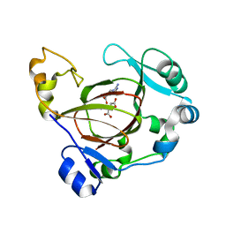
Complex structure of JMJD5 and substrate
Descriptor: (2S)-2-amino-5-[(N-methylcarbamimidoyl)amino]pentanoic acid, 2-OXOGLUTARIC ACID, Lysine-specific demethylase 8, ...
Authors:Liu, H.L, Wang, Y, Wang, C, Zhang, G.Y.
Deposit date:2015-12-14
Release date:2016-12-14
Last modified:2023-09-27
Method:X-RAY DIFFRACTION (2.42 Å)
Cite:to be published
To Be Published
4EV8
DownloadVisualize
BU of 4ev8 by Molmil
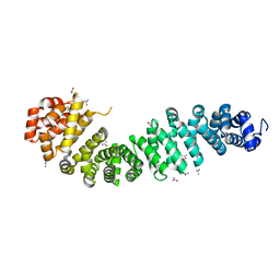
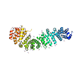
Crystal structure of mouse catenin beta-59 in 2.4M urea.
Descriptor: Catenin beta-1, UREA
Authors:Wang, C, Zhang, G.Y.
Deposit date:2012-04-26
Release date:2013-05-01
Last modified:2023-09-13
Method:X-RAY DIFFRACTION (1.9 Å)
Cite:Crystal structure of mouse catenin beta-59 in 2.4M urea.
TO BE PUBLISHED
4EVA
DownloadVisualize
BU of 4eva by Molmil
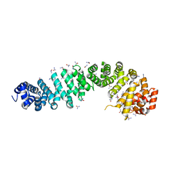
Crystal Structure of Mouse Catenin beta-59 in 5.6M urea
Descriptor: Catenin beta-1, UREA
Authors:Wang, C, Zhang, G.Y.
Deposit date:2012-04-26
Release date:2013-05-01
Last modified:2024-02-28
Method:X-RAY DIFFRACTION (2.003 Å)
Cite:Crystal Structure of Mouse Catenin beta-59 in 5.6M urea
TO BE PUBLISHED
4EVT
DownloadVisualize
BU of 4evt by Molmil
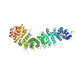
Crystal Structure of Mouse Catenin beta-59 in 8.3M urea
Descriptor: Catenin beta-1, UREA
Authors:Wang, C, Zhang, G.Y.
Deposit date:2012-04-26
Release date:2013-05-01
Last modified:2024-02-28
Method:X-RAY DIFFRACTION (2.339 Å)
Cite:Crystal Structure of Mouse Catenin beta-59 in 8.3M urea
TO BE PUBLISHED
4EV9
DownloadVisualize
BU of 4ev9 by Molmil
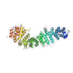
Crystal Structure of Mouse Catenin beta-59 in 4.0M urea
Descriptor: Catenin beta-1, UREA
Authors:Wang, C, Zhang, G.Y.
Deposit date:2012-04-26
Release date:2013-05-01
Last modified:2024-02-28
Method:X-RAY DIFFRACTION (2.1 Å)
Cite:Crystal Structure of Mouse Catenin beta-59 in 4.0M urea
TO BE PUBLISHED
4EVP
DownloadVisualize
BU of 4evp by Molmil
Crystal Structure of Mouse Catenin beta-59 in 7.2M urea
Descriptor: Catenin beta-1, UREA
Authors:Wang, C, Zhang, G.Y.
Deposit date:2012-04-26
Release date:2013-05-01
Last modified:2024-02-28
Method:X-RAY DIFFRACTION (2.255 Å)
Cite:Crystal Structure of Mouse Catenin beta-59 in 7.2M urea
TO BE PUBLISHED
4QU1
DownloadVisualize
BU of 4qu1 by Molmil
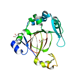
Crystal structure of human JMJD5 jmj-c domain
Descriptor: 2-OXOGLUTARIC ACID, GLYCEROL, Lysine-specific demethylase 8, ...
Authors:Liu, H.L, Wang, C, Zhang, G.Y.
Deposit date:2014-07-10
Release date:2015-07-15
Last modified:2024-02-28
Method:X-RAY DIFFRACTION (1.57 Å)
Cite:The Histone Arginine Demethylase JMJD5
To be Published
4QU2
DownloadVisualize
BU of 4qu2 by Molmil
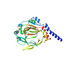
The structure of JMJD7 with alpha-KG
Descriptor: 2-OXOGLUTARIC ACID, JmjC domain-containing protein 7, NICKEL (II) ION
Authors:Liu, H.L, Wang, C, Zhang, G.Y.
Deposit date:2014-07-10
Release date:2015-07-15
Last modified:2024-02-28
Method:X-RAY DIFFRACTION (2.7 Å)
Cite:The structure of JMJD7 with alpha-KG
To be Published
4QSZ
DownloadVisualize
BU of 4qsz by Molmil
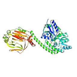
Crystal structure of mouse JMJd7 fused with maltose-binding protein
Descriptor: CITRATE ANION, Maltose-binding periplasmic protein, JmjC domain-containing protein 7 chimera, ...
Authors:Liu, H, Wang, C, Zhang, G.Y.
Deposit date:2014-07-06
Release date:2015-07-08
Last modified:2020-07-29
Method:X-RAY DIFFRACTION (2.86 Å)
Cite:Crystal structure of mouse JMJd7 fused with maltose-binding protein
To be Published
8U77
DownloadVisualize
BU of 8u77 by Molmil
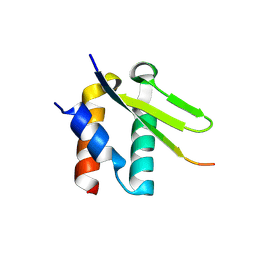
Crystal structure of Taf14 in complex with Yng1
Descriptor: Protein YNG1, Transcription initiation factor TFIID subunit 14
Authors:Nguyen, M.C, Wei, P.C, Zhang, G.Y, Kutateladze, T.G.
Deposit date:2023-09-14
Release date:2024-08-21
Method:X-RAY DIFFRACTION (1.93 Å)
Cite:Molecular insight into interactions between the Taf14, Yng1 and Sas3 subunits of the NuA3 complex.
Nat Commun, 15, 2024
1CLK
DownloadVisualize
BU of 1clk by Molmil
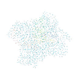
CRYSTAL STRUCTURE OF STREPTOMYCES DIASTATICUS NO.7 STRAIN M1033 XYLOSE ISOMERASE AT 1.9 A RESOLUTION WITH PSEUDO-I222 SPACE GROUP
Descriptor: COBALT (II) ION, MAGNESIUM ION, XYLOSE ISOMERASE
Authors:Niu, L, Teng, M, Zhu, X, Gong, W.
Deposit date:1999-04-29
Release date:2000-05-03
Last modified:2023-08-09
Method:X-RAY DIFFRACTION (1.9 Å)
Cite:Structure of xylose isomerase from Streptomyces diastaticus no. 7 strain M1033 at 1.85 A resolution.
Acta Crystallogr.,Sect.D, 56, 2000

225158

PDB entries from 2024-09-18

PDB statisticsPDBj update infoContact PDBjnumon