3RLZ
 
 | |
4YBH
 | |
1QX2
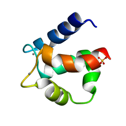
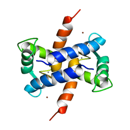
 | | X-ray Structure of Calcium-loaded Calbindomodulin (A Calbindin D9k Re-engineered to Undergo a Conformational Opening) at 1.44 A Resolution | | 分子名称: | CALCIUM ION, Vitamin D-dependent calcium-binding protein, intestinal, ... | | 著者 | Bunick, C.G, Nelson, M.R, Mangahas, S, Mizoue, L.S, Bunick, G.J, Chazin, W.J. | | 登録日 | 2003-09-04 | | 公開日 | 2004-05-25 | | 最終更新日 | 2021-10-27 | | 実験手法 | X-RAY DIFFRACTION (1.44 Å) | | 主引用文献 | Designing Sequence to Control Protein Function in an EF-Hand Protein
J.Am.Chem.Soc., 126, 2004
|
|
1QLK
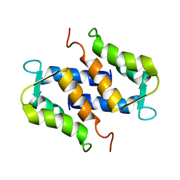
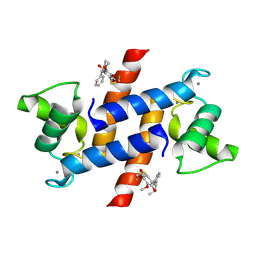
 | | SOLUTION STRUCTURE OF CA(2+)-LOADED RAT S100B (BETABETA) NMR, 20 STRUCTURES | | 分子名称: | CALCIUM ION, S-100 PROTEIN | | 著者 | Drohat, A.C, Baldisseri, D.M, Rustandi, R.R, Weber, D.J. | | 登録日 | 1997-09-26 | | 公開日 | 1998-11-11 | | 最終更新日 | 2024-05-22 | | 実験手法 | SOLUTION NMR | | 主引用文献 | Solution structure of calcium-bound rat S100B(betabeta) as determined by nuclear magnetic resonance spectroscopy,.
Biochemistry, 37, 1998
|
|
1QLS
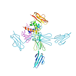
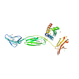
 | | S100C (S100A11),OR CALGIZZARIN, IN COMPLEX WITH ANNEXIN I N-TERMINUS | | 分子名称: | ANNEXIN I, CALCIUM ION, S100C PROTEIN | | 著者 | Rety, S, Sopkova, J, Renouard, M, Osterloh, D, Gerke, V, Russo-Marie, F, Lewit-Bentley, A. | | 登録日 | 1999-09-15 | | 公開日 | 2000-02-25 | | 最終更新日 | 2023-12-13 | | 実験手法 | X-RAY DIFFRACTION (2.3 Å) | | 主引用文献 | Structural Basis of the Ca2+ Dependent Association between S100C (S100A11) and its Target, the N-Terminal Part of Annexin I
Structure, 8, 2000
|
|
1SYM
 | |
2H61
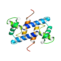
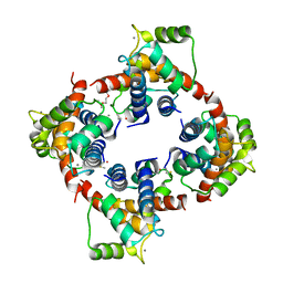 | | X-ray structure of human Ca2+-loaded S100B | | 分子名称: | CALCIUM ION, Protein S100-B, TETRAETHYLENE GLYCOL | | 著者 | Ostendorp, T, Heizmann, C.W, Kroneck, P.M.H, Fritz, G. | | 登録日 | 2006-05-30 | | 公開日 | 2007-06-05 | | 最終更新日 | 2023-08-30 | | 実験手法 | X-RAY DIFFRACTION (1.9 Å) | | 主引用文献 | Structural and functional insights into RAGE activation by multimeric S100B.
Embo J., 26, 2007
|
|
1UWO
 | |
4PE7
 | | Crystal Structure of Calcium-loaded S100B bound to SC1982 | | 分子名称: | (1beta,6beta,7beta,8alpha,9beta,10alpha,13alpha,14R,16beta)-1,6,7,14-tetrahydroxy-7,20-epoxykauran-15-one, CALCIUM ION, Protein S100-B | | 著者 | Cavalier, M.C, Pierce, A.D, Wilder, P.T, Neau, D, Toth, E.A, Weber, D.J. | | 登録日 | 2014-04-22 | | 公開日 | 2014-10-15 | | 最終更新日 | 2023-12-27 | | 実験手法 | X-RAY DIFFRACTION (1.652 Å) | | 主引用文献 | Covalent Small Molecule Inhibitors of Ca(2+)-Bound S100B.
Biochemistry, 53, 2014
|
|
4P2Y
 | |
4PE1
 | | Crystal Structure of Calcium-loaded S100B bound to SC124 | | 分子名称: | CALCIUM ION, DIETHYLCARBAMODITHIOIC ACID, Protein S100-B | | 著者 | Cavalier, M.C, Pierce, A.D, Wilder, P.T, Neau, D, Toth, E.A, Weber, D.J. | | 登録日 | 2014-04-22 | | 公開日 | 2014-10-15 | | 最終更新日 | 2023-09-27 | | 実験手法 | X-RAY DIFFRACTION (1.576 Å) | | 主引用文献 | Covalent Small Molecule Inhibitors of Ca(2+)-Bound S100B.
Biochemistry, 53, 2014
|
|
4PE0
 | | Crystal Structure of Calcium-loaded S100B bound to SBi4434 | | 分子名称: | 2-[(2-hydroxyethyl)sulfanyl]naphthalene-1,4-dione, CALCIUM ION, Protein S100-B | | 著者 | Cavalier, M.C, Pierce, P.D, Wilder, P.T, Neau, D, Toth, E.A, Weber, D.J. | | 登録日 | 2014-04-22 | | 公開日 | 2014-11-05 | | 最終更新日 | 2023-09-27 | | 実験手法 | X-RAY DIFFRACTION (1.08 Å) | | 主引用文献 | Covalent Small Molecule Inhibitors of Ca(2+)-Bound S100B.
Biochemistry, 53, 2014
|
|
4PDZ
 | | Crystal Structure of Calcium-loaded S100B bound to SBi4172 | | 分子名称: | 1,2-dimethoxy-12-methyl[1,3]benzodioxolo[5,6-c]phenanthridin-12-ium, CALCIUM ION, Protein S100-B | | 著者 | Cavalier, M.C, Pierce, A.D, Wilder, P.T, Neau, D, Toth, E.A, Weber, D.J. | | 登録日 | 2014-04-22 | | 公開日 | 2014-10-15 | | 最終更新日 | 2023-09-27 | | 実験手法 | X-RAY DIFFRACTION (1.73 Å) | | 主引用文献 | Covalent Small Molecule Inhibitors of Ca(2+)-Bound S100B.
Biochemistry, 53, 2014
|
|
4PE4
 | | Crystal Structure of Calcium-loaded S100B bound to SC1475 | | 分子名称: | 2,3-dimethoxy-5-[(1S)-1-phenylpropyl]benzene-1,4-diol, CALCIUM ION, Protein S100-B | | 著者 | Cavalier, M.C, Pierce, A.D, Wilder, P.T, Neau, D, Toth, E.A, Weber, D.J. | | 登録日 | 2014-04-22 | | 公開日 | 2014-10-15 | | 最終更新日 | 2023-12-27 | | 実験手法 | X-RAY DIFFRACTION (2.178 Å) | | 主引用文献 | Covalent Small Molecule Inhibitors of Ca(2+)-Bound S100B.
Biochemistry, 53, 2014
|
|
3GK2
 | | X-ray structure of bovine SBi279,Ca(2+)-S100B | | 分子名称: | (Z)-2-[2-(4-methylpiperazin-1-yl)benzyl]diazenecarbothioamide, CACODYLATE ION, CALCIUM ION, ... | | 著者 | Charpentier, T.H, Weber, D.J, Toth, E.A. | | 登録日 | 2009-03-09 | | 公開日 | 2009-06-09 | | 最終更新日 | 2023-09-06 | | 実験手法 | X-RAY DIFFRACTION (1.984 Å) | | 主引用文献 | Small molecules bound to unique sites in the target protein binding cleft of calcium-bound S100B as characterized by nuclear magnetic resonance and X-ray crystallography.
Biochemistry, 48, 2009
|
|
3GK4
 | | X-ray structure of bovine SBi523,Ca(2+)-S100B | | 分子名称: | CALCIUM ION, Protein S100-B, ethyl 5-{[(1R)-1-(ethoxycarbonyl)-2-oxopropyl]sulfanyl}-1,2-dihydro[1,2,3]triazolo[1,5-a]quinazoline-3-carboxylate | | 著者 | Charpentier, T.H, Weber, D.J, Toth, E.A. | | 登録日 | 2009-03-09 | | 公開日 | 2009-06-09 | | 最終更新日 | 2023-09-06 | | 実験手法 | X-RAY DIFFRACTION (1.9 Å) | | 主引用文献 | Small molecules bound to unique sites in the target protein binding cleft of calcium-bound S100B as characterized by nuclear magnetic resonance and X-ray crystallography.
Biochemistry, 48, 2009
|
|
3GK1
 | | X-ray structure of bovine SBi132,Ca(2+)-S100B | | 分子名称: | 2-[(5-hex-1-yn-1-ylfuran-2-yl)carbonyl]-N-methylhydrazinecarbothioamide, CACODYLATE ION, CALCIUM ION, ... | | 著者 | Charpentier, T.H, Weber, D.J, Toth, E.A. | | 登録日 | 2009-03-09 | | 公開日 | 2009-06-09 | | 最終更新日 | 2023-09-06 | | 実験手法 | X-RAY DIFFRACTION (2.1 Å) | | 主引用文献 | Small molecules bound to unique sites in the target protein binding cleft of calcium-bound S100B as characterized by nuclear magnetic resonance and X-ray crystallography.
Biochemistry, 48, 2009
|
|
3HCM
 | | Crystal structure of human S100B in complex with S45 | | 分子名称: | (3R)-3-[3-(4-chlorophenyl)-1,2,4-oxadiazol-5-yl]piperidine, ACETATE ION, CALCIUM ION, ... | | 著者 | Mangani, S, Cesari, L. | | 登録日 | 2009-05-06 | | 公開日 | 2010-02-02 | | 最終更新日 | 2023-11-01 | | 実験手法 | X-RAY DIFFRACTION (2 Å) | | 主引用文献 | Fragmenting the S100B-p53 Interaction: Combined Virtual/Biophysical Screening Approaches to Identify Ligands
Chemmedchem, 5, 2010
|
|
3IQQ
 | | X-ray structure of bovine TRTK12-Ca(2+)-S100B | | 分子名称: | CALCIUM ION, Protein S100-B, TRTK12 peptide, ... | | 著者 | Charpentier, T.H, Weber, D.J, Toth, E.A. | | 登録日 | 2009-08-20 | | 公開日 | 2010-02-02 | | 最終更新日 | 2023-09-06 | | 実験手法 | X-RAY DIFFRACTION (2.01 Å) | | 主引用文献 | The Effects of CapZ Peptide (TRTK-12) Binding to S100B-Ca(2+) as Examined by NMR and X-ray Crystallography
J.Mol.Biol., 396, 2010
|
|
3IQO
 | |
3ICB
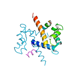
 | | THE REFINED STRUCTURE OF VITAMIN D-DEPENDENT CALCIUM-BINDING PROTEIN FROM BOVINE INTESTINE. MOLECULAR DETAILS, ION BINDING, AND IMPLICATIONS FOR THE STRUCTURE OF OTHER CALCIUM-BINDING PROTEINS | | 分子名称: | CALCIUM ION, CALCIUM-BINDING PROTEIN, SULFATE ION | | 著者 | Szebenyi, D.M.E, Moffat, K. | | 登録日 | 1986-09-09 | | 公開日 | 1986-10-24 | | 最終更新日 | 2024-02-21 | | 実験手法 | X-RAY DIFFRACTION (2.3 Å) | | 主引用文献 | The refined structure of vitamin D-dependent calcium-binding protein from bovine intestine. Molecular details, ion binding, and implications for the structure of other calcium-binding proteins.
J.Biol.Chem., 261, 1986
|
|
1XYD
 | | NMR Solution Structure of Rat Zinc-Calcium-S100B, 20 Structures | | 分子名称: | CALCIUM ION, S-100 protein, beta chain, ... | | 著者 | Wilder, P.T, Varney, K.M, Weber, D.J. | | 登録日 | 2004-11-09 | | 公開日 | 2005-06-07 | | 最終更新日 | 2024-05-22 | | 実験手法 | SOLUTION NMR | | 主引用文献 | Solution Structure of Zinc- and Calcium-Bound Rat S100B as Determined by Nuclear Magnetic Resonance Spectroscopy
Biochemistry, 44, 2005
|
|
1ZFS
 | |
3CR5
 | |
3CR2
 | |


