6AS9
 
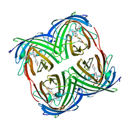
 | | Filamentous Assembly of Green Fluorescent Protein Supported by a C-terminal fusion of 18-residues, viewed in space group P212121 form 2 | | 分子名称: | (4S)-2-METHYL-2,4-PENTANEDIOL, ACETATE ION, Green fluorescent protein | | 著者 | Sawaya, M.R, Heller, D.M, McPartland, L, Hochschild, A, Eisenberg, D.S. | | 登録日 | 2017-08-23 | | 公開日 | 2018-05-30 | | 最終更新日 | 2019-11-20 | | 実験手法 | X-RAY DIFFRACTION (1.75 Å) | | 主引用文献 | Atomic insights into the genesis of cellular filaments by globular proteins.
Nat. Struct. Mol. Biol., 25, 2018
|
|
3I19
 | |
6B9C
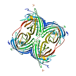
 | | Superfolder Green Fluorescent Protein with 4-nitro-L-phenylalanine at the chromophore (position 66) | | 分子名称: | CARBON DIOXIDE, Green fluorescent protein | | 著者 | Phillips-Piro, C.M, Brewer, S.H, Olenginski, G.M, Piacentini, J. | | 登録日 | 2017-10-10 | | 公開日 | 2018-10-17 | | 最終更新日 | 2023-11-15 | | 実験手法 | X-RAY DIFFRACTION (1.695 Å) | | 主引用文献 | Structural and spectrophotometric investigation of two unnatural amino-acid altered chromophores in the superfolder green fluorescent protein
Acta Crystallogr.,Sect.D, 2021
|
|
6B7R
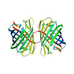
 | | Truncated strand 11-less green fluorescent protein | | 分子名称: | 2-[N-CYCLOHEXYLAMINO]ETHANE SULFONIC ACID, Green fluorescent protein | | 著者 | Deng, A, Boxer, S.G. | | 登録日 | 2017-10-05 | | 公開日 | 2017-12-27 | | 最終更新日 | 2023-11-15 | | 実験手法 | X-RAY DIFFRACTION (1.73 Å) | | 主引用文献 | Structural Insight into the Photochemistry of Split Green Fluorescent Proteins: A Unique Role for a His-Tag.
J. Am. Chem. Soc., 140, 2018
|
|
3IP2
 | | Crystal structure of red fluorescent protein Neptune at pH 7.0 | | 分子名称: | Neptune red fluorescent protein | | 著者 | Lin, M.Z, McKeown, M.R, Ng, H.L, Aguilera, T.A, Shaner, N.C, Ma, W, Adams, S.R, Campbell, R.E, Alber, T, Tsien, R.Y. | | 登録日 | 2009-08-15 | | 公開日 | 2009-12-15 | | 最終更新日 | 2023-11-22 | | 実験手法 | X-RAY DIFFRACTION (1.6 Å) | | 主引用文献 | Autofluorescent proteins with excitation in the optical window for intravital imaging in mammals.
Chem.Biol., 16, 2009
|
|
3IR8
 | | Red fluorescent protein mKeima at pH 7.0 | | 分子名称: | Large stokes shift fluorescent protein | | 著者 | Henderson, J.N, Osborn, M.F, Koon, N, Gepshtein, R, Huppert, D, Remington, S.J. | | 登録日 | 2009-08-21 | | 公開日 | 2009-09-08 | | 最終更新日 | 2023-11-15 | | 実験手法 | X-RAY DIFFRACTION (1.63 Å) | | 主引用文献 | Excited state proton transfer in the red fluorescent protein mKeima.
J.Am.Chem.Soc., 131, 2009
|
|
6CIU
 | | Structure of a Thr-rich interface in an Azami Green tetramer | | 分子名称: | Azami-Green | | 著者 | Oi, C, Lim, C.S, Knecht, K.M, Xiong, Y, Regan, L. | | 登録日 | 2018-02-25 | | 公開日 | 2018-09-26 | | 最終更新日 | 2023-11-15 | | 実験手法 | X-RAY DIFFRACTION (1.7 Å) | | 主引用文献 | A threonine zipper that mediates protein-protein interactions: Structure and prediction.
Protein Sci., 27, 2018
|
|
6D39
 | |
3K1K
 | | Green fluorescent protein bound to enhancer nanobody | | 分子名称: | Enhancer, Green Fluorescent Protein | | 著者 | Kirchhofer, A, Helma, J, Schmidthals, K, Frauer, C, Cui, S, Karcher, A, Pellis, M, Muyldermans, S, Delucci, C.C, Cardoso, M.C, Leonhardt, H, Hopfner, K.-P, Rothbauer, U. | | 登録日 | 2009-09-28 | | 公開日 | 2009-12-08 | | 最終更新日 | 2023-11-15 | | 実験手法 | X-RAY DIFFRACTION (2.15 Å) | | 主引用文献 | Modulation of protein properties in living cells using nanobodies
Nat.Struct.Mol.Biol., 17, 2010
|
|
3KCS
 | | Crystal structure of PAmCherry1 in the dark state | | 分子名称: | PAmCherry1 protein | | 著者 | Malashkevich, V.N, Subach, F.V, Zencheck, W.D, Xiao, H, Filonov, G.S, Almo, S.C, Verkhusha, V.V. | | 登録日 | 2009-10-21 | | 公開日 | 2009-11-17 | | 最終更新日 | 2018-01-24 | | 実験手法 | X-RAY DIFFRACTION (1.5 Å) | | 主引用文献 | Photoactivation mechanism of PAmCherry based on crystal structures of the protein in the dark and fluorescent states.
Proc.Natl.Acad.Sci.USA, 106, 2009
|
|
6D38
 | |
3KCT
 | | CRYSTAL STRUCTURE OF PAmCherry1 in the photoactivated state | | 分子名称: | PAmCherry1 protein | | 著者 | Malashkevich, V.N, Subach, F.V, Zencheck, W.D, Xiao, H, Filonov, G.S, Almo, S.C, Verkhusha, V.V. | | 登録日 | 2009-10-21 | | 公開日 | 2009-11-17 | | 最終更新日 | 2018-01-24 | | 実験手法 | X-RAY DIFFRACTION (1.65 Å) | | 主引用文献 | Photoactivation mechanism of PAmCherry based on crystal structures of the protein in the dark and fluorescent states.
Proc.Natl.Acad.Sci.USA, 106, 2009
|
|
6DQ1
 | |
6DEJ
 | |
6DQ0
 | | sfGFP D133 mutated to 4-nitro-L-phenylalanine | | 分子名称: | 1,2-ETHANEDIOL, SODIUM ION, superfolder green fluorescent protein | | 著者 | Phillips-Piro, C.M, Maurici, N, Lee, B. | | 登録日 | 2018-06-10 | | 公開日 | 2018-10-17 | | 最終更新日 | 2023-11-15 | | 実験手法 | X-RAY DIFFRACTION (2.048 Å) | | 主引用文献 | Crystal structures of green fluorescent protein with the unnatural amino acid 4-nitro-L-phenylalanine.
Acta Crystallogr F Struct Biol Commun, 74, 2018
|
|
3LA1
 | |
3LF4
 | |
3LVD
 | |
3LF3
 | |
6EFR
 | | Crystal Structure of iNicSnFR 1.0 | | 分子名称: | iNicSnFR 1.0, a genetically encoded nicotine biosensor,Green fluorescent protein | | 著者 | Shivange, A.V, Borden, P.M. | | 登録日 | 2018-08-17 | | 公開日 | 2019-01-23 | | 最終更新日 | 2023-11-15 | | 実験手法 | X-RAY DIFFRACTION (2.4 Å) | | 主引用文献 | Nicotinic Drugs in the Endoplasmic Reticulum: Beginning the Inside-out Pathway of Addiction and Therapy
J.Gen.Physiol., 2019
|
|
3LVC
 | |
3LVA
 | |
3MGF
 | | Crystal Structure of Monomeric Kusabira-Orange (MKO), Orange-Emitting GFP-like Protein, at pH 7.5 | | 分子名称: | Fluorescent protein | | 著者 | Ebisawa, T, Yamamura, A, Ohtsuka, J, Kameda, Y, Hayakawa, K, Nagata, K, Tanokura, M. | | 登録日 | 2010-04-06 | | 公開日 | 2011-03-16 | | 最終更新日 | 2023-11-15 | | 実験手法 | X-RAY DIFFRACTION (1.8 Å) | | 主引用文献 | Crystal Structure of Monomeric Kusabira-Orange (MKO), Orange-Emitting GFP-like Protein, at pH 7.5
To be Published
|
|
8YDO
 | |
8UBG
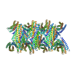 | | DpHF19 filament | | 分子名称: | DpHF19,Green fluorescent protein (Fragment) | | 著者 | Lynch, E.M, Shen, H, Kollman, J.M, Baker, D. | | 登録日 | 2023-09-22 | | 公開日 | 2024-04-10 | | 実験手法 | ELECTRON MICROSCOPY (3.5 Å) | | 主引用文献 | De novo design of pH-responsive self-assembling helical protein filaments.
Nat Nanotechnol, 2024
|
|


