Loading
PDBj
MenuPDBj@FacebookPDBj@TwitterPDBj@YouTubewwPDB FoundationwwPDB
RCSB PDBPDBeBMRBAdv. SearchSearch help
PDB: 7 results

2KY8
DownloadVisualize
BU of 2ky8 by Molmil
Solution structure and dynamic analysis of chicken MBD2 methyl binding domain bound to a target methylated DNA sequence
Descriptor: DNA (5'-D(*GP*AP*GP*CP*(5CM)P*GP*AP*TP*(TED)P*CP*C)-3'), DNA (5'-D(*GP*GP*AP*AP*TP*(5CM)P*GP*GP*CP*(TED)P*C)-3'), MANGANESE (II) ION, ...
Authors:Williams Jr, D.C, Scarsdale Jr, J.N.
Deposit date:2010-05-18
Release date:2011-05-18
Last modified:2020-02-05
Method:SOLUTION NMR
Cite:Solution structure and dynamic analysis of chicken MBD2 methyl binding domain bound to a target-methylated DNA sequence.
Nucleic Acids Res., 39, 2011
2L2L
DownloadVisualize
BU of 2l2l by Molmil
Solution structure of the coiled-coil complex between MBD2 and p66alpha
Descriptor: Methyl-CpG-binding domain protein 2, Transcriptional repressor p66-alpha
Authors:Williams Jr, D.C, Scarsdale Jr, N.
Deposit date:2010-08-20
Release date:2011-05-04
Last modified:2020-02-05
Method:SOLUTION NMR
Cite:p66Alpha-MBD2 coiled-coil interaction and recruitment of Mi-2 are critical for globin gene silencing by the MBD2-NuRD complex.
Proc.Natl.Acad.Sci.USA, 108, 2011
2MB7
DownloadVisualize
BU of 2mb7 by Molmil
Solution structure of MBD3 methylcytosine binding domain
Descriptor: Methyl-CpG-binding domain protein 3
Authors:Williams Jr, D.C, Scarsdale, J.N.
Deposit date:2013-07-26
Release date:2013-12-11
Last modified:2023-06-14
Method:SOLUTION NMR
Cite:Probing the Dynamic Distribution of Bound States for Methylcytosine-binding Domains on DNA.
J.Biol.Chem., 289, 2014
2N2Y
DownloadVisualize
BU of 2n2y by Molmil
Solution structure of the meiosis-expressed gene 1 (Meig1)
Descriptor: Meiosis-expressed gene 1 protein
Authors:Williams Jr, D.C, Walavalkar, N.M, Buchwald, W.A.
Deposit date:2015-05-16
Release date:2016-01-20
Last modified:2023-06-14
Method:SOLUTION NMR
Cite:Dissecting the structural basis of MEIG1 interaction with PACRG.
Sci Rep, 6, 2016
1CL0
DownloadVisualize
BU of 1cl0 by Molmil
CRYSTAL STRUCTURE OF REDUCED THIOREDOXIN REDUCTASE FROM ESCHERICHIA COLI.
Descriptor: FLAVIN-ADENINE DINUCLEOTIDE, THIOREDOXIN REDUCTASE
Authors:Lennon, B.W, Williams Jr, C.H, Ludwig, M.L.
Deposit date:1999-05-04
Release date:1999-12-03
Last modified:2023-08-09
Method:X-RAY DIFFRACTION (2.5 Å)
Cite:Crystal structure of reduced thioredoxin reductase from Escherichia coli: structural flexibility in the isoalloxazine ring of the flavin adenine dinucleotide cofactor.
Protein Sci., 8, 1999
1F6M
DownloadVisualize
BU of 1f6m by Molmil
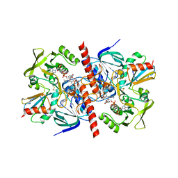
CRYSTAL STRUCTURE OF A COMPLEX BETWEEN THIOREDOXIN REDUCTASE, THIOREDOXIN, AND THE NADP+ ANALOG, AADP+
Descriptor: 3-AMINOPYRIDINE-ADENINE DINUCLEOTIDE PHOSPHATE, FLAVIN-ADENINE DINUCLEOTIDE, THIOREDOXIN 1, ...
Authors:Lennon, B.W, Williams Jr, C.H, Ludwig, M.L.
Deposit date:2000-06-22
Release date:2000-08-30
Last modified:2024-03-13
Method:X-RAY DIFFRACTION (2.95 Å)
Cite:Twists in catalysis: alternating conformations of Escherichia coli thioredoxin reductase.
Science, 289, 2000
2JZH
DownloadVisualize
BU of 2jzh by Molmil
structure of IIB domain of the mannose transporter of E. coli
Descriptor: PTS system mannose-specific EIIAB component
Authors:Komlosh, M, Williams Jr, D.C.
Deposit date:2008-01-08
Release date:2008-02-19
Last modified:2022-03-16
Method:SOLUTION NMR
Cite:Solution NMR Structures of Productive and Non-productive Complexes between the A and B Domains of the Cytoplasmic Subunit of the Mannose Transporter of the Escherichia coli Phosphotransferase System.
J.Biol.Chem., 283, 2008

218853

PDB entries from 2024-04-24

PDB statisticsPDBj update infoContact PDBjnumon