Loading
PDBj
MenuPDBj@FacebookPDBj@TwitterPDBj@YouTubewwPDB FoundationwwPDB
RCSB PDBPDBeBMRBAdv. SearchSearch help
PDB: 491 results

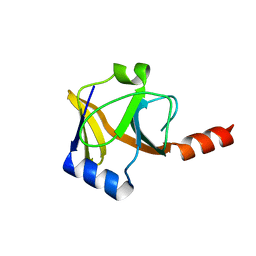
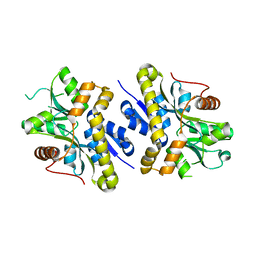
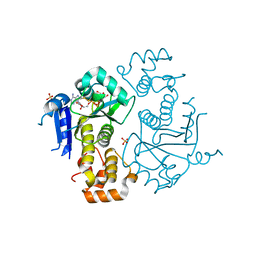
2GUW
DownloadVisualize
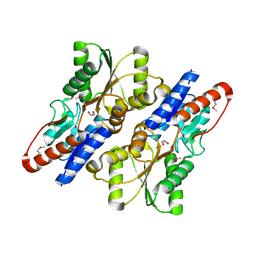
BU of 2guw by Molmil
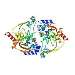
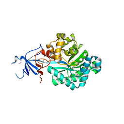
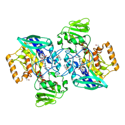
Crystal structure of AMP Nucleosidase from Salmonella typhimurium LT2
Descriptor: AMP nucleosidase
Authors:Rao, K.N, Swaminathan, S, Burley, S.K, New York SGX Research Center for Structural Genomics (NYSGXRC)
Deposit date:2006-05-01
Release date:2006-06-06
Last modified:2024-02-14
Method:X-RAY DIFFRACTION (2.64 Å)
Cite:Crystal structure of AMP Nucleosidase from Salmonella typhimurium LT2
To be Published
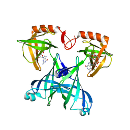
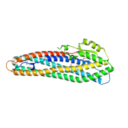
1TXZ
DownloadVisualize
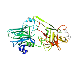
BU of 1txz by Molmil
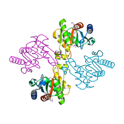
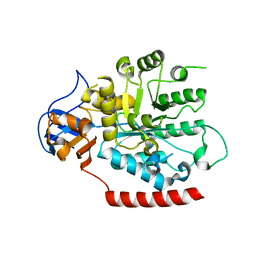
Crystal structure of yeast ymx7, an ADP-ribose-1''-monophosphatase, complexed with ADP-ribose
Descriptor: 1,2-ETHANEDIOL, ADENOSINE-5-DIPHOSPHORIBOSE, Hypothetical 32.1 kDa protein in ADH3-RCA1 intergenic region, ...
Authors:Kumaran, D, Swaminathan, S, Burley, S.K, New York SGX Research Center for Structural Genomics (NYSGXRC)
Deposit date:2004-07-06
Release date:2004-11-30
Last modified:2023-08-23
Method:X-RAY DIFFRACTION (2.05 Å)
Cite:Structure and mechanism of ADP-ribose-1''-monophosphatase (Appr-1''-pase), a ubiquitous cellular processing enzyme.
Protein Sci., 14, 2005
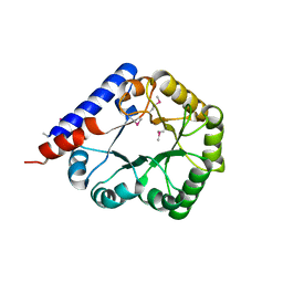
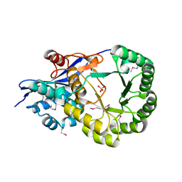
2NYG
DownloadVisualize
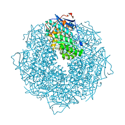
BU of 2nyg by Molmil
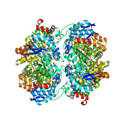
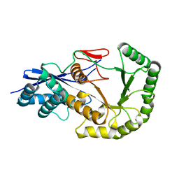
Crystal structure of YokD protein from Bacillus subtilis
Descriptor: COENZYME A, YokD protein
Authors:Madegowda, M, Eswaramoorthy, S, Burley, S.K, Swaminathan, S, New York SGX Research Center for Structural Genomics (NYSGXRC)
Deposit date:2006-11-20
Release date:2006-12-12
Last modified:2023-12-27
Method:X-RAY DIFFRACTION (2.6 Å)
Cite:Crystal structure of YokD protein from Bacillus subtilis
To be Published
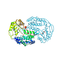
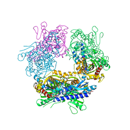
2HAF
DownloadVisualize
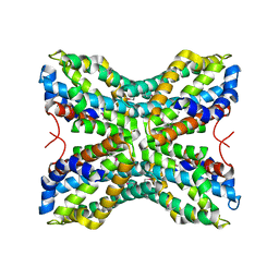
BU of 2haf by Molmil
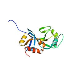
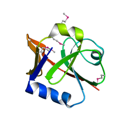
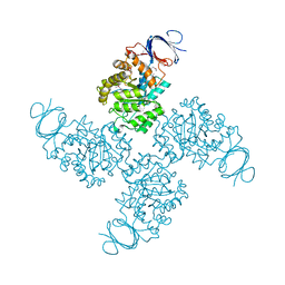
Crystal structure of a putative translation repressor from Vibrio cholerae
Descriptor: Putative translation repressor
Authors:Sugadev, R, Seetharaman, J, Kumaran, D, Swaminathan, S, Burley, S.K, New York SGX Research Center for Structural Genomics (NYSGXRC)
Deposit date:2006-06-12
Release date:2006-07-04
Last modified:2024-02-14
Method:X-RAY DIFFRACTION (2.88 Å)
Cite:Crystal structure of a putative translation repressor from Vibrio cholerae
To be Published
2HAE
DownloadVisualize
BU of 2hae by Molmil
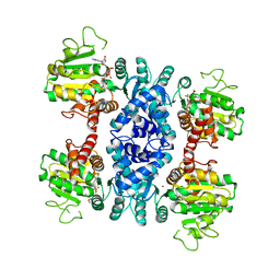
Crystal structure of a putative malic enzyme (malate oxidoreductase)
Descriptor: Malate oxidoreductase, NICOTINAMIDE-ADENINE-DINUCLEOTIDE, ZINC ION
Authors:Seetharaman, J, Swaminathan, S, Burley, S.K, New York SGX Research Center for Structural Genomics (NYSGXRC)
Deposit date:2006-06-12
Release date:2006-07-04
Last modified:2023-08-30
Method:X-RAY DIFFRACTION (3.13 Å)
Cite:Crystal structure of a putative malic enzyme (malate oxidoreductase)
To be Published
3NZG
DownloadVisualize
BU of 3nzg by Molmil
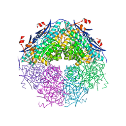
Crystal structure of a putative racemase with Mg ion
Descriptor: GLYCEROL, MAGNESIUM ION, Putative racemase
Authors:Eswaramoorthy, S, Raparia, E, Burley, S.K, Swaminathan, S, New York SGX Research Center for Structural Genomics (NYSGXRC)
Deposit date:2010-07-16
Release date:2010-08-11
Last modified:2023-11-22
Method:X-RAY DIFFRACTION (2 Å)
Cite:Crystal structure of a putative racemase with Mg ion
TO BE PUBLISHED
2OOF
DownloadVisualize
BU of 2oof by Molmil
The crystal structure of 4-imidazolone-5-propanoate amidohydrolase from environmental sample
Descriptor: 4-imidazolone-5-propanoate amidohydrolase, FE (III) ION
Authors:Tyagi, R, Eswaramoorthy, S, Burley, S.K, Swaminathan, S, New York SGX Research Center for Structural Genomics (NYSGXRC)
Deposit date:2007-01-25
Release date:2007-02-20
Last modified:2023-12-27
Method:X-RAY DIFFRACTION (2.2 Å)
Cite:The crystal structure of 4-imidazolone-5-propanoate amidohydrolase from environmental sample
To be Published
2PB9
DownloadVisualize
BU of 2pb9 by Molmil
Crystal structure of C-terminal domain of phosphomethylpyrimidine kinase
Descriptor: PHOSPHATE ION, Phosphomethylpyrimidine kinase
Authors:Eswaramoorthy, S, Burley, S.K, Swaminathan, S, New York SGX Research Center for Structural Genomics (NYSGXRC)
Deposit date:2007-03-28
Release date:2007-04-10
Last modified:2021-02-03
Method:X-RAY DIFFRACTION (2.7 Å)
Cite:Crystal structure of C-terminal domain of phosphomethylpyrimidine kinase
To be Published
3OVG
DownloadVisualize
BU of 3ovg by Molmil
The crystal structure of an amidohydrolase from Mycoplasma synoviae with Zn ion bound
Descriptor: PHOSPHATE ION, ZINC ION, amidohydrolase
Authors:Zhang, Z, Kumaran, D, Burley, S.K, Swaminathan, S, New York SGX Research Center for Structural Genomics (NYSGXRC)
Deposit date:2010-09-16
Release date:2010-10-13
Last modified:2023-12-06
Method:X-RAY DIFFRACTION (2.059 Å)
Cite:The crystal structure of an amidohydrolase from Mycoplasma synoviae with Zn ion bound
To be Published
2P2E
DownloadVisualize
BU of 2p2e by Molmil
Crystal structure of a putative Fe-S biosynthesis protein from Lactobacillus salivarius with novel protein fold
Descriptor: Putative Fe-S biosynthesis protein
Authors:Agarwal, R, Burley, S.K, Swaminathan, S, New York SGX Research Center for Structural Genomics (NYSGXRC)
Deposit date:2007-03-07
Release date:2007-03-20
Last modified:2021-02-03
Method:X-RAY DIFFRACTION (2.48 Å)
Cite:Crystal structure of a putative Fe-S biosynthesis protein from Lactobacillus salivarius with novel protein fold
To be Published
1VQW
DownloadVisualize
BU of 1vqw by Molmil
Crystal structure of a protein with similarity to flavin-containing monooxygenases and to mammalian dimethylalanine monooxygenases
Descriptor: 4-(2-HYDROXYETHYL)-1-PIPERAZINE ETHANESULFONIC ACID, FLAVIN-ADENINE DINUCLEOTIDE, PROTEIN WITH SIMILARITY TO FLAVIN-CONTAINING MONOOXYGENASES AND TO MAMMALIAN DIMETHYLALANINE MONOOXYGENASES
Authors:Eswaramoorthy, S, Swaminathan, S, Burley, S.K, New York SGX Research Center for Structural Genomics (NYSGXRC)
Deposit date:2005-01-05
Release date:2005-01-11
Last modified:2023-12-27
Method:X-RAY DIFFRACTION (2.4 Å)
Cite:Mechanism of action of a flavin-containing monooxygenase.
Proc.Natl.Acad.Sci.Usa, 103, 2006
2PAJ
DownloadVisualize
BU of 2paj by Molmil
Crystal structure of an amidohydrolase from an environmental sample of Sargasso sea
Descriptor: ZINC ION, putative cytosine/guanine deaminase
Authors:Agarwal, R, Burley, S.K, Swaminathan, S, New York SGX Research Center for Structural Genomics (NYSGXRC)
Deposit date:2007-03-27
Release date:2007-04-03
Last modified:2024-02-21
Method:X-RAY DIFFRACTION (2.7 Å)
Cite:Discovery and structure determination of the orphan enzyme isoxanthopterin deaminase.
Biochemistry, 49, 2010
2PGE
DownloadVisualize
BU of 2pge by Molmil
Crystal structure of MenC from Desulfotalea psychrophila LSv54
Descriptor: MenC
Authors:Agarwal, R, Burley, S.K, Swaminathan, S, New York SGX Research Center for Structural Genomics (NYSGXRC)
Deposit date:2007-04-09
Release date:2007-04-24
Last modified:2024-02-21
Method:X-RAY DIFFRACTION (1.6 Å)
Cite:Loss of quaternary structure is associated with rapid sequence divergence in the OSBS family.
Proc.Natl.Acad.Sci.USA, 111, 2014
2QS8
DownloadVisualize
BU of 2qs8 by Molmil
Crystal structure of a Xaa-Pro dipeptidase with bound methionine in the active site
Descriptor: MAGNESIUM ION, METHIONINE, Xaa-Pro Dipeptidase
Authors:Kumaran, D, Burley, S.K, Swaminathan, S, New York SGX Research Center for Structural Genomics (NYSGXRC)
Deposit date:2007-07-30
Release date:2007-08-21
Last modified:2021-02-03
Method:X-RAY DIFFRACTION (2.33 Å)
Cite:Functional annotation of two new carboxypeptidases from the amidohydrolase superfamily of enzymes.
Biochemistry, 48, 2009
2QGO
DownloadVisualize
BU of 2qgo by Molmil
Crystal structure of a putative Fe-S biosynthesis protein from Lactobacillus acidophilus
Descriptor: Putative Fe-S biosynthesis protein
Authors:Agarwal, R, Burley, S.K, Swaminathan, S, New York SGX Research Center for Structural Genomics (NYSGXRC)
Deposit date:2007-06-29
Release date:2007-07-31
Last modified:2024-02-21
Method:X-RAY DIFFRACTION (2.04 Å)
Cite:Structural analysis of Fe-S cluster proteins.
To be Published
2RDE
DownloadVisualize
BU of 2rde by Molmil
Crystal structure of VCA0042 complexed with c-di-GMP
Descriptor: 9,9'-[(2R,3R,3aS,5S,7aR,9R,10R,10aS,12S,14aR)-3,5,10,12-tetrahydroxy-5,12-dioxidooctahydro-2H,7H-difuro[3,2-d:3',2'-j][1,3,7,9,2,8]tetraoxadiphosphacyclododecine-2,9-diyl]bis(2-amino-1,9-dihydro-6H-purin-6-one), Uncharacterized protein VCA0042
Authors:Benach, J, Swaminathan, S.S, Tamayo, R, Seetharaman, J, Handelman, S, Forouhar, F, Neely, H, Camilli, A, Hunt, J.F, Northeast Structural Genomics Consortium (NESG)
Deposit date:2007-09-22
Release date:2007-10-30
Last modified:2023-08-30
Method:X-RAY DIFFRACTION (1.92 Å)
Cite:The structural basis of cyclic diguanylate signal transduction by PilZ domains.
Embo J., 26, 2007
3R79
DownloadVisualize
BU of 3r79 by Molmil
Crystal structure of an uncharactertized protein from Agrobacterium tumefaciens
Descriptor: ACETATE ION, PRASEODYMIUM ION, PYRIDOXAL-5'-PHOSPHATE, ...
Authors:Eswaramoorthy, S, Almo, S.C, Swaminathan, S, New York Structural Genomics Research Consortium (NYSGRC)
Deposit date:2011-03-22
Release date:2011-04-06
Last modified:2012-02-22
Method:X-RAY DIFFRACTION (1.9 Å)
Cite:Crystal structure of an uncharactertized protein from Agrobacterium tumefaciens
To be Published
3ROS
DownloadVisualize
BU of 3ros by Molmil
Crystal structure of NAD-dependent aldehyde dehydrogenase from Lactobacillus acidophilus
Descriptor: NAD-dependent aldehyde dehydrogenase, SULFATE ION
Authors:Eswaramoorthy, S, Almo, S.C, Swaminathan, S, New York Structural Genomics Research Consortium (NYSGRC)
Deposit date:2011-04-26
Release date:2011-05-18
Last modified:2011-07-13
Method:X-RAY DIFFRACTION (1.88 Å)
Cite:Crystal structure of NAD-dependent aldehyde dehydrogenase from Lactobacillus acidophilus
To be Published
3ROT
DownloadVisualize
BU of 3rot by Molmil
Crystal structure of ABC sugar transporter (periplasmic sugar binding protein) from Legionella pneumophila
Descriptor: ABC sugar transporter, periplasmic sugar binding protein, GLYCEROL
Authors:Eswaramoorthy, S, Almo, S.C, Swaminathan, S, New York Structural Genomics Research Consortium (NYSGRC)
Deposit date:2011-04-26
Release date:2011-05-18
Last modified:2011-07-13
Method:X-RAY DIFFRACTION (1.91 Å)
Cite:Crystal structure of ABC sugar transporter (periplasmic sugar binding protein) from Legionella pneumophila
To be Published
1YXW
DownloadVisualize
BU of 1yxw by Molmil
A common binding site for disialyllactose and a tri-peptide in the C-fragment of tetanus neurotoxin
Descriptor: GLUTAMIC ACID, TRYPTOPHAN, TYROSINE, ...
Authors:Jayaraman, S, Eswaramoorthy, S, Kumaran, D, Swaminathan, S.
Deposit date:2005-02-22
Release date:2005-03-15
Last modified:2023-08-23
Method:X-RAY DIFFRACTION (2.2 Å)
Cite:Common binding site for disialyllactose and tri-peptide in C-fragment of tetanus neurotoxin
Proteins, 61, 2005
3SSZ
DownloadVisualize
BU of 3ssz by Molmil
The crystal structure of Mandelate racemase/muconate lactonizing enzyme from Rhodobacteraceae bacterium
Descriptor: Mandelate racemase/muconate lactonizing enzyme, N-terminal domain protein, SULFATE ION
Authors:Zhang, Z, Chamala, S, Evans, B, Foti, R, Gizzi, A, Hillerich, B, Kar, A, LaFleur, J, Seidel, R, Villigas, G, Zencheck, W, Almo, S.C, Swaminathan, S, New York Structural Genomics Research Consortium (NYSGRC)
Deposit date:2011-07-08
Release date:2011-08-17
Last modified:2023-09-13
Method:X-RAY DIFFRACTION (2.392 Å)
Cite:The crystal structure of Mandelate racemase/muconate lactonizing enzyme from Rhodobacteraceae bacterium
To be Published
1TYH
DownloadVisualize
BU of 1tyh by Molmil
Crystal Structure of Transcriptional Activator tenA from Bacillus subtilis
Descriptor: Transcriptional activator tenA
Authors:Eswaramoorthy, S, Swaminathan, S, Burley, S.K, New York SGX Research Center for Structural Genomics (NYSGXRC)
Deposit date:2004-07-07
Release date:2004-07-20
Last modified:2021-02-03
Method:X-RAY DIFFRACTION (2.54 Å)
Cite:Crystal Structure of Transcriptional Activator tenA from Bacillus subtilis
To be Published
2NXO
DownloadVisualize
BU of 2nxo by Molmil
Crystal structure of protein SCO4506 from Streptomyces coelicolor, Pfam DUF178
Descriptor: Hypothetical protein SCO4506
Authors:Tyagi, R, Eswaramoorthy, S, Burley, S.K, Swaminathan, S, New York SGX Research Center for Structural Genomics (NYSGXRC)
Deposit date:2006-11-17
Release date:2006-12-19
Last modified:2023-12-27
Method:X-RAY DIFFRACTION (2.04 Å)
Cite:The crystal structure of a hypothetical protein SCO4506 (gene ID: Q9L0T8) from Streptomyces coelicolor to 2.04 Angstrom resolution
To be Published
2NRJ
DownloadVisualize
BU of 2nrj by Molmil
Crystal Structure of Hemolysin binding component from Bacillus cereus
Descriptor: Hbl B protein
Authors:Madegowda, M, Eswaramoorthy, S, Burley, S.K, Swaminathan, S, New York SGX Research Center for Structural Genomics (NYSGXRC)
Deposit date:2006-11-02
Release date:2006-11-14
Last modified:2023-12-27
Method:X-RAY DIFFRACTION (2.03 Å)
Cite:X-ray crystal structure of the B component of Hemolysin BL from Bacillus cereus
Proteins, 71, 2008
2OQH
DownloadVisualize
BU of 2oqh by Molmil
Crystal structure of an isomerase from Streptomyces coelicolor A3(2)
Descriptor: Putative isomerase, SULFATE ION
Authors:Agarwal, R, Burley, S.K, Swaminathan, S, New York SGX Research Center for Structural Genomics (NYSGXRC)
Deposit date:2007-01-31
Release date:2007-02-20
Last modified:2023-12-27
Method:X-RAY DIFFRACTION (1.98 Å)
Cite:Crystal structure of an isomerase from Streptomyces coelicolor
To be Published

220472

PDB entries from 2024-05-29

PDB statisticsPDBj update infoContact PDBjnumon