Loading
PDBj
MenuPDBj@FacebookPDBj@TwitterPDBj@YouTubewwPDB FoundationwwPDB
RCSB PDBPDBeBMRBAdv. SearchSearch help
Search by PDB author
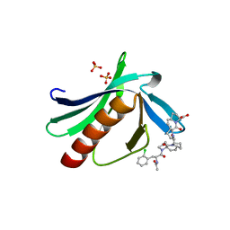
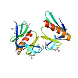
5N9C
DownloadVisualize
BU of 5n9c by Molmil
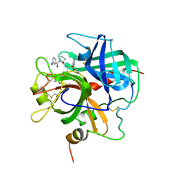
ENAH EVH1 in complex with Ac-[2-Cl-F]-PP-[ProM-1]-OH
Descriptor: Ac-[2-Cl-F]-PP-[ProM-1]-OH, GLYCEROL, NITRATE ION, ...
Authors:Barone, M, Roske, Y.
Deposit date:2017-02-24
Release date:2017-06-14
Last modified:2024-01-31
Method:X-RAY DIFFRACTION (1.16 Å)
Cite:Designed nanomolar small-molecule inhibitors of Ena/VASP EVH1 interaction impair invasion and extravasation of breast cancer cells.
Proc.Natl.Acad.Sci.USA, 117, 2020
5NCF
DownloadVisualize
BU of 5ncf by Molmil
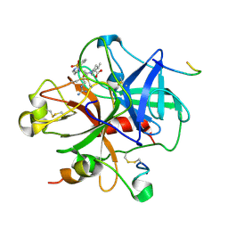
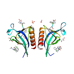
ENAH EVH1 in complex with Ac-[2-Cl-F]-[ProM-2]-[ProM-4]-OH
Descriptor: (1~{S},4~{S},7~{R},10~{R})-14-[(3~{S},6~{R},8~{a}~{S})-1'-[(2~{S})-2-acetamido-3-(2-chlorophenyl)propanoyl]-5-oxidanylidene-spiro[1,2,3,8~{a}-tetrahydroindolizine-6,2'-pyrrolidine]-3-yl]carbonyl-2-oxidanylidene-3,14-diazatricyclo[8.4.0.0^{3,7}]tetradec-8-ene-4-carboxylic acid, GLYCEROL, NITRATE ION, ...
Authors:Barone, M, Roske, Y.
Deposit date:2017-03-04
Release date:2017-06-14
Last modified:2024-01-31
Method:X-RAY DIFFRACTION (1.4 Å)
Cite:Designed nanomolar small-molecule inhibitors of Ena/VASP EVH1 interaction impair invasion and extravasation of breast cancer cells.
Proc.Natl.Acad.Sci.USA, 117, 2020
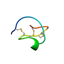
3CTI
DownloadVisualize
BU of 3cti by Molmil
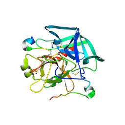
RELAXATION MATRIX REFINEMENT OF THE SOLUTION STRUCTURE OF SQUASH TRYPSIN INHIBITOR
Descriptor: TRYPSIN INHIBITOR
Authors:Nilges, M, Habazettl, J, Bruenger, A.T, Holak, T.A.
Deposit date:1991-03-27
Release date:1992-04-15
Last modified:2022-03-16
Method:SOLUTION NMR
Cite:Relaxation matrix refinement of the solution structure of squash trypsin inhibitor.
J.Mol.Biol., 219, 1991
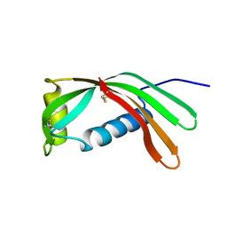
1CEW
DownloadVisualize
BU of 1cew by Molmil
THE 2.0 ANGSTROMS X-RAY CRYSTAL STRUCTURE OF CHICKEN EGG WHITE CYSTATIN AND ITS POSSIBLE MODE OF INTERACTION WITH CYSTEINE PROTEINASES
Descriptor: CYSTATIN
Authors:Bode, W, Musil, D, Huber, R.
Deposit date:1993-04-21
Release date:1994-01-31
Last modified:2011-09-07
Method:X-RAY DIFFRACTION (2 Å)
Cite:The 2.0 A X-ray crystal structure of chicken egg white cystatin and its possible mode of interaction with cysteine proteinases.
EMBO J., 7, 1988
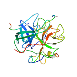
1FPH
DownloadVisualize
BU of 1fph by Molmil
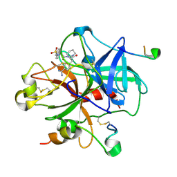
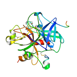
THE INTERACTION OF THROMBIN WITH FIBRINOGEN: A STRUCTURAL BASIS FOR ITS SPECIFICITY
Descriptor: ALPHA-THROMBIN (LARGE SUBUNIT), ALPHA-THROMBIN (SMALL SUBUNIT), FIBRINOPEPTIDE A, ...
Authors:Stubbs, M.T, Bode, W.
Deposit date:1993-04-21
Release date:1994-01-31
Last modified:2012-12-12
Method:X-RAY DIFFRACTION (2.5 Å)
Cite:The interaction of thrombin with fibrinogen. A structural basis for its specificity.
Eur.J.Biochem., 206, 1992
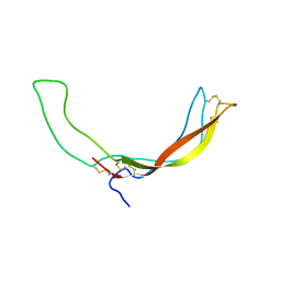
2KD3
DownloadVisualize
BU of 2kd3 by Molmil
NMR structure of the Wnt modulator protein Sclerostin
Descriptor: Sclerostin
Authors:Weidauer, S.E, Mueller, T.D.
Deposit date:2009-01-02
Release date:2009-02-10
Last modified:2022-03-16
Method:SOLUTION NMR
Cite:NMR structure of the Wnt modulator protein Sclerostin
Biochem.Biophys.Res.Commun., 380, 2009
5N9P
DownloadVisualize
BU of 5n9p by Molmil
ENAH EVH1 in complex with Ac-[2-Cl-F]-PP-[ProM-1]-NH2
Descriptor: Ac-[2-Cl-F]-PP-[ProM-1]-NH2, CHLORIDE ION, Protein enabled homolog
Authors:Barone, M, Roske, Y.
Deposit date:2017-02-26
Release date:2017-06-14
Last modified:2024-01-31
Method:X-RAY DIFFRACTION (1.8 Å)
Cite:Designed nanomolar small-molecule inhibitors of Ena/VASP EVH1 interaction impair invasion and extravasation of breast cancer cells.
Proc.Natl.Acad.Sci.USA, 117, 2020
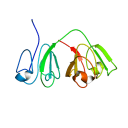
2M3U
DownloadVisualize
BU of 2m3u by Molmil
Solution-state NMR structure of cataract-related human gamma(S)-crystallin point variant G18V
Descriptor: Beta-crystallin S
Authors:Brubaker, W.D, Martin, R.W.
Deposit date:2013-01-25
Release date:2013-11-13
Last modified:2024-05-01
Method:SOLUTION NMR
Cite:Preferential and Specific Binding of Human alpha B-Crystallin to a Cataract-Related Variant of gamma S-Crystallin.
Structure, 21, 2013
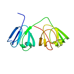
2M3T
DownloadVisualize
BU of 2m3t by Molmil
Solution-state NMR structure of wild-type human gamma(S)-crystallin
Descriptor: Beta-crystallin S
Authors:Brubaker, W.D, Martin, R.W.
Deposit date:2013-01-25
Release date:2013-11-13
Last modified:2024-05-01
Method:SOLUTION NMR
Cite:Preferential and Specific Binding of Human alpha B-Crystallin to a Cataract-Related Variant of gamma S-Crystallin.
Structure, 21, 2013
1PPB
DownloadVisualize
BU of 1ppb by Molmil
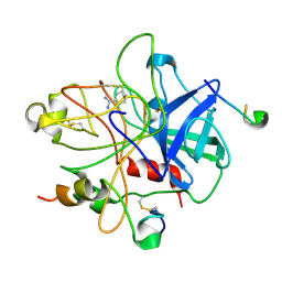
THE REFINED 1.9 ANGSTROMS CRYSTAL STRUCTURE OF HUMAN ALPHA-THROMBIN: INTERACTION WITH D-PHE-PRO-ARG CHLOROMETHYLKETONE AND SIGNIFICANCE OF THE TYR-PRO-PRO-TRP INSERTION SEGMENT
Descriptor: ALPHA-THROMBIN (LARGE SUBUNIT), ALPHA-THROMBIN (SMALL SUBUNIT), D-phenylalanyl-N-[(2S,3S)-6-{[amino(iminio)methyl]amino}-1-chloro-2-hydroxyhexan-3-yl]-L-prolinamide
Authors:Bode, W.
Deposit date:1991-10-24
Release date:1994-01-31
Last modified:2013-02-27
Method:X-RAY DIFFRACTION (1.92 Å)
Cite:The refined 1.9 A crystal structure of human alpha-thrombin: interaction with D-Phe-Pro-Arg chloromethylketone and significance of the Tyr-Pro-Pro-Trp insertion segment.
EMBO J., 8, 1989
1DWC
DownloadVisualize
BU of 1dwc by Molmil
CRYSTALLOGRAPHIC ANALYSIS AT 3.0-ANGSTROMS RESOLUTION OF THE BINDING TO HUMAN THROMBIN OF FOUR ACTIVE SITE-DIRECTED INHIBITORS
Descriptor: ALPHA-THROMBIN (LARGE SUBUNIT), ALPHA-THROMBIN (SMALL SUBUNIT), HIRUDIN IIIA, ...
Authors:Banner, D.W, Hadvary, P.
Deposit date:1992-08-19
Release date:1994-01-31
Last modified:2012-12-12
Method:X-RAY DIFFRACTION (3 Å)
Cite:Crystallographic analysis at 3.0-A resolution of the binding to human thrombin of four active site-directed inhibitors.
J.Biol.Chem., 266, 1991
1DWD
DownloadVisualize
BU of 1dwd by Molmil
CRYSTALLOGRAPHIC ANALYSIS AT 3.0-ANGSTROMS RESOLUTION OF THE BINDING TO HUMAN THROMBIN OF FOUR ACTIVE SITE-DIRECTED INHIBITORS
Descriptor: 1-[N-(naphthalen-2-ylsulfonyl)glycyl-4-carbamimidoyl-D-phenylalanyl]piperidine, ALPHA-THROMBIN (LARGE SUBUNIT), ALPHA-THROMBIN (SMALL SUBUNIT), ...
Authors:Banner, D.W, Hadvary, P.
Deposit date:1992-08-19
Release date:1994-01-31
Last modified:2012-12-12
Method:X-RAY DIFFRACTION (3 Å)
Cite:Crystallographic analysis at 3.0-A resolution of the binding to human thrombin of four active site-directed inhibitors.
J.Biol.Chem., 266, 1991
1DWB
DownloadVisualize
BU of 1dwb by Molmil
CRYSTALLOGRAPHIC ANALYSIS AT 3.0-ANGSTROMS RESOLUTION OF THE BINDING TO HUMAN THROMBIN OF FOUR ACTIVE SITE-DIRECTED INHIBITORS
Descriptor: ALPHA-THROMBIN (LARGE SUBUNIT), ALPHA-THROMBIN (SMALL SUBUNIT), BENZAMIDINE, ...
Authors:Banner, D.W, Hadvary, P.
Deposit date:1992-08-19
Release date:1994-01-31
Last modified:2011-07-13
Method:X-RAY DIFFRACTION (3.16 Å)
Cite:Crystallographic analysis at 3.0-A resolution of the binding to human thrombin of four active site-directed inhibitors.
J.Biol.Chem., 266, 1991
1DWE
DownloadVisualize
BU of 1dwe by Molmil
Crystallographic analysis at 3.0-Angstroms resolution of the binding to human thrombin of four active site-directed inhibitors
Descriptor: ALPHA-THROMBIN heavy chain, ALPHA-THROMBIN light chain, D-phenylalanyl-N-[(2S,3S)-6-{[amino(iminio)methyl]amino}-1-chloro-2-hydroxyhexan-3-yl]-L-prolinamide, ...
Authors:Banner, D.W, Hadvary, P.
Deposit date:1992-08-19
Release date:1994-01-31
Last modified:2013-02-27
Method:X-RAY DIFFRACTION (3 Å)
Cite:Crystallographic analysis at 3.0-A resolution of the binding to human thrombin of four active site-directed inhibitors.
J.Biol.Chem., 266, 1991
2JM5
DownloadVisualize
BU of 2jm5 by Molmil
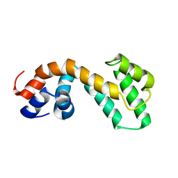
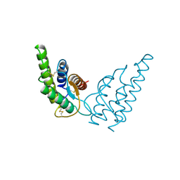
Solution Structure of the RGS domain from human RGS18
Descriptor: Regulator of G-protein signaling 18
Authors:Higman, V.A, Leidert, M, Bray, J, Elkins, J, Soundararajan, M, Doyle, D.A, Gileadi, C, Phillips, C, Schoch, G, Yang, X, Brockmann, C, Schmieder, P, Diehl, A, Sundstrom, M, Arrowsmith, C, Weigelt, J, Edwards, A, Oschkinat, H, Ball, L.J, Structural Genomics Consortium (SGC)
Deposit date:2006-10-11
Release date:2006-10-24
Last modified:2023-12-20
Method:SOLUTION NMR
Cite:Structural diversity in the RGS domain and its interaction with heterotrimeric G protein alpha-subunits.
Proc.Natl.Acad.Sci.Usa, 105, 2008
1DE7
DownloadVisualize
BU of 1de7 by Molmil
INTERACTION OF FACTOR XIII ACTIVATION PEPTIDE WITH ALPHA-THROMBIN: CRYSTAL STRUCTURE OF THE ENZYME-SUBSTRATE COMPLEX
Descriptor: ALPHA-THROMBIN (HEAVY CHAIN), ALPHA-THROMBIN (LIGHT CHAIN), FACTOR XIII ACTIVATION PEPTIDE (28-37), ...
Authors:Sadasivan, C, Yee, V.C.
Deposit date:1999-11-13
Release date:2000-12-13
Last modified:2023-08-09
Method:X-RAY DIFFRACTION (2 Å)
Cite:Interaction of the factor XIII activation peptide with alpha -thrombin. Crystal structure of its enzyme-substrate analog complex.
J.Biol.Chem., 275, 2000
1HIJ
DownloadVisualize
BU of 1hij by Molmil
INTERLEUKIN-4 MUTANT WITH ARG 88 REPLACED WITH GLN (R88Q)
Descriptor: INTERLEUKIN-4
Authors:Mueller, T, Buehner, M.
Deposit date:1995-06-01
Release date:1996-01-29
Last modified:2021-11-03
Method:X-RAY DIFFRACTION (3 Å)
Cite:Human interleukin-4 and variant R88Q: phasing X-ray diffraction data by molecular replacement using X-ray and nuclear magnetic resonance models.
J.Mol.Biol., 247, 1995
1HIK
DownloadVisualize
BU of 1hik by Molmil
INTERLEUKIN-4 (WILD-TYPE)
Descriptor: INTERLEUKIN-4
Authors:Mueller, T, Buehner, M.
Deposit date:1995-06-01
Release date:1996-01-29
Last modified:2011-07-13
Method:X-RAY DIFFRACTION (2.6 Å)
Cite:Human interleukin-4 and variant R88Q: phasing X-ray diffraction data by molecular replacement using X-ray and nuclear magnetic resonance models.
J.Mol.Biol., 247, 1995
2I59
DownloadVisualize
BU of 2i59 by Molmil
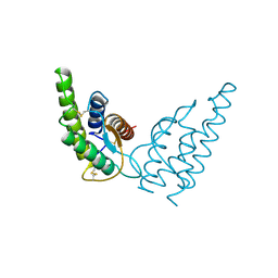
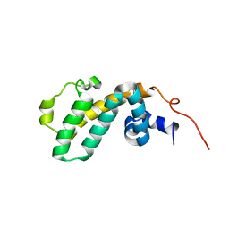
Solution structure of RGS10
Descriptor: Regulator of G-protein signaling 10
Authors:Fedorov, O, Higman, V.A, Diehl, A, Leidert, M, Lemak, A, Schmieder, P, Oschkinat, H, Elkins, J, Soundarajan, M, Doyle, D.A, Arrowsmith, C, Sundstrom, M, Weigelt, J, Edwards, A, Ball, L.J, Structural Genomics Consortium (SGC)
Deposit date:2006-08-24
Release date:2006-10-31
Last modified:2023-06-14
Method:SOLUTION NMR
Cite:Structural diversity in the RGS domain and its interaction with heterotrimeric G protein alpha-subunits.
Proc.Natl.Acad.Sci.Usa, 105, 2008
2JNU
DownloadVisualize
BU of 2jnu by Molmil
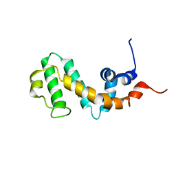
Solution structure of the RGS domain of human RGS14
Descriptor: Regulator of G-protein signaling 14
Authors:Dowler, E.F, Diehl, A, Bray, J, Elkins, J, Soundararajan, M, Doyle, D.A, Gileadi, C, Phillips, C, Schoch, G.A, Yang, X, Brockmann, C, Leidert, M, Rehbein, K, Schmieder, P, Kuhne, R, Higman, V.A, Sundstrom, M, Arrowsmith, C, Weigelt, J, Edwards, A, Oschkinat, H, Ball, L.J, Structural Genomics Consortium (SGC)
Deposit date:2007-02-02
Release date:2007-02-27
Last modified:2023-12-20
Method:SOLUTION NMR
Cite:Structural diversity in the RGS domain and its interaction with heterotrimeric G protein alpha-subunits.
Proc.Natl.Acad.Sci.Usa, 105, 2008
1N1X
DownloadVisualize
BU of 1n1x by Molmil
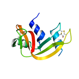
Crystal Structure Analysis of the monomeric [S-carboxyamidomethyl-Cys31, S-carboxyamidomethyl-Cys32] Bovine seminal ribonuclease
Descriptor: Ribonuclease, seminal
Authors:Sica, F, Di Fiore, A, Zagari, A, Mazzarella, L.
Deposit date:2002-10-21
Release date:2003-08-26
Last modified:2011-07-13
Method:X-RAY DIFFRACTION (1.45 Å)
Cite:The unswapped chain of bovine seminal ribonuclease: Crystal structure of the free and liganded monomeric derivative
Proteins, 52, 2003
1PVW
DownloadVisualize
BU of 1pvw by Molmil
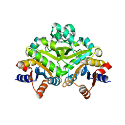
3,4-dihydroxy-2-butanone 4-phosphate synthase from M. jannaschii
Descriptor: 3,4-dihydroxy-2-butanone 4-phosphate synthase, CALCIUM ION, PHOSPHATE ION, ...
Authors:Steinbacher, S, Schiffmann, S, Richter, G, Huber, R, Bacher, A, Fischer, M.
Deposit date:2003-06-29
Release date:2003-11-04
Last modified:2024-03-13
Method:X-RAY DIFFRACTION (2.45 Å)
Cite:Structure of 3,4-Dihydroxy-2-butanone 4-Phosphate Synthase from Methanococcus jannaschii in Complex with Divalent Metal Ions and the Substrate Ribulose 5-Phosphate: IMPLICATIONS FOR THE CATALYTIC MECHANISM
J.Biol.Chem., 278, 2003
1PVY
DownloadVisualize
BU of 1pvy by Molmil
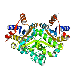
3,4-dihydroxy-2-butanone 4-phosphate synthase from M. jannaschii in complex with ribulose 5-phosphate
Descriptor: 3,4-dihydroxy-2-butanone 4-phosphate synthase, CALCIUM ION, RIBULOSE-5-PHOSPHATE, ...
Authors:Steinbacher, S, Schiffmann, S, Richter, G, Huber, R, Bacher, A, Fischer, M.
Deposit date:2003-06-29
Release date:2003-11-04
Last modified:2023-10-25
Method:X-RAY DIFFRACTION (1.7 Å)
Cite:Structure of 3,4-Dihydroxy-2-butanone 4-Phosphate Synthase from Methanococcus jannaschii in Complex with Divalent Metal Ions and the Substrate Ribulose 5-Phosphate: IMPLICATIONS FOR THE CATALYTIC MECHANISM
J.Biol.Chem., 278, 2003

219140

PDB entries from 2024-05-01

PDB statisticsPDBj update infoContact PDBjnumon