Loading
PDBj
MenuPDBj@FacebookPDBj@TwitterPDBj@YouTubewwPDB FoundationwwPDB
RCSB PDBPDBeBMRBAdv. SearchSearch help
Search by PDB author
2GG1
DownloadVisualize
BU of 2gg1 by Molmil
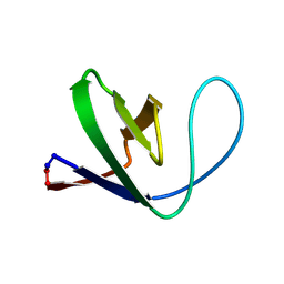
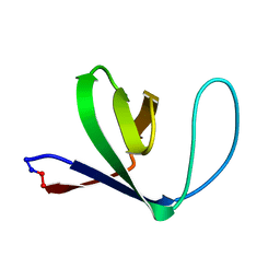
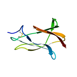
NMR solution structure of domain III of the E-protein of tick-borne Langat flavivirus (includes RDC restraints)
Descriptor: Genome polyprotein
Authors:Mukherjee, M, Dutta, K, White, M.A, Cowburn, D, Fox, R.O.
Deposit date:2006-03-23
Release date:2006-04-25
Last modified:2023-11-29
Method:SOLUTION NMR
Cite:NMR solution structure and backbone dynamics of domain III of the E protein of tick-borne Langat flavivirus suggests a potential site for molecular recognition.
Protein Sci., 15, 2006
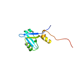
2KJD
DownloadVisualize
BU of 2kjd by Molmil
Solution structure of extended PDZ2 domain from NHERF1 (150-270)
Descriptor: Sodium/hydrogen exchange regulatory cofactor NHE-RF1
Authors:Bhattacharya, S, Cowburn, D, Bu, Z.
Deposit date:2009-05-27
Release date:2009-12-29
Last modified:2022-03-16
Method:SOLUTION NMR
Cite:A dynamic intramolecular conformational switch autoregulates the scaffolding protein NHERF1
To be Published
2KRG
DownloadVisualize
BU of 2krg by Molmil
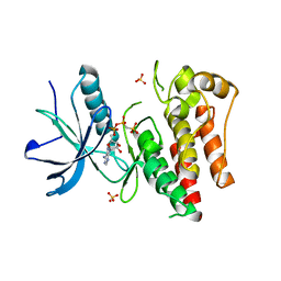
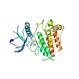
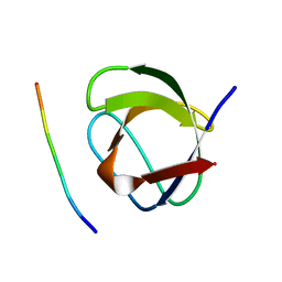
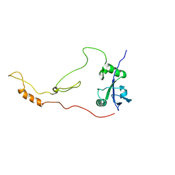
Solution Structure of human sodium/ hydrogen exchange regulatory factor 1(150-358)
Descriptor: Na(+)/H(+) exchange regulatory cofactor NHE-RF1
Authors:Bhattacharya, S, Dai, Z, Li, J, Baxter, S, Callaway, D.J.E, Cowburn, D, Bu, Z.
Deposit date:2009-12-17
Release date:2009-12-29
Last modified:2023-07-26
Method:SOLUTION NMR
Cite:A conformational switch in the scaffolding protein NHERF1 controls autoinhibition and complex formation.
J.Biol.Chem., 285, 2010
1AYB
DownloadVisualize
BU of 1ayb by Molmil
CRYSTAL STRUCTURES OF PEPTIDE COMPLEXES OF THE AMINO-TERMINAL SH2 DOMAIN OF THE SYP TYROSINE PHOSPHATASE
Descriptor: PEPTIDE IRS-1-895, PROTEIN-TYROSINE PHOSPHATASE SYP (N-TERMINAL SH2 DOMAIN)
Authors:Lee, C.-H, Kuriyan, J.
Deposit date:1994-05-15
Release date:1994-08-31
Last modified:2011-07-13
Method:X-RAY DIFFRACTION (3 Å)
Cite:Crystal structures of peptide complexes of the amino-terminal SH2 domain of the Syp tyrosine phosphatase.
Structure, 2, 1994
1AYD
DownloadVisualize
BU of 1ayd by Molmil
CRYSTAL STRUCTURES OF PEPTIDE COMPLEXES OF THE AMINO-TERMINAL SH2 DOMAIN OF THE SYP TYROSINE PHOSPHATASE
Descriptor: PROTEIN-TYROSINE PHOSPHATASE SYP (N-TERMINAL SH2 DOMAIN)
Authors:Lee, C.-H, Kuriyan, J.
Deposit date:1994-05-15
Release date:1994-08-31
Last modified:2024-02-07
Method:X-RAY DIFFRACTION (2.2 Å)
Cite:Crystal structures of peptide complexes of the amino-terminal SH2 domain of the Syp tyrosine phosphatase.
Structure, 2, 1994
1AYC
DownloadVisualize
BU of 1ayc by Molmil
CRYSTAL STRUCTURES OF PEPTIDE COMPLEXES OF THE AMINO-TERMINAL SH2 DOMAIN OF THE SYP TYROSINE PHOSPHATASE
Descriptor: PEPTIDE PDGFR-740, PROTEIN-TYROSINE PHOSPHATASE SYP (N-TERMINAL SH2 DOMAIN)
Authors:Lee, C.-H, Kuriyan, J.
Deposit date:1994-05-15
Release date:1994-08-31
Last modified:2011-07-13
Method:X-RAY DIFFRACTION (2.3 Å)
Cite:Crystal structures of peptide complexes of the amino-terminal SH2 domain of the Syp tyrosine phosphatase.
Structure, 2, 1994
1AYA
DownloadVisualize
BU of 1aya by Molmil
CRYSTAL STRUCTURES OF PEPTIDE COMPLEXES OF THE AMINO-TERMINAL SH2 DOMAIN OF THE SYP TYROSINE PHOSPHATASE
Descriptor: PEPTIDE PDGFR-1009, PROTEIN-TYROSINE PHOSPHATASE SYP (N-TERMINAL SH2 DOMAIN)
Authors:Lee, C.-H, Kuriyan, J.
Deposit date:1994-05-15
Release date:1994-08-31
Last modified:2011-07-13
Method:X-RAY DIFFRACTION (2.05 Å)
Cite:Crystal structures of peptide complexes of the amino-terminal SH2 domain of the Syp tyrosine phosphatase.
Structure, 2, 1994
1SHB
DownloadVisualize
BU of 1shb by Molmil
CRYSTAL STRUCTURE OF THE PHOSPHOTYROSINE RECOGNITION DOMAIN SH2 OF V-SRC COMPLEXED WITH TYROSINE-PHOSPHORYLATED PEPTIDES
Descriptor: PHOSPHOPEPTIDE B, V-SRC SH2 DOMAIN
Authors:Waksman, G, Kuriyan, J.
Deposit date:1992-08-18
Release date:1993-10-31
Last modified:2011-07-13
Method:X-RAY DIFFRACTION (2 Å)
Cite:Crystal structure of the phosphotyrosine recognition domain SH2 of v-src complexed with tyrosine-phosphorylated peptides.
Nature, 358, 1992
1SHA
DownloadVisualize
BU of 1sha by Molmil
CRYSTAL STRUCTURE OF THE PHOSPHOTYROSINE RECOGNITION DOMAIN SH2 OF V-SRC COMPLEXED WITH TYROSINE-PHOSPHORYLATED PEPTIDES
Descriptor: PHOSPHOPEPTIDE A, V-SRC SH2 DOMAIN
Authors:Waksman, G, Kuriyan, J.
Deposit date:1992-08-18
Release date:1993-10-31
Last modified:2011-07-13
Method:X-RAY DIFFRACTION (1.5 Å)
Cite:Crystal structure of the phosphotyrosine recognition domain SH2 of v-src complexed with tyrosine-phosphorylated peptides.
Nature, 358, 1992
1HCS
DownloadVisualize
BU of 1hcs by Molmil
NMR STRUCTURE OF THE HUMAN SRC SH2 DOMAIN COMPLEX
Descriptor: ACETYL-PYEEIE-OH, HUMAN SRC
Authors:Gampe Junior, R.T, Xu, R.X.
Deposit date:1994-09-02
Release date:1995-09-15
Last modified:2022-02-23
Method:SOLUTION NMR
Cite:Solution structure of the human pp60c-src SH2 domain complexed with a phosphorylated tyrosine pentapeptide.
Biochemistry, 34, 1995
1HCT
DownloadVisualize
BU of 1hct by Molmil
NMR STRUCTURE OF THE HUMAN SRC SH2 DOMAIN COMPLEX
Descriptor: ACETYL-PYEEIE-OH, HUMAN SRC
Authors:Gampe Junior, R.T, Xu, R.X.
Deposit date:1994-09-02
Release date:1995-09-15
Last modified:2022-02-23
Method:SOLUTION NMR
Cite:Solution structure of the human pp60c-src SH2 domain complexed with a phosphorylated tyrosine pentapeptide.
Biochemistry, 34, 1995
4J99
DownloadVisualize
BU of 4j99 by Molmil
Crystal Structure of FGF Receptor 2 (FGFR2) Kinase Domain Harboring the Gain-of-Function K659T Mutation.
Descriptor: Fibroblast growth factor receptor 2, MAGNESIUM ION, PHOSPHOMETHYLPHOSPHONIC ACID ADENYLATE ESTER, ...
Authors:Chen, H, Mohammadi, M.
Deposit date:2013-02-15
Release date:2013-08-07
Last modified:2023-09-20
Method:X-RAY DIFFRACTION (1.8474 Å)
Cite:Cracking the Molecular Origin of Intrinsic Tyrosine Kinase Activity through Analysis of Pathogenic Gain-of-Function Mutations.
Cell Rep, 4, 2013
4J95
DownloadVisualize
BU of 4j95 by Molmil
Crystal Structure of FGF Receptor 2 (FGFR2) Kinase Domain Harboring the Pathogenic K659N Mutation Responsible for an Unclassified Craniosynostosis Syndrome in Space Group C2.
Descriptor: Fibroblast growth factor receptor 2, PHOSPHOMETHYLPHOSPHONIC ACID ADENYLATE ESTER, SULFATE ION
Authors:Chen, H, Mohammadi, M.
Deposit date:2013-02-15
Release date:2013-08-07
Last modified:2023-09-20
Method:X-RAY DIFFRACTION (2.3767 Å)
Cite:Cracking the Molecular Origin of Intrinsic Tyrosine Kinase Activity through Analysis of Pathogenic Gain-of-Function Mutations.
Cell Rep, 4, 2013
4J97
DownloadVisualize
BU of 4j97 by Molmil
Crystal Structure of FGF Receptor 2 (FGFR2) Kinase Domain Harboring the Pathogenic Gain-of-Function K659E Mutation Identified in Endometrial Cancer.
Descriptor: Fibroblast growth factor receptor 2, PHOSPHOMETHYLPHOSPHONIC ACID ADENYLATE ESTER, SULFATE ION
Authors:Chen, H, Mohammadi, M.
Deposit date:2013-02-15
Release date:2013-08-07
Last modified:2023-09-20
Method:X-RAY DIFFRACTION (2.5482 Å)
Cite:Cracking the Molecular Origin of Intrinsic Tyrosine Kinase Activity through Analysis of Pathogenic Gain-of-Function Mutations.
Cell Rep, 4, 2013
4J98
DownloadVisualize
BU of 4j98 by Molmil
Crystal Structure of FGF Receptor 2 (FGFR2) Kinase Domain Harboring the Gain-of-Function K659Q Mutation.
Descriptor: Fibroblast growth factor receptor 2, PHOSPHOMETHYLPHOSPHONIC ACID ADENYLATE ESTER, SULFATE ION
Authors:Huang, Z, Chen, H, Mohammadi, M.
Deposit date:2013-02-15
Release date:2013-08-07
Last modified:2023-09-20
Method:X-RAY DIFFRACTION (2.3067 Å)
Cite:Cracking the Molecular Origin of Intrinsic Tyrosine Kinase Activity through Analysis of Pathogenic Gain-of-Function Mutations.
Cell Rep, 4, 2013
4J96
DownloadVisualize
BU of 4j96 by Molmil
Crystal Structure of FGF Receptor 2 (FGFR2) Kinase Domain Harboring the Pathogenic Gain-of-Function K659M Mutation Identified in Cervical Cancer.
Descriptor: CITRATE ANION, Fibroblast growth factor receptor 2, MAGNESIUM ION, ...
Authors:Chen, H, Mohammadi, M.
Deposit date:2013-02-15
Release date:2013-08-07
Last modified:2023-09-20
Method:X-RAY DIFFRACTION (2.2972 Å)
Cite:Cracking the Molecular Origin of Intrinsic Tyrosine Kinase Activity through Analysis of Pathogenic Gain-of-Function Mutations.
Cell Rep, 4, 2013
1M3A
DownloadVisualize
BU of 1m3a by Molmil
Solution structure of a circular form of the truncated N-terminal SH3 domain from oncogene protein c-Crk.
Descriptor: Proto-oncogene C-crk
Authors:Schumann, F.H, Varadan, R, Tayakuniyil, P.P, Hall, J.B, Camarero, J.A, Fushman, D.
Deposit date:2002-06-27
Release date:2003-08-05
Last modified:2021-10-27
Method:SOLUTION NMR
Cite:Changing protein backbone topology: Structural and dynamic consequences of the backbone cyclization in SH3 domain
To be Published
1M30
DownloadVisualize
BU of 1m30 by Molmil
Solution structure of N-terminal SH3 domain from oncogene protein c-Crk
Descriptor: Proto-oncogene C-crk
Authors:Schumann, F.H, Varadan, R, Tayakuniyil, P.P, Hall, J.B, Camarero, J.A, Fushman, D.
Deposit date:2002-06-26
Release date:2003-08-05
Last modified:2021-10-27
Method:SOLUTION NMR
Cite:Changing protein backbone topology: Structural and dynamic consequences of the backbone cyclization in SH3 domain
To be Published
1M3C
DownloadVisualize
BU of 1m3c by Molmil
Solution structure of a circular form of the N-terminal SH3 domain (E132C, E133G, R191G mutant) from oncogene protein c-Crk
Descriptor: Proto-oncogene C-crk
Authors:Schumann, F.H, Varadan, R, Tayakuniyil, P.P, Hall, J.B, Camarero, J.A, Fushman, D.
Deposit date:2002-06-27
Release date:2003-08-05
Last modified:2021-10-27
Method:SOLUTION NMR
Cite:Changing protein backbone topology: Structural and dynamic consequences of the backbone cyclization in SH3 domain
To be Published
1M3B
DownloadVisualize
BU of 1m3b by Molmil
Solution structure of a circular form of the N-terminal SH3 domain (A134C, E135G, R191G mutant) from oncogene protein c-Crk.
Descriptor: Proto-oncogene C-crk
Authors:Schumann, F.H, Varadan, R, Tayakuniyil, P.P, Hall, J.B, Camarero, J.A, Fushman, D.
Deposit date:2002-06-27
Release date:2003-08-05
Last modified:2021-10-27
Method:SOLUTION NMR
Cite:Changing protein backbone topology: Structural and dynamic consequences of the backbone cyclization in SH3 domain
To be Published
1SPS
DownloadVisualize
BU of 1sps by Molmil
BINDING OF A HIGH AFFINITY PHOSPHOTYROSYL PEPTIDE TO THE SRC SH2 DOMAIN: CRYSTAL STRUCTURES OF THE COMPLEXED AND PEPTIDE-FREE FORMS
Descriptor: PEPTIDE YEEI, SRC SH2 DOMAIN
Authors:Waksman, G, Kuriyan, J.
Deposit date:1993-03-05
Release date:1994-05-31
Last modified:2011-07-13
Method:X-RAY DIFFRACTION (2.7 Å)
Cite:Binding of a high affinity phosphotyrosyl peptide to the Src SH2 domain: crystal structures of the complexed and peptide-free forms.
Cell(Cambridge,Mass.), 72, 1993
1SPR
DownloadVisualize
BU of 1spr by Molmil
BINDING OF A HIGH AFFINITY PHOSPHOTYROSYL PEPTIDE TO THE SRC SH2 DOMAIN: CRYSTAL STRUCTURES OF THE COMPLEXED AND PEPTIDE-FREE FORMS
Descriptor: PHOSPHATE ION, SRC TYROSINE KINASE SH2 DOMAIN
Authors:Waksman, G, Kuriyan, J.
Deposit date:1993-03-05
Release date:1994-05-31
Last modified:2024-02-14
Method:X-RAY DIFFRACTION (2.5 Å)
Cite:Binding of a high affinity phosphotyrosyl peptide to the Src SH2 domain: crystal structures of the complexed and peptide-free forms.
Cell(Cambridge,Mass.), 72, 1993
1CKB
DownloadVisualize
BU of 1ckb by Molmil
STRUCTURAL BASIS FOR THE SPECIFIC INTERACTION OF LYSINE-CONTAINING PROLINE-RICH PEPTIDES WITH THE N-TERMINAL SH3 DOMAIN OF C-CRK
Descriptor: C-CRK N-TERMINAL SH3 DOMAIN, SOS PEPTIDE (PRO-PRO-PRO-VAL-PRO-PRO-ARG-ARG-ARG-ARG)
Authors:Wu, X, Kuriyan, J.
Deposit date:1995-01-24
Release date:1995-05-08
Last modified:2024-02-07
Method:X-RAY DIFFRACTION (1.9 Å)
Cite:Structural basis for the specific interaction of lysine-containing proline-rich peptides with the N-terminal SH3 domain of c-Crk.
Structure, 3, 1995
1CKA
DownloadVisualize
BU of 1cka by Molmil
STRUCTURAL BASIS FOR THE SPECIFIC INTERACTION OF LYSINE-CONTAINING PROLINE-RICH PEPTIDES WITH THE N-TERMINAL SH3 DOMAIN OF C-CRK
Descriptor: C-CRK N-TERMINAL SH3 DOMAIN, C3G PEPTIDE (PRO-PRO-PRO-ALA-LEU-PRO-PRO-LYS-LYS-ARG)
Authors:Wu, X, Kuriyan, J.
Deposit date:1995-01-24
Release date:1995-05-08
Last modified:2024-02-07
Method:X-RAY DIFFRACTION (1.5 Å)
Cite:Structural basis for the specific interaction of lysine-containing proline-rich peptides with the N-terminal SH3 domain of c-Crk.
Structure, 3, 1995
1SKJ
DownloadVisualize
BU of 1skj by Molmil
COCRYSTAL STRUCTURE OF UREA-SUBSTITUTED PHOSPHOPEPTIDE COMPLEX
Descriptor: 4-[3-CARBOXYMETHYL-3-(4-PHOSPHONOOXY-BENZYL)-UREIDO]-4-[(3-CYCLOHEXYL-PROPYL)-METHYL-CARBAMOYL]BUTYRIC ACID, PP60 V-SRC TYROSINE KINASE TRANSFORMING PROTEIN
Authors:Holland, D.R, Rubin, J.R.
Deposit date:1997-09-18
Release date:1998-02-25
Last modified:2023-08-09
Method:X-RAY DIFFRACTION (2 Å)
Cite:Design, synthesis, and cocrystal structure of a nonpeptide Src SH2 domain ligand.
J.Med.Chem., 40, 1997

219140

PDB entries from 2024-05-01

PDB statisticsPDBj update infoContact PDBjnumon