 Movie
Movie Controller

[English] 日本語
 Yorodumi
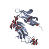
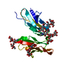
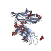
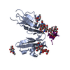
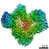
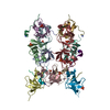
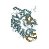
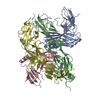
- PDB-2y7q: THE HIGH-AFFINITY COMPLEX BETWEEN IGE AND ITS RECEPTOR FC EPSILON RI -
+ Open data

- Basic information
| Entry | Database: PDB / ID: 2y7q | |||||||||
|---|---|---|---|---|---|---|---|---|---|---|
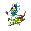
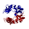
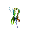
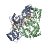
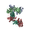
| Title | THE HIGH-AFFINITY COMPLEX BETWEEN IGE AND ITS RECEPTOR FC EPSILON RI | |||||||||
 Components Components |
| |||||||||
 Keywords Keywords |  IMMUNE SYSTEM / ALLERGY / ANTIBODY / IGE-BINDING PROTEIN / HIGH-AFFINITY RECEPTOR / IMMUNOGLOBULIN C REGION IMMUNE SYSTEM / ALLERGY / ANTIBODY / IGE-BINDING PROTEIN / HIGH-AFFINITY RECEPTOR / IMMUNOGLOBULIN C REGION | |||||||||
| Function / homology |  Function and homology information Function and homology informationhigh-affinity IgE receptor activity / IgE B cell receptor complex / adaptive immune memory response / primary adaptive immune response / type I hypersensitivity / B cell antigen processing and presentation / Fc receptor-mediated immune complex endocytosis / eosinophil degranulation / IgE immunoglobulin complex / macrophage activation ...high-affinity IgE receptor activity / IgE B cell receptor complex / adaptive immune memory response / primary adaptive immune response / type I hypersensitivity / B cell antigen processing and presentation / Fc receptor-mediated immune complex endocytosis / eosinophil degranulation / IgE immunoglobulin complex / macrophage activation / IgE binding / type 2 immune response / Fc epsilon receptor (FCERI) signaling / antibody-dependent cellular cytotoxicity / mast cell degranulation / B cell proliferation / macrophage differentiation / immunoglobulin mediated immune response / Role of LAT2/NTAL/LAB on calcium mobilization / immunoglobulin complex, circulating / immunoglobulin receptor binding / FCERI mediated Ca+2 mobilization / complement activation, classical pathway / antigen binding / FCERI mediated MAPK activation / B cell receptor signaling pathway / FCERI mediated NF-kB activation / antibacterial humoral response / Interleukin-4 and Interleukin-13 signaling / adaptive immune response / cell surface receptor signaling pathway / inflammatory response / immune response / external side of plasma membrane / cell surface / extracellular space / extracellular region / plasma membraneSimilarity search - Function | |||||||||
| Biological species |  HOMO SAPIENS (human) HOMO SAPIENS (human) | |||||||||
| Method | X-RAY DIFFRACTION / SYNCHROTRON / MOLECULAR REPLACEMENT / Resolution: 3.4 Å | |||||||||
 Authors Authors | Davies, A.M. / Holdom, M.D. / Nettleship, J.E. / Beavil, A.J. / Owens, R.J. / Sutton, B.J. | |||||||||
 Citation Citation | Journal: Nat.Struct.Mol.Biol. / Year: 2011 Title: Conformational Changes in Ige Contribute to its Uniquely Slow Dissociation Rate from Receptor Fceri Authors: Holdom, M.D. / Davies, A.M. / Nettleship, J.E. / Bagby, S.C. / Dhaliwal, B. / Girardi, E. / Hunt, J. / Gould, H.J. / Beavil, A.J. / Mcdonnell, J.M. / Owens, R.J. / Sutton, B.J. #1: Journal: Nat.Immunol. / Year: 2002Title: The Crystal Structure of Ige Fc Reveals an Asymmetrically Bent Conformation. Authors: Wan, T. / Beavil, R.L. / Fabiane, S.M. / Beavil, A.J. / Sohi, M.K. / Keown, M. / Young, R.J. / Henry, A.J. / Owens, R.J. / Gould, H.J. / Sutton, B.J. | |||||||||
| History |
|
- Structure visualization
| Structure viewer | Molecule: MolmilJmol/JSmol |
|---|
- Downloads & links
-Download
| PDBx/mmCIF format | 2y7q.cif.gz | 142.2 KB | Display | PDBx/mmCIF format |
|---|---|---|---|---|
| PDB format | pdb2y7q.ent.gz | 106.9 KB | Display | PDB format |
| PDBx/mmJSON format | 2y7q.json.gz | Tree view | PDBx/mmJSON format | |
| Others |  Other downloads Other downloads |
-Validation report
| Arichive directory | https://data.pdbj.org/pub/pdb/validation_reports/y7/2y7qftp://data.pdbj.org/pub/pdb/validation_reports/y7/2y7q | HTTPS FTP |
|---|
-Related structure data
| Related structure data |  2wqrC 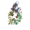 1f6aS  1o0vS S: Starting model for refinement C: citing same article ( |
|---|---|
| Similar structure data |


-Links
 PDBj
PDBj


- Assembly
| Deposited unit | 
| ||||||||||||||||||||||||||||||||||||||||||||||||||||||||||||||||||||||||
|---|---|---|---|---|---|---|---|---|---|---|---|---|---|---|---|---|---|---|---|---|---|---|---|---|---|---|---|---|---|---|---|---|---|---|---|---|---|---|---|---|---|---|---|---|---|---|---|---|---|---|---|---|---|---|---|---|---|---|---|---|---|---|---|---|---|---|---|---|---|---|---|---|---|
| 1 |
| ||||||||||||||||||||||||||||||||||||||||||||||||||||||||||||||||||||||||
| Unit cell |
| ||||||||||||||||||||||||||||||||||||||||||||||||||||||||||||||||||||||||
| Noncrystallographic symmetry (NCS) | NCS domain:
NCS domain segments:
NCS ensembles :
|
-Components
| #1: Protein | Mass: 21722.098 Da / Num. of mol.: 1 / Fragment: SOLUBLE EXTRACELLULAR DOMAINS, RESIDUES 26-201 / Mutation: YES Source method: isolated from a genetically manipulated source Source: (gene. exp.) HOMO SAPIENS (human) / Cell line (production host): HEK293S / Production host: HOMO SAPIENS (human) / References: UniProt: P12319 | ||||||||
|---|---|---|---|---|---|---|---|---|---|
| #2: Protein | Mass: 36354.781 Da / Num. of mol.: 2 Fragment: FC FRAGMENT COMPRISING DOMAINS CEPSILON2-4, RESIDUES 104-427 Mutation: YES Source method: isolated from a genetically manipulated source Source: (gene. exp.) HOMO SAPIENS (human) / Cell line (production host): MOUSE MYELOMA NS0 / Production host:  MUS MUSCULUS (house mouse) / References: UniProt: P01854 MUS MUSCULUS (house mouse) / References: UniProt: P01854#3: Polysaccharide | alpha-D-mannopyranose-(1-3)-beta-D-mannopyranose-(1-4)-2-acetamido-2-deoxy-beta-D-glucopyranose-(1- ...alpha-D-mannopyranose-(1-3)-beta-D-mannopyranose-(1-4)-2-acetamido-2-deoxy-beta-D-glucopyranose-(1-4)-2-acetamido-2-deoxy-beta-D-glucopyranose | / Mass: 748.682 Da / Num. of mol.: 1Source method: isolated from a genetically manipulated source #4: Sugar | N-Acetylglucosamine  Type: D-saccharide, beta linking / Mass: 221.208 Da / Num. of mol.: 3 Type: D-saccharide, beta linking / Mass: 221.208 Da / Num. of mol.: 3Source method: isolated from a genetically manipulated source Formula: C8H15NO6 Compound details | ENGINEERED RESIDUE IN CHAIN A, ASN 99 TO ALA ENGINEERED RESIDUE IN CHAIN A, ASN 160 TO ALA ...ENGINEERED | Sequence details | CHAIN A RESIDUES -2 TO 0 ARTEFACT FROM PHLSEC VECTOR CHAIN A RESIDUES 177-179 ARTEFACT FROM PHLSEC ...CHAIN A RESIDUES -2 TO 0 ARTEFACT FROM PHLSEC VECTOR CHAIN A RESIDUES 177-179 ARTEFACT FROM PHLSEC VECTOR CHAIN A RESIDUES 180-185 C-TERMINAL HIS TAG CHAIN B RESIDUES 222-223 VECTOR LEADER SEQUENCE CHAIN D RESIDUES 222-223 VECTOR LEADER SEQUENCE SEQUENCE DISCREPANC | |
-Experimental details
-Experiment
| Experiment | Method: X-RAY DIFFRACTION / Number of used crystals: 1 |
|---|
- Sample preparation
| Crystal | Density Matthews: 3.78 Å3/Da / Density % sol: 48 % / Description: NONE |
|---|---|
| Crystal grow | Method: vapor diffusion, sitting drop / pH: 7 Details: SITTING DROP VAPOR DIFFUSION. RESERVOIR SOLUTION CONTAINED 2.8M SODIUM ACETATE PH 7. |
-Data collection
| Diffraction | Mean temperature: 100 K |
|---|---|
| Diffraction source | Source: SYNCHROTRON / Site: ESRF  / Beamline: ID29 / Wavelength: 0.9791 / Beamline: ID29 / Wavelength: 0.9791 |
| Detector | Type: ADSC CCD / Detector: CCD |
| Radiation | Protocol: SINGLE WAVELENGTH / Monochromatic (M) / Laue (L): M / Scattering type: x-ray |
| Radiation wavelength | Wavelength: 0.9791 Å / Relative weight: 1 |
| Reflection | Resolution: 3.4→37 Å / Num. obs: 16204 / % possible obs: 99.9 % / Observed criterion σ(I): 0 / Redundancy: 5.9 % / Rmerge(I) obs: 0.25 / Net I/σ(I): 8.5 |
| Reflection shell | Resolution: 3.4→3.49 Å / Redundancy: 6.1 % / Mean I/σ(I) obs: 1.6 / % possible all: 99.9 |
- Processing
| Software |
| |||||||||||||||||||||||||||||||||||||||||||||||||||||||||||||||||||||||||||||
|---|---|---|---|---|---|---|---|---|---|---|---|---|---|---|---|---|---|---|---|---|---|---|---|---|---|---|---|---|---|---|---|---|---|---|---|---|---|---|---|---|---|---|---|---|---|---|---|---|---|---|---|---|---|---|---|---|---|---|---|---|---|---|---|---|---|---|---|---|---|---|---|---|---|---|---|---|---|---|
| Refinement | Method to determine structure: MOLECULAR REPLACEMENT Starting model: PDB ENTRIES 1F6A AND 1O0V Resolution: 3.4→37.68 Å / SU ML: 0.45 / σ(F): 1.35 / Phase error: 27.55 / Stereochemistry target values: ML / Details: DISORDERED REGIONS WERE NOT MODELED.
| |||||||||||||||||||||||||||||||||||||||||||||||||||||||||||||||||||||||||||||
| Solvent computation | Shrinkage radii: 0.9 Å / VDW probe radii: 1.11 Å / Solvent model: FLAT BULK SOLVENT MODEL / Bsol: 91.81 Å2 / ksol: 0.35 e/Å3 | |||||||||||||||||||||||||||||||||||||||||||||||||||||||||||||||||||||||||||||
| Displacement parameters |
| |||||||||||||||||||||||||||||||||||||||||||||||||||||||||||||||||||||||||||||
| Refinement step | Cycle: LAST / Resolution: 3.4→37.68 Å
| |||||||||||||||||||||||||||||||||||||||||||||||||||||||||||||||||||||||||||||
| Refine LS restraints |
| |||||||||||||||||||||||||||||||||||||||||||||||||||||||||||||||||||||||||||||
| Refine LS restraints NCS |
| |||||||||||||||||||||||||||||||||||||||||||||||||||||||||||||||||||||||||||||
| LS refinement shell |
|
