 Movie
Movie Controller

[English] 日本語
 Yorodumi
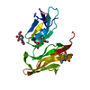
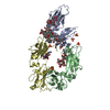
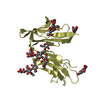
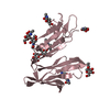
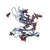
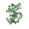
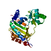
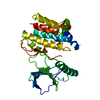
- PDB-1j89: HUMAN HIGH AFFINITY FC RECEPTOR FC(EPSILON)RI(ALPHA), TETRAGONAL ... -
+ Open data

- Basic information
| Entry | Database: PDB / ID: 1j89 | |||||||||
|---|---|---|---|---|---|---|---|---|---|---|
| Title | HUMAN HIGH AFFINITY FC RECEPTOR FC(EPSILON)RI(ALPHA), TETRAGONAL CRYSTAL FORM 2 | |||||||||
 Components Components | HIGH AFFINITY IMMUNOGLOBULIN EPSILON RECEPTOR ALPHA-SUBUNIT | |||||||||
 Keywords Keywords |  IMMUNE SYSTEM / Fc Receptor / IgE receptor / Glycoprotein IMMUNE SYSTEM / Fc Receptor / IgE receptor / Glycoprotein | |||||||||
| Function / homology |  Function and homology information Function and homology informationhigh-affinity IgE receptor activity / type I hypersensitivity / eosinophil degranulation / IgE binding / type 2 immune response / Fc epsilon receptor (FCERI) signaling / mast cell degranulation / regulation of immune response / Role of LAT2/NTAL/LAB on calcium mobilization / FCERI mediated Ca+2 mobilization ...high-affinity IgE receptor activity / type I hypersensitivity / eosinophil degranulation / IgE binding / type 2 immune response / Fc epsilon receptor (FCERI) signaling / mast cell degranulation / regulation of immune response / Role of LAT2/NTAL/LAB on calcium mobilization / FCERI mediated Ca+2 mobilization / FCERI mediated MAPK activation / FCERI mediated NF-kB activation / transmembrane signaling receptor activity / cell surface receptor signaling pathway / cell surface / plasma membraneSimilarity search - Function | |||||||||
| Biological species |  Homo sapiens (human) Homo sapiens (human) | |||||||||
| Method | X-RAY DIFFRACTION / SYNCHROTRON / FOURIER SYNTHESIS / Resolution: 4.1 Å | |||||||||
 Authors Authors | Garman, S.C. / Sechi, S. / Kinet, J.P. / Jardetzky, T.S. | |||||||||
 Citation Citation | Journal: J.Mol.Biol. / Year: 2001 Title: The analysis of the human high affinity IgE receptor Fc epsilon Ri alpha from multiple crystal forms. Authors: Garman, S.C. / Sechi, S. / Kinet, J.P. / Jardetzky, T.S. | |||||||||
| History |
|
- Structure visualization
| Structure viewer | Molecule: MolmilJmol/JSmol |
|---|
- Downloads & links
-Download
| PDBx/mmCIF format | 1j89.cif.gz | 181.2 KB | Display | PDBx/mmCIF format |
|---|---|---|---|---|
| PDB format | pdb1j89.ent.gz | 152.2 KB | Display | PDB format |
| PDBx/mmJSON format | 1j89.json.gz | Tree view | PDBx/mmJSON format | |
| Others |  Other downloads Other downloads |
-Validation report
| Arichive directory | https://data.pdbj.org/pub/pdb/validation_reports/j8/1j89ftp://data.pdbj.org/pub/pdb/validation_reports/j8/1j89 | HTTPS FTP |
|---|
-Related structure data
| Related structure data | 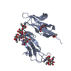 1j86C 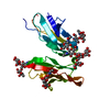 1j87C  1j88SC C: citing same article ( S: Starting model for refinement |
|---|---|
| Similar structure data |


-Links
 PDBj
PDBj


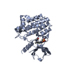
- Assembly
| Deposited unit | 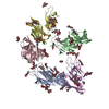
| ||||||||||
|---|---|---|---|---|---|---|---|---|---|---|---|
| 1 | 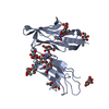
| ||||||||||
| 2 | 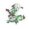
| ||||||||||
| 3 | 
| ||||||||||
| 4 | 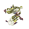
| ||||||||||
| 5 | 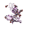
| ||||||||||
| Unit cell |
| ||||||||||
| Details | The biological assembly is a protein monomer with attached carbohydrate |
-Components
| #1: Protein | Mass: 19950.135 Da / Num. of mol.: 5 / Fragment: EXTRACELLULAR FRAGMENT Source method: isolated from a genetically manipulated source Details: GLYCOSYLATED PROTEIN, CHAIN A BY SUGARS F, B BY SUGARS G, C BY SUGARS H, D BY SUGARS I, E BY SUGARS J Source: (gene. exp.) Homo sapiens (human) / Cell line (production host): LDLD.LEC1 / Organ (production host): OVARY / Production host:   Cricetulus griseus (Chinese hamster) / References: UniProt: P12319 Cricetulus griseus (Chinese hamster) / References: UniProt: P12319#2: Polysaccharide | 2-acetamido-2-deoxy-beta-D-glucopyranose-(1-4)-2-acetamido-2-deoxy-beta-D-glucopyranose / Mass: 424.401 Da / Num. of mol.: 10Source method: isolated from a genetically manipulated source #3: Polysaccharide | beta-D-mannopyranose-(1-4)-2-acetamido-2-deoxy-beta-D-glucopyranose-(1-4)-2-acetamido-2-deoxy-beta- ...beta-D-mannopyranose-(1-4)-2-acetamido-2-deoxy-beta-D-glucopyranose-(1-4)-2-acetamido-2-deoxy-beta-D-glucopyranose / Mass: 586.542 Da / Num. of mol.: 5Source method: isolated from a genetically manipulated source #4: Sugar | ChemComp-NAG / N-Acetylglucosamine  Type: D-saccharide, beta linking / Mass: 221.208 Da / Num. of mol.: 20 Type: D-saccharide, beta linking / Mass: 221.208 Da / Num. of mol.: 20Source method: isolated from a genetically manipulated source Formula: C8H15NO6 |
|---|
-Experimental details
-Experiment
| Experiment | Method: X-RAY DIFFRACTION / Number of used crystals: 1 |
|---|
- Sample preparation
| Crystal | Density Matthews: 4.21 Å3/Da / Density % sol: 60 % | |||||||||||||||||||||||||
|---|---|---|---|---|---|---|---|---|---|---|---|---|---|---|---|---|---|---|---|---|---|---|---|---|---|---|
| Crystal grow | Temperature: 293 K / Method: vapor diffusion, hanging drop / pH: 5.6 Details: PEG 10000, Ammonium Citrate, Sodium Chloride, pH 5.6. VAPOR DIFFUSION, HANGING DROP at 293K | |||||||||||||||||||||||||
| Crystal grow | *PLUS Temperature: 23 ℃ / pH: 8.5 / Details: Garman, S.C., (2000) Nature, 406, 259. | |||||||||||||||||||||||||
| Components of the solutions | *PLUS
|
-Data collection
| Diffraction | Mean temperature: 110 K |
|---|---|
| Diffraction source | Source: SYNCHROTRON / Site: CHESS  / Beamline: A1 / Wavelength: 0.92 Å / Beamline: A1 / Wavelength: 0.92 Å |
| Detector | Type: CUSTOM-MADE / Detector: CCD / Date: Jan 1, 1995 |
| Radiation | Protocol: SINGLE WAVELENGTH / Monochromatic (M) / Laue (L): M / Scattering type: x-ray |
| Radiation wavelength | Wavelength: 0.92 Å / Relative weight: 1 |
| Reflection | Resolution: 4.1→40 Å / Num. all: 11510 / Num. obs: 11510 / % possible obs: 86.7 % / Observed criterion σ(F): 0 / Observed criterion σ(I): 0 / Redundancy: 2.2 % / Biso Wilson estimate: 204.8 Å2 / Rmerge(I) obs: 0.058 / Net I/σ(I): 12.4 |
| Reflection shell | Resolution: 4.1→4.25 Å / Redundancy: 1.9 % / Rmerge(I) obs: 0.3 / % possible all: 83.2 |
| Reflection | *PLUS Lowest resolution: 40 Å / Num. measured all: 25057 |
| Reflection shell | *PLUS % possible obs: 83.2 % / Rmerge(I) obs: 0.3 / Mean I/σ(I) obs: 1.9 |
- Processing
| Software |
| |||||||||||||||||||||||||
|---|---|---|---|---|---|---|---|---|---|---|---|---|---|---|---|---|---|---|---|---|---|---|---|---|---|---|
| Refinement | Method to determine structure: FOURIER SYNTHESIS Starting model: PDB ENTRY 1J88 Resolution: 4.1→36.5 Å / Rfactor Rfree error: 0.012 / Data cutoff high absF: 2780561.23 / Data cutoff low absF: 0 / Isotropic thermal model: GROUP / Cross valid method: THROUGHOUT / σ(F): 0 / σ(I): 0 / Stereochemistry target values: Engh & Huber Details: Synchrotron beam failed during data collection, causing poor data completeness and redundancy. 300 kcal/mol/A^2 NCS restraints applied to all atoms.
| |||||||||||||||||||||||||
| Solvent computation | Solvent model: FLAT MODEL / Bsol: 300 Å2 / ksol: 0.34 e/Å3 | |||||||||||||||||||||||||
| Displacement parameters | Biso mean: 199.3 Å2
| |||||||||||||||||||||||||
| Refine analyze |
| |||||||||||||||||||||||||
| Refinement step | Cycle: LAST / Resolution: 4.1→36.5 Å
| |||||||||||||||||||||||||
| Refine LS restraints |
| |||||||||||||||||||||||||
| Refine LS restraints NCS | Rms dev position: 0.04 Å / Weight Biso : 2 / Weight position: 300 | |||||||||||||||||||||||||
| LS refinement shell | Resolution: 4.1→4.36 Å / Rfactor Rfree error: 0.043 / Total num. of bins used: 6
| |||||||||||||||||||||||||
| Xplor file |
| |||||||||||||||||||||||||
| Software | *PLUS Name: CNS / Version: 0.9 / Classification: refinement | |||||||||||||||||||||||||
| Refinement | *PLUS σ(F): 0 / % reflection Rfree: 5.1 % | |||||||||||||||||||||||||
| Solvent computation | *PLUS | |||||||||||||||||||||||||
| Displacement parameters | *PLUS | |||||||||||||||||||||||||
| Refine LS restraints | *PLUS
| |||||||||||||||||||||||||
| LS refinement shell | *PLUS Rfactor Rfree: 0.369 / % reflection Rfree: 4 % / Rfactor Rwork: 0.312 |
