 Movie
Movie Controller

[English] 日本語
 Yorodumi
- PDB-1o0v: The crystal structure of IgE Fc reveals an asymmetrically bent co... -
+ Open data

- Basic information
| Entry | Database: PDB / ID: 1o0v | ||||||||||||
|---|---|---|---|---|---|---|---|---|---|---|---|---|---|
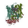
| Title | The crystal structure of IgE Fc reveals an asymmetrically bent conformation | ||||||||||||
 Components Components | Immunoglobulin heavy chain epsilon-1 | ||||||||||||
 Keywords Keywords |  IMMUNE SYSTEM / IgE Fc / Immunoglobulin E IMMUNE SYSTEM / IgE Fc / Immunoglobulin E | ||||||||||||
| Function / homology |  Function and homology information Function and homology informationIgE B cell receptor complex / adaptive immune memory response / primary adaptive immune response / type I hypersensitivity / B cell antigen processing and presentation / Fc receptor-mediated immune complex endocytosis / eosinophil degranulation / IgE immunoglobulin complex / macrophage activation / type 2 immune response ...IgE B cell receptor complex / adaptive immune memory response / primary adaptive immune response / type I hypersensitivity / B cell antigen processing and presentation / Fc receptor-mediated immune complex endocytosis / eosinophil degranulation / IgE immunoglobulin complex / macrophage activation / type 2 immune response / Fc epsilon receptor (FCERI) signaling / antibody-dependent cellular cytotoxicity / mast cell degranulation / B cell proliferation / macrophage differentiation / Role of LAT2/NTAL/LAB on calcium mobilization / immunoglobulin complex, circulating / immunoglobulin receptor binding / FCERI mediated Ca+2 mobilization / complement activation, classical pathway / antigen binding / FCERI mediated MAPK activation / B cell receptor signaling pathway / FCERI mediated NF-kB activation / antibacterial humoral response / Interleukin-4 and Interleukin-13 signaling / adaptive immune response / inflammatory response / immune response / extracellular space / extracellular region / plasma membraneSimilarity search - Function | ||||||||||||
| Biological species |  Homo sapiens (human) Homo sapiens (human) | ||||||||||||
| Method | X-RAY DIFFRACTION / SYNCHROTRON / MAD / Resolution: 2.6 Å | ||||||||||||
 Authors Authors | Wan, T. / Beavil, R.L. / Fabiane, S.M. / Beavil, A.J. / Sohi, M.K. / Keown, M. / Young, R.J. / Henry, A.J. / Owens, R.J. / Gould, H.J. / Sutton, B.J. | ||||||||||||
 Citation Citation | Journal: NAT.IMMUNOL. / Year: 2002 Title: The crystal structure of IgE Fc reveals an asymmetrically bent conformation Authors: Wan, T. / Beavil, R.L. / Fabiane, S.M. / Beavil, A.J. / Sohi, M.K. / Keown, M. / Young, R.J. / Henry, A.J. / Owens, R.J. / Gould, H.J. / Sutton, B.J. | ||||||||||||
| History |
|
- Structure visualization
| Structure viewer | Molecule: MolmilJmol/JSmol |
|---|
- Downloads & links
-Download
| PDBx/mmCIF format | 1o0v.cif.gz | 146.9 KB | Display | PDBx/mmCIF format |
|---|---|---|---|---|
| PDB format | pdb1o0v.ent.gz | 115.5 KB | Display | PDB format |
| PDBx/mmJSON format | 1o0v.json.gz | Tree view | PDBx/mmJSON format | |
| Others |  Other downloads Other downloads |
-Validation report
| Arichive directory | https://data.pdbj.org/pub/pdb/validation_reports/o0/1o0vftp://data.pdbj.org/pub/pdb/validation_reports/o0/1o0v | HTTPS FTP |
|---|
-Related structure data
| Similar structure data |
|---|


-Links
 PDBj
PDBj


- Assembly
| Deposited unit | 
| ||||||||
|---|---|---|---|---|---|---|---|---|---|
| 1 |
| ||||||||
| Unit cell |
|
-Components
-Antibody , 1 types, 2 molecules AB
| #1: Antibody | Mass: 36338.758 Da / Num. of mol.: 2 / Fragment: Fc portion, residues 251-573 / Mutation: C225A, N265Q, N371Q Source method: isolated from a genetically manipulated source Source: (gene. exp.) Homo sapiens (human) / Gene: IgE(ND) / Cell line (production host): mouse myeloma ns0 / Production host:  Mus musculus (house mouse) / References: GenBank: 386807, UniProt: P01854*PLUS Mus musculus (house mouse) / References: GenBank: 386807, UniProt: P01854*PLUS |
|---|
-Sugars , 2 types, 2 molecules
| #2: Polysaccharide | alpha-D-mannopyranose-(1-3)-[alpha-D-mannopyranose-(1-6)]alpha-D-mannopyranose-(1-6)-[alpha-D- ...alpha-D-mannopyranose-(1-3)-[alpha-D-mannopyranose-(1-6)]alpha-D-mannopyranose-(1-6)-[alpha-D-mannopyranose-(1-3)]beta-D-mannopyranose-(1-4)-2-acetamido-2-deoxy-beta-D-glucopyranose-(1-4)-2-acetamido-2-deoxy-beta-D-glucopyranose / Mass: 1235.105 Da / Num. of mol.: 1 Source method: isolated from a genetically manipulated source |
|---|---|
| #3: Polysaccharide | alpha-D-mannopyranose-(1-3)-[beta-D-mannopyranose-(1-6)]alpha-D-mannopyranose-(1-6)-[alpha-D- ...alpha-D-mannopyranose-(1-3)-[beta-D-mannopyranose-(1-6)]alpha-D-mannopyranose-(1-6)-[alpha-D-mannopyranose-(1-3)]beta-D-mannopyranose-(1-4)-2-acetamido-2-deoxy-beta-D-glucopyranose-(1-4)-2-acetamido-2-deoxy-beta-D-glucopyranose / Mass: 1235.105 Da / Num. of mol.: 1 Source method: isolated from a genetically manipulated source |
-Non-polymers , 3 types, 250 molecules 


| #4: Chemical | ChemComp-SO4 / Sulfate Mass: 96.063 Da / Num. of mol.: 4 / Source method: obtained synthetically / Formula: SO4 Mass: 96.063 Da / Num. of mol.: 4 / Source method: obtained synthetically / Formula: SO4#5: Chemical | Glycerol Mass: 92.094 Da / Num. of mol.: 2 / Source method: obtained synthetically / Formula: C3H8O3 Mass: 92.094 Da / Num. of mol.: 2 / Source method: obtained synthetically / Formula: C3H8O3#6: Water | ChemComp-HOH / | WaterMass: 18.015 Da / Num. of mol.: 244 / Source method: isolated from a natural source / Formula: H2O |
|---|
-Experimental details
-Experiment
| Experiment | Method: X-RAY DIFFRACTION / Number of used crystals: 1 |
|---|
- Sample preparation
| Crystal | Density Matthews: 2.62 Å3/Da / Density % sol: 52.98 % | ||||||||||||||||||||||||||||||||||||||||||||||||||||||||
|---|---|---|---|---|---|---|---|---|---|---|---|---|---|---|---|---|---|---|---|---|---|---|---|---|---|---|---|---|---|---|---|---|---|---|---|---|---|---|---|---|---|---|---|---|---|---|---|---|---|---|---|---|---|---|---|---|---|
| Crystal grow | Temperature: 295 K / Method: vapor diffusion, sitting drop / pH: 8.5 Details: 42.5% saturated ammonium sulphate, 100mM Tris ph 8.5, VAPOR DIFFUSION, SITTING DROP, temperature 295K | ||||||||||||||||||||||||||||||||||||||||||||||||||||||||
| Crystal grow | *PLUS Method: vapor diffusion, hanging drop | ||||||||||||||||||||||||||||||||||||||||||||||||||||||||
| Components of the solutions | *PLUS
|
-Data collection
| Diffraction | Mean temperature: 100 K |
|---|---|
| Diffraction source | Source: SYNCHROTRON / Site: NSLS  / Beamline: X12C / Wavelength: 1.1 Å / Beamline: X12C / Wavelength: 1.1 Å |
| Detector | Type: BRANDEIS - B4 / Detector: CCD / Date: Oct 24, 2000 / Details: toroidal mirror |
| Radiation | Monochromator: Si(111) / Protocol: SINGLE WAVELENGTH / Monochromatic (M) / Laue (L): M / Scattering type: x-ray |
| Radiation wavelength | Wavelength: 1.1 Å / Relative weight: 1 |
| Reflection | Resolution: 2.6→54 Å / Num. all: 24126 / Num. obs: 24126 / % possible obs: 99.8 % / Observed criterion σ(F): 2 / Observed criterion σ(I): 2 / Redundancy: 6.3 % / Biso Wilson estimate: 53.1 Å2 / Rmerge(I) obs: 0.063 / Rsym value: 0.063 / Net I/σ(I): 8.8 |
| Reflection shell | Resolution: 2.6→2.67 Å / Redundancy: 5.6 % / Rmerge(I) obs: 0.387 / Mean I/σ(I) obs: 2 / Num. unique all: 1686 / Rsym value: 0.353 / % possible all: 99.5 |
| Reflection | *PLUS Lowest resolution: 54 Å |
- Processing
| Software |
| ||||||||||||||||||||||||||||||||||||
|---|---|---|---|---|---|---|---|---|---|---|---|---|---|---|---|---|---|---|---|---|---|---|---|---|---|---|---|---|---|---|---|---|---|---|---|---|---|
| Refinement | Method to determine structure: MAD / Resolution: 2.6→54 Å / Isotropic thermal model: isotropic / Cross valid method: THROUGHOUT / σ(F): 0 / σ(I): 0 / Stereochemistry target values: Engh & Huber
| ||||||||||||||||||||||||||||||||||||
| Solvent computation | Bsol: 75.07 Å2 / ksol: 0.4 e/Å3 | ||||||||||||||||||||||||||||||||||||
| Displacement parameters | Biso mean: 46 Å2 | ||||||||||||||||||||||||||||||||||||
| Refine analyze |
| ||||||||||||||||||||||||||||||||||||
| Refinement step | Cycle: LAST / Resolution: 2.6→54 Å
| ||||||||||||||||||||||||||||||||||||
| Refine LS restraints |
| ||||||||||||||||||||||||||||||||||||
| LS refinement shell | Resolution: 2.6→2.64 Å /
| ||||||||||||||||||||||||||||||||||||
| Refinement | *PLUS Lowest resolution: 54 Å / % reflection Rfree: 5 % / Rfactor Rwork: 0.217 | ||||||||||||||||||||||||||||||||||||
| Solvent computation | *PLUS | ||||||||||||||||||||||||||||||||||||
| Displacement parameters | *PLUS | ||||||||||||||||||||||||||||||||||||
| Refine LS restraints | *PLUS
|
