 Movie
Movie Controller


+ Open data

- Basic information
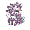
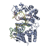
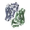
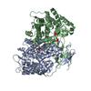
| Entry | Database: PDB / ID: 1b43 | ||||||
|---|---|---|---|---|---|---|---|
| Title | FEN-1 FROM P. FURIOSUS | ||||||
 Components Components | PROTEIN (FEN-1) | ||||||
 Keywords Keywords |  TRANSFERASE / NUCLEASE / DNA REPAIR / DNA REPLICATION TRANSFERASE / NUCLEASE / DNA REPAIR / DNA REPLICATION | ||||||
| Function / homology |  Function and homology information Function and homology information5'-flap endonuclease activity / DNA replication, removal of RNA primer / 5'-3' exonuclease activity / RNA-DNA hybrid ribonuclease activity / manganese ion binding / Hydrolases; Acting on ester bonds / DNA repair / magnesium ion binding / DNA binding / cytoplasmSimilarity search - Function | ||||||
| Biological species |   Pyrococcus furiosus (archaea) Pyrococcus furiosus (archaea) | ||||||
| Method | X-RAY DIFFRACTION / SYNCHROTRON / MIRAS / Resolution: 2 Å | ||||||
 Authors Authors | Hosfield, D.J. / Mol, C.D. / Shen, B. / Tainer, J.A. | ||||||
 Citation Citation | Journal: Cell(Cambridge,Mass.) / Year: 1998 Title: Structure of the DNA repair and replication endonuclease and exonuclease FEN-1: coupling DNA and PCNA binding to FEN-1 activity. Authors: Hosfield, D.J. / Mol, C.D. / Shen, B. / Tainer, J.A. | ||||||
| History |
|
- Structure visualization
| Structure viewer | Molecule: MolmilJmol/JSmol |
|---|
- Downloads & links
-Download
| PDBx/mmCIF format | 1b43.cif.gz | 151.2 KB | Display | PDBx/mmCIF format |
|---|---|---|---|---|
| PDB format | pdb1b43.ent.gz | 120.5 KB | Display | PDB format |
| PDBx/mmJSON format | 1b43.json.gz | Tree view | PDBx/mmJSON format | |
| Others |  Other downloads Other downloads |
-Validation report
| Arichive directory | https://data.pdbj.org/pub/pdb/validation_reports/b4/1b43ftp://data.pdbj.org/pub/pdb/validation_reports/b4/1b43 | HTTPS FTP |
|---|
-Related structure data
| Similar structure data |
|---|


-Links
 PDBj
PDBj

- Assembly
| Deposited unit | 
| ||||||||
|---|---|---|---|---|---|---|---|---|---|
| 1 |
| ||||||||
| Unit cell |
| ||||||||
| Noncrystallographic symmetry (NCS) | NCS oper: (Code: given Matrix: (0.1574, -0.9873, -0.0205), Vector : |
-Components
| #1: Protein | Mass: 38798.816 Da / Num. of mol.: 2 Source method: isolated from a genetically manipulated source Details: RESIDUE 1 IN THE STRUCTURE IS THE FIRST ORDERED RESIDUE, WHICH CORRESPONDS TO GLY2 IN THE SEQUENCE. Source: (gene. exp.) Pyrococcus furiosus (archaea) / Plasmid: PET15-B / Species (production host): Escherichia coli / Production host:  Escherichia coli BL21(DE3) (bacteria) / Strain (production host): BL21(DE3) / References: UniProt: O93634 Escherichia coli BL21(DE3) (bacteria) / Strain (production host): BL21(DE3) / References: UniProt: O93634#2: Water | ChemComp-HOH / | Water Mass: 18.015 Da / Num. of mol.: 413 / Source method: isolated from a natural source / Formula: H2O Mass: 18.015 Da / Num. of mol.: 413 / Source method: isolated from a natural source / Formula: H2O |
|---|
-Experimental details
-Experiment
| Experiment | Method: X-RAY DIFFRACTION / Number of used crystals: 1 |
|---|
- Sample preparation
| Crystal | Density Matthews: 3.1 Å3/Da / Density % sol: 60 % | |||||||||||||||||||||||||||||||||||
|---|---|---|---|---|---|---|---|---|---|---|---|---|---|---|---|---|---|---|---|---|---|---|---|---|---|---|---|---|---|---|---|---|---|---|---|---|
| Crystal grow | pH: 6.5 Details: 60 % AMMONIUM SULFATE 100 MM IMIDAZOLE MALATE 50 MM MGCL2, pH 6.5 | |||||||||||||||||||||||||||||||||||
| Crystal grow | *PLUS Method: vapor diffusion, hanging dropDetails: drop consists of equal volume of protein and precipitant solutions | |||||||||||||||||||||||||||||||||||
| Components of the solutions | *PLUS
|
-Data collection
| Diffraction | Mean temperature: 100 K |
|---|---|
| Diffraction source | Source: SYNCHROTRON / Site: SSRL  / Beamline: BL9-1 / Wavelength: 0.98 / Beamline: BL9-1 / Wavelength: 0.98 |
| Radiation | Protocol: SINGLE WAVELENGTH / Monochromatic (M) / Laue (L): M / Scattering type: x-ray |
| Radiation wavelength | Wavelength: 0.98 Å / Relative weight: 1 |
| Reflection | Resolution: 2→20 Å / Num. obs: 62063 / % possible obs: 96 % / Redundancy: 3.4 % / Biso Wilson estimate: 62.3 Å2 / Rsym value: 0.038 / Net I/σ(I): 15 |
| Reflection shell | Resolution: 2→2.07 Å / Redundancy: 3 % / Mean I/σ(I) obs: 3 / Rsym value: 0.346 / % possible all: 88.3 |
| Reflection | *PLUS Num. measured all: 208396 / Rmerge(I) obs: 0.038 |
| Reflection shell | *PLUS % possible obs: 88.3 % / Rmerge(I) obs: 0.346 |
- Processing
| Software |
| ||||||||||||||||||||||||||||||||||||||||||||||||||||||||||||
|---|---|---|---|---|---|---|---|---|---|---|---|---|---|---|---|---|---|---|---|---|---|---|---|---|---|---|---|---|---|---|---|---|---|---|---|---|---|---|---|---|---|---|---|---|---|---|---|---|---|---|---|---|---|---|---|---|---|---|---|---|---|
| Refinement | Method to determine structure: MIRAS / Resolution: 2→20 Å / Cross valid method: THROUGHOUT / σ(F): 0
| ||||||||||||||||||||||||||||||||||||||||||||||||||||||||||||
| Refinement step | Cycle: LAST / Resolution: 2→20 Å
| ||||||||||||||||||||||||||||||||||||||||||||||||||||||||||||
| Refine LS restraints |
| ||||||||||||||||||||||||||||||||||||||||||||||||||||||||||||
| LS refinement shell | Resolution: 2→2.09 Å / Total num. of bins used: 10
| ||||||||||||||||||||||||||||||||||||||||||||||||||||||||||||
| Xplor file | Serial no: 1 / Param file: PARAM19X.PRO / Topol file: TOPH19X.PAR |
