 ムービー
ムービー コントローラー
 構造ビューア
 EMN検索について


-検索条件
-検索結果
検索 (著者・登録者: cambillau & c)の結果54件中、1から50件目までを表示しています
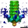
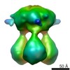
EMDB-11224: 
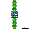
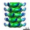
negative staining 3D reconstruction of p2 virion baseplate in activated conformation (3D class with open Tal trimer)
EMDB-11225:
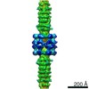
negative staining 3D reconstruction of p2 virion baseplate in activated conformation (3D class with closed Tal trimer)
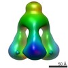
EMDB-11226:
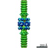
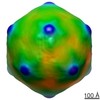
Negative staining 3D reconstruction of p2 virion baseplate in activated conformation
PDB-6zig:
Topological model of the p2 virion baseplate in activated conformation (closed Tal trimer)
PDB-6zih:
Topological model of p2 virion baseplate in activated conformation
PDB-6zjj:
Topological model of p2 virion baseplate in resting conformation
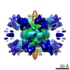
EMDB-10913:
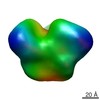
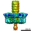
Structure of the phage STP1 host adhesion device (baseplate) by negative staining
EMDB-4900:
Cryo-EM structure of St1Cas9-sgRNA-tDNA20-AcrIIA6 monomeric assembly.
EMDB-4901:
Cryo-EM structure of St1Cas9-sgRNA-tDNA20-AcrIIA6 dimeric assembly.
EMDB-4902:
Cryo-EM structure of St1Cas9-sgRNA-tDNA59-ntPAM complex.
EMDB-4904:
Cryo-EM structure of St1Cas9-sgRNA-AcrIIA6-tDNA59-ntPAM complex.

PDB-6rj9:
Cryo-EM structure of St1Cas9-sgRNA-tDNA20-AcrIIA6 monomeric assembly.
PDB-6rja:
Cryo-EM structure of St1Cas9-sgRNA-tDNA20-AcrIIA6 dimeric assembly.
PDB-6rjd:
Cryo-EM structure of St1Cas9-sgRNA-tDNA59-ntPAM complex.
PDB-6rjg:
Cryo-EM structure of St1Cas9-sgRNA-AcrIIA6-tDNA59-ntPAM complex.
EMDB-9341:
Structure of the type VI secretion system TssK-TssF-TssG baseplate subcomplex revealed by cryo-electron microscopy - full map sharpened
EMDB-9342:
Structure of the type VI secretion system TssK-TssF-TssG baseplate subcomplex revealed by cryo-electron microscopy - TssK focused map
EMDB-9343:
Structure of the type VI secretion system TssK-TssF-TssG baseplate subcomplex revealed by cryo-electron microscopy - TssK focused map
PDB-6n38:
Structure of the type VI secretion system TssK-TssF-TssG baseplate subcomplex revealed by cryo-electron microscopy - full map sharpened
EMDB-3641:
Unraveling the self-assembly of Pseudomonas aeruginosa XcpQ secretin periplasmic domain provides new molecular insights into T2SS secreton architecture & dynamics
EMDB-3649:
Unraveling the self-assembly of Pseudomonas aeruginosa XcpQ secretin periplasmic domain provides new molecular insights into T2SS secreton architecture & dynamics
EMDB-4150:
Structure of the baseplate of Siphophage J-1
EMDB-3282:
Negative stain electron microscopy structure of TssA from EAEC type VI secretion system
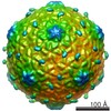
EMDB-2816:
Electron cryoEM structure of lactococcal siphophage 1358 virion
EMDB-2817:
Electron cryoEM structure of lactococcal siphophage 1358 virion
EMDB-2819:
Electron cryoEM structure of lactococcal siphophage 1358 virion
EMDB-2820:
Electron cryoEM structure of lactococcal siphophage 1358 virion
EMDB-2928:
Negative stain structure of a type 6 secretion system membrane core complex

EMDB-2927:
Negative stain structure of a type 6 secretion system membrane core complex
EMDB-2698:
The cryoEM structure of Monalysin Toxin
EMDB-2647:
electron cryo-microscopy of 1358 Lactococcus phage mature empty capsid
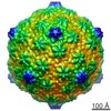
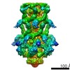
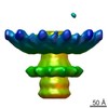
EMDB-2459:
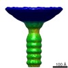
Structure and Host Adhesion Mechanism of Virulent Lactococcal Phage p2
EMDB-2462:
Structure and Host Adhesion Mechanism of Virulent Lactococcal Phage p2
EMDB-2463:
The electron cryo-miscroscopy reconstruction of the connector of lactococcal phage p2
EMDB-2464:
The electron miscroscopy reconstruction of the tail of lactococcal phage p2
EMDB-5739:
Electron microscopy map of the T6SS TssK component
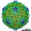
EMDB-2335:
The capsid of mycobacteriophage Araucaria
EMDB-2336:
The connector of mycobacteriophage Araucaria
EMDB-2337:
The tail of mycobacteriophage Araucaria
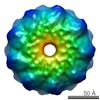
EMDB-2338:
The baseplate of mycobacteriophage Araucaria
EMDB-2340:
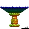
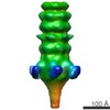
The electron microscopy reconstruction of the baseplate of lactococcal phage Tuc2009.
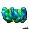
EMDB-2343:
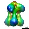
The electron microscopy reconstruction of the BppU-CtAL tripod of lactococcal phage Tuc2009.
EMDB-2345:
The electron microscopy reconstruction of the tripod of lactococcal phage Tuc2009.

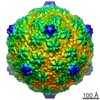
EMDB-2133:
The Structure of Lactococcal Phage TP901-1 by electron microscopy: the capsid
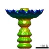
EMDB-2227:
The Structure of Lactococcal Phage TP901-1 by electron microscopy: the connector
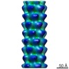
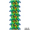
EMDB-2228:
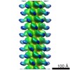
The Structure of Lactococcal Phage TP901-1 by electron microscopy: the helical tail
EMDB-1792:
EM map of TP901-1 BppU-BppL complex

EMDB-1900:
Cryo-EM map of the SPP1 bacteriophage gp19.1-gp21(1-552) complex
EMDB-1779:
EM structure of bacteriophage SPP1 distal tail protein (GP 19.1): a baseplate hub paradigm in gram positive infecting phages
EMDB-1793:
The three-dimensional reconstruction of the baseplate of wild-type TP901-1
ページ:
 wwPDBはEMDBデータモデルのバージョン3へ移行します
