 Movie
Movie Controller

[English] 日本語
 Yorodumi
- EMDB-11224: negative staining 3D reconstruction of p2 virion baseplate in act... -
+ Open data

- Basic information
| Entry | Database: EMDB / ID: EMD-11224 | |||||||||
|---|---|---|---|---|---|---|---|---|---|---|
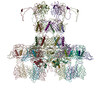
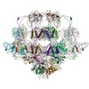
| Title | negative staining 3D reconstruction of p2 virion baseplate in activated conformation (3D class with open Tal trimer) | |||||||||
 Map data Map data | negative staining 3D reconstruction of p2 virion baseplate bound to VHH5 (3D class with open Tal trimer) | |||||||||
 Sample Sample |
| |||||||||
| Biological species |  Lactococcus virus P2 Lactococcus virus P2 | |||||||||
| Method | single particle reconstruction / negative staining / Resolution: 35.4 Å | |||||||||
 Authors Authors | Spinelli S / Cambillau C / Goulet A | |||||||||
 Citation Citation | Journal: Viruses / Year: 2020 Title: Structural Insights into Lactococcal Siphophage p2 Baseplate Activation Mechanism. Authors: Silvia Spinelli / Denise Tremblay / Sylvain Moineau / Christian Cambillau / Adeline Goulet /   Abstract: Virulent phages infecting , an industry-relevant bacterium, pose a significant risk to the quality of the fermented milk products. Phages of the Skunavirus genus are by far the most isolated ...Virulent phages infecting , an industry-relevant bacterium, pose a significant risk to the quality of the fermented milk products. Phages of the Skunavirus genus are by far the most isolated lactococcal phages in the cheese environments and phage p2 is the model siphophage for this viral genus. The baseplate of phage p2, which is used to recognize its host, was previously shown to display two conformations by X-ray crystallography, a rested state and an activated state ready to bind to the host. The baseplate became only activated and opened in the presence of Ca. However, such an activated state was not previously observed in the virion. Here, using nanobodies binding to the baseplate, we report on the negative staining electron microscopy structure of the activated form of the baseplate directly observed in the p2 virion, that is compatible with the activated baseplate crystal structure. Analyses of this new structure also established the presence of a second distal tail (Dit) hexamer as a component of the baseplate, the topology of which differs largely from the first one. We also observed an uncoupling between the baseplate activation and the tail tip protein (Tal) opening, suggesting an infection mechanism more complex than previously expected. | |||||||||
| History |
|
- Structure visualization
| Movie |
 Movie viewer Movie viewer |
|---|---|
| Structure viewer | EM map: SurfViewMolmilJmol/JSmol |
| Supplemental images |
 UCSF Chimera
- Downloads & links
-EMDB archive
| Map data | emd_11224.map.gz | 2.6 MB | EMDB map data format | |
|---|---|---|---|---|
| Header (meta data) | emd-11224-v30.xmlemd-11224.xml | 9.4 KB 9.4 KB | Display Display | EMDB header |
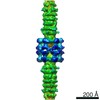
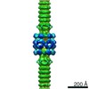
| Images | 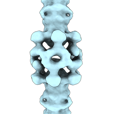 emd_11224.png emd_11224.png | 98.8 KB | ||
| Archive directory |  http://ftp.pdbj.org/pub/emdb/structures/EMD-11224ftp://ftp.pdbj.org/pub/emdb/structures/EMD-11224 http://ftp.pdbj.org/pub/emdb/structures/EMD-11224ftp://ftp.pdbj.org/pub/emdb/structures/EMD-11224 | HTTPS FTP |
-Validation report
| Summary document | emd_11224_validation.pdf.gz | 192.4 KB | Display | EMDB validaton report |
|---|---|---|---|---|
| Full document | emd_11224_full_validation.pdf.gz | 191.5 KB | Display | |
| Data in XML | emd_11224_validation.xml.gz | 6.5 KB | Display | |
| Arichive directory | https://ftp.pdbj.org/pub/emdb/validation_reports/EMD-11224ftp://ftp.pdbj.org/pub/emdb/validation_reports/EMD-11224 | HTTPS FTP |
-Related structure data
-Links
| EMDB pages | EMDB (EBI/PDBe) / EMDataResource |
|---|
-Map
| File | Download / File: emd_11224.map.gz / Format: CCP4 / Size: 64 MB / Type: IMAGE STORED AS FLOATING POINT NUMBER (4 BYTES) | ||||||||||||||||||||||||||||||||||||||||||||||||||||||||||||
|---|---|---|---|---|---|---|---|---|---|---|---|---|---|---|---|---|---|---|---|---|---|---|---|---|---|---|---|---|---|---|---|---|---|---|---|---|---|---|---|---|---|---|---|---|---|---|---|---|---|---|---|---|---|---|---|---|---|---|---|---|---|
| Annotation | negative staining 3D reconstruction of p2 virion baseplate bound to VHH5 (3D class with open Tal trimer) | ||||||||||||||||||||||||||||||||||||||||||||||||||||||||||||
| Voxel size | X=Y=Z: 3.46 Å | ||||||||||||||||||||||||||||||||||||||||||||||||||||||||||||
| Density |
| ||||||||||||||||||||||||||||||||||||||||||||||||||||||||||||
| Symmetry | Space group: 1 | ||||||||||||||||||||||||||||||||||||||||||||||||||||||||||||
| Details | EMDB XML:
CCP4 map header:
| ||||||||||||||||||||||||||||||||||||||||||||||||||||||||||||
-Supplemental data
- Sample components
-Entire : Lactococcus virus P2
| Entire | Name: Lactococcus virus P2 |
|---|---|
| Components |
|
-Supramolecule #1: Lactococcus virus P2
| Supramolecule | Name: Lactococcus virus P2 / type: virus / ID: 1 / Parent: 0 / Macromolecule list: #1-#3 / NCBI-ID: 254252 / Sci species name: Lactococcus virus P2 / Virus type: VIRION / Virus isolate: STRAIN / Virus enveloped: No / Virus empty: No |
|---|---|
| Host (natural) | Organism:  Lactococcus lactis (lactic acid bacteria) Lactococcus lactis (lactic acid bacteria) |
-Experimental details
-Structure determination
| Method | negative staining |
|---|---|
 Processing Processing | single particle reconstruction |
| Aggregation state | particle |
-Sample preparation
| Buffer | pH: 7.5 |
|---|---|
| Staining | Type: NEGATIVE / Material: uranyl acetate |
- Electron microscopy
| Microscope | FEI TECNAI SPIRIT |
|---|---|
| Image recording | Film or detector model: OTHER / Average electron dose: 25.0 e/Å2 |
| Electron beam | Acceleration voltage: 120 kV / Electron source: LAB6 |
| Electron optics | Illumination mode: SPOT SCAN / Imaging mode: BRIGHT FIELD |
| Experimental equipment |  Model: Tecnai Spirit / Image courtesy: FEI Company |
+Image processing
-Atomic model buiding 1
| Refinement | Protocol: RIGID BODY FIT |
|---|
