 Movie
Movie Controller
 Structure viewers
 About Yorodumi Papers


+Search query
-Structure paper
| Title | Visualizing a complete Siphoviridae member by single-particle electron microscopy: the structure of lactococcal phage TP901-1. |
|---|---|
| Journal, issue, pages | J Virol, Vol. 87, Issue 2, Page 1061-1068, Year 2013 |
| Publish date | Nov 7, 2012 |
 Authors Authors | Cecilia Bebeacua / Livia Lai / Christina Skovgaard Vegge / Lone Brøndsted / Marin van Heel / David Veesler / Christian Cambillau /  |
| PubMed Abstract | Tailed phages are genome delivery machines exhibiting unequaled efficiency acquired over more than 3 billion years of evolution. Siphophages from the P335 and 936 families infect the Gram-positive ...Tailed phages are genome delivery machines exhibiting unequaled efficiency acquired over more than 3 billion years of evolution. Siphophages from the P335 and 936 families infect the Gram-positive bacterium Lactococcus lactis using receptor-binding proteins anchored to the host adsorption apparatus (baseplate). Crystallographic and electron microscopy (EM) studies have shed light on the distinct adsorption strategies used by phages of these two families, suggesting that they might also rely on different infection mechanisms. Here, we report electron microscopy reconstructions of the whole phage TP901-1 (P335 species) and propose a composite EM model of this gigantic molecular machine. Our results suggest conservation of structural proteins among tailed phages and add to the growing body of evidence pointing to a common evolutionary origin for these virions. Finally, we propose that host adsorption apparatus architectures have evolved in correlation with the nature of the receptors used during infection. |
 External links External links | J Virol / PubMed:23135714 / PubMed Central |
| Methods | EM (single particle) |
| Resolution | 15.0 - 20.0 Å |
| Structure data |  EMDB-2133: 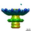 EMDB-2227: 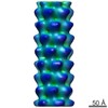 EMDB-2228: |
