 Movie
Movie Controller


+ Open data

- Basic information
| Entry | Database: EMDB / ID: EMD-4150 | |||||||||
|---|---|---|---|---|---|---|---|---|---|---|
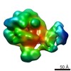
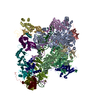
| Title | Structure of the baseplate of Siphophage J-1 | |||||||||
 Map data Map data | This is the baseplate of siphophage J-1 (Lactobacillus casei BL23 phage) with a part of the tail | |||||||||
 Sample Sample |
| |||||||||
| Biological species |  Lactobacillus phage J-1 (virus) Lactobacillus phage J-1 (virus) | |||||||||
| Method |  single particle reconstruction / negative staining / Resolution: 20.0 Å single particle reconstruction / negative staining / Resolution: 20.0 Å | |||||||||
 Authors Authors | Cambillau C / Spinelli S / Dieterle ME / Piuri M | |||||||||
 Citation Citation | Journal: Mol Microbiol / Year: 2017 Title: Evolved distal tail carbohydrate binding modules of Lactobacillus phage J-1: a novel type of anti-receptor widespread among lactic acid bacteria phages. Authors: Maria-Eugenia Dieterle / Silvia Spinelli / Irina Sadovskaya / Mariana Piuri / Christian Cambillau /   Abstract: Bacteriophage replication requires specific host-recognition. Some siphophages harbour a large complex, the baseplate, at the tip of their non-contractile tail. This baseplate holds receptor binding ...Bacteriophage replication requires specific host-recognition. Some siphophages harbour a large complex, the baseplate, at the tip of their non-contractile tail. This baseplate holds receptor binding proteins (RBPs) that can recognize the host cell-wall polysaccharide (CWPS) and specifically attach the phage to its host. While most phages possess a dedicated RBP, the phage J-1 that infects Lactobacillus casei seemed to lack one. It has been shown that the phage J-1 distal tail protein (Dit) plays a role in host recognition and that its sequence comprises two inserted modules compared with 'classical' Dits. The first insertion is similar to carbohydrate-binding modules (CBMs), whereas the second insertion remains undocumented. Here, we determined the structure of the second insertion and found it also similar to several CBMs. Expressed insertion CBM2, but not CBM1, binds to L. casei cells and neutralize phage attachment to the bacterial cell wall and the isolated and purified CWPS of L. casei BL23 prevents CBM2 attachment to the host. Electron microscopy single particle reconstruction of the J-1 virion baseplate revealed that CBM2 is projected at the periphery of Dit to optimally bind the CWPS receptor. Taken together, these results identify J-1 evolved Dit as the phage RBP. | |||||||||
| History |
|
- Structure visualization
| Movie |
 Movie viewer Movie viewer |
|---|---|
| Structure viewer | EM map: SurfViewMolmilJmol/JSmol |
| Supplemental images |

- Downloads & links
-EMDB archive
| Map data | emd_4150.map.gz | 157.7 KB | EMDB map data format | |
|---|---|---|---|---|
| Header (meta data) | emd-4150-v30.xmlemd-4150.xml | 9 KB 9 KB | Display Display | EMDB header |
| Images | 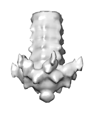 emd_4150.png emd_4150.png | 37.3 KB | ||
| Archive directory |  http://ftp.pdbj.org/pub/emdb/structures/EMD-4150ftp://ftp.pdbj.org/pub/emdb/structures/EMD-4150 http://ftp.pdbj.org/pub/emdb/structures/EMD-4150ftp://ftp.pdbj.org/pub/emdb/structures/EMD-4150 | HTTPS FTP |
-Related structure data

-Links
| EMDB pages | EMDB (EBI/PDBe) / EMDataResource |
|---|
-Map
| File | Download / File: emd_4150.map.gz / Format: CCP4 / Size: 214.8 KB / Type: IMAGE STORED AS FLOATING POINT NUMBER (4 BYTES) | ||||||||||||||||||||||||||||||||||||||||||||||||||||||||||||
|---|---|---|---|---|---|---|---|---|---|---|---|---|---|---|---|---|---|---|---|---|---|---|---|---|---|---|---|---|---|---|---|---|---|---|---|---|---|---|---|---|---|---|---|---|---|---|---|---|---|---|---|---|---|---|---|---|---|---|---|---|---|
| Annotation | This is the baseplate of siphophage J-1 (Lactobacillus casei BL23 phage) with a part of the tail | ||||||||||||||||||||||||||||||||||||||||||||||||||||||||||||
| Voxel size | X=Y=Z: 4.83 Å | ||||||||||||||||||||||||||||||||||||||||||||||||||||||||||||
| Density |
| ||||||||||||||||||||||||||||||||||||||||||||||||||||||||||||
| Symmetry | Space group: 1 | ||||||||||||||||||||||||||||||||||||||||||||||||||||||||||||
| Details | EMDB XML:
CCP4 map header:
| ||||||||||||||||||||||||||||||||||||||||||||||||||||||||||||
-Supplemental data
- Sample components
-Entire : virion's baseplate
| Entire | Name: virion's baseplate |
|---|---|
| Components |
|
-Supramolecule #1: virion's baseplate
| Supramolecule | Name: virion's baseplate / type: complex / ID: 1 / Parent: 0 |
|---|---|
| Source (natural) | Organism: Lactobacillus phage J-1 (virus) |
-Experimental details
-Structure determination
| Method | negative staining |
|---|---|
 Processing Processing | single particle reconstruction |
| Aggregation state | particle |
-Sample preparation
| Buffer | pH: 7 |
|---|---|
| Staining | Type: NEGATIVE / Material: 1% uranyl acetate Details: stained with 10 microL of 1% uranyl acetate for 30 sec |
| Details | The baseplate was boxed at the virion's tip |
- Electron microscopy
| Microscope | FEI TECNAI SPIRIT |
|---|---|
| Electron beam | Acceleration voltage: 120 kV / Electron source: LAB6 |
| Electron optics | Illumination mode: OTHER / Imaging mode: BRIGHT FIELDBright-field microscopy |
| Image recording | Film or detector model: FEI EAGLE (2k x 2k) / Average electron dose: 2.0 e/Å2 |
| Experimental equipment |  Model: Tecnai Spirit / Image courtesy: FEI Company |
-Image processing
| Particle selection | Number selected: 865 |
|---|---|
| Initial angle assignment | Type: OTHER / Software - Name: Xmipp (ver. 3.1) |
| Final 3D classification | Number classes: 3 / Software - Name: Xmipp (ver. 3.1) |
| Final angle assignment | Type: OTHER / Software - Name: Xmipp (ver. 3.1) |
| Final reconstruction | Number classes used: 3 / Applied symmetry - Point group: C6 (6 fold cyclic) / Resolution.type: BY AUTHOR / Resolution: 20.0 Å / Resolution method: FSC 0.5 CUT-OFF / Software - Name: Xmipp (ver. 3.1) / Number images used: 865 |
-Atomic model buiding 1
| Refinement | Space: REAL / Protocol: OTHER |
|---|
