Journal: Microb Biotechnol / Year: 2020 Title: Revisiting the host adhesion determinants of Streptococcus thermophilus siphophages. Authors: Katherine Lavelle / Adeline Goulet / Brian McDonnell / Silvia Spinelli / Douwe van Sinderen / Jennifer Mahony / Christian Cambillau / Abstract: Available 3D structures of bacteriophage modules combined with predictive bioinformatic algorithms enabled the identification of adhesion modules in 57 siphophages infecting Streptococcus ...Available 3D structures of bacteriophage modules combined with predictive bioinformatic algorithms enabled the identification of adhesion modules in 57 siphophages infecting Streptococcus thermophilus (St). We identified several carbohydrate-binding modules (CBMs) in so-called evolved distal tail (Dit) and tail-associated lysozyme (Tal) proteins of St phage baseplates. We examined the open reading frame (ORF) downstream of the Tal-encoding ORF and uncovered the presence of a putative p2-like receptor-binding protein (RBP). A 21 Å resolution electron microscopy structure of the baseplate of cos-phage STP1 revealed the presence of six elongated electron densities, surrounding the core of the baseplate, that harbour the p2-like RBPs at their tip. To verify the functionality of these modules, we expressed GFP- or mCherry-coupled Tal and putative RBP CBMs and observed by fluorescence microscopy that both modules bind to their corresponding St host, the putative RBP CBM with higher affinity than the Tal-associated one. The large number of CBM functional domains in St phages suggests that they play a contributory role in the infection process, a feature that we previously described in lactococcal phages and beyond, possibly representing a universal feature of the siphophage host-recognition apparatus.
History
Deposition
Apr 24, 2020
-
Header (metadata) release
Jun 24, 2020
-
Map release
Jun 24, 2020
-
Update
Oct 14, 2020
-
Current status
Oct 14, 2020
Processing site: PDBe / Status: Released
-
Structure visualization
Movie
Surface view with section colored by density value
In the structure databanks used in Yorodumi, some data are registered as the other names, "COVID-19 virus" and "2019-nCoV". Here are the details of the virus and the list of structure data.
Jan 31, 2019. EMDB accession codes are about to change! (news from PDBe EMDB page)
EMDB accession codes are about to change! (news from PDBe EMDB page)
The allocation of 4 digits for EMDB accession codes will soon come to an end. Whilst these codes will remain in use, new EMDB accession codes will include an additional digit and will expand incrementally as the available range of codes is exhausted. The current 4-digit format prefixed with “EMD-” (i.e. EMD-XXXX) will advance to a 5-digit format (i.e. EMD-XXXXX), and so on. It is currently estimated that the 4-digit codes will be depleted around Spring 2019, at which point the 5-digit format will come into force.
The EM Navigator/Yorodumi systems omit the EMD- prefix.
Related info.:Q: What is EMD? / ID/Accession-code notation in Yorodumi/EM Navigator
Yorodumi is a browser for structure data from EMDB, PDB, SASBDB, etc.
This page is also the successor to EM Navigator detail page, and also detail information page/front-end page for Omokage search.
The word "yorodu" (or yorozu) is an old Japanese word meaning "ten thousand". "mi" (miru) is to see.
Related info.:EMDB / PDB / SASBDB / Comparison of 3 databanks / Yorodumi Search / Aug 31, 2016. New EM Navigator & Yorodumi / Yorodumi Papers / Jmol/JSmol / Function and homology information / Changes in new EM Navigator and Yorodumi
 Movie
Movie Controller

 Yorodumi
 Open data
 Basic information
 Map data
 Sample
 Streptococcus phage STP1 (virus)
 single particle reconstruction /
  Authors
 Ireland,
  France, 3 items
  Citation
 Structure visualization
 Movie viewer
 Downloads & links
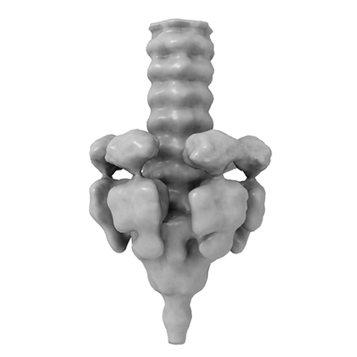 emd_10913.png
 http://ftp.pdbj.org/pub/emdb/structures/EMD-10913

 Sample components
 Processing
 Electron microscopy
