Loading
PDBj
MenuPDBj@FacebookPDBj@TwitterPDBj@YouTubewwPDB FoundationwwPDB
RCSB PDBPDBeBMRBAdv. SearchSearch help

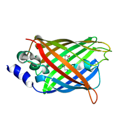
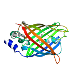
3DQ9
DownloadVisualize
BU of 3dq9 by Molmil
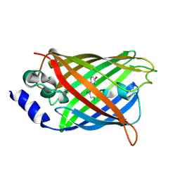
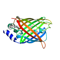
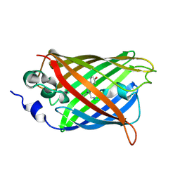
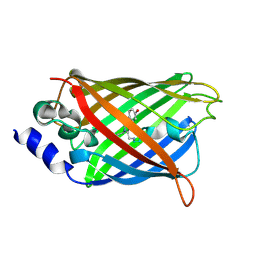
Structure of the Yellow Fluorescent Protein Citrine Frozen at 1500 Atmospheres Number 1: Structure 15 in a Series of 26 High Pressure Structures
Descriptor: Green fluorescent protein
Authors:Barstow, B, Kim, C.U.
Deposit date:2008-07-09
Release date:2008-09-23
Last modified:2023-11-15
Method:X-RAY DIFFRACTION (1.4 Å)
Cite:Alteration of citrine structure by hydrostatic pressure explains the accompanying spectral shift.
Proc.Natl.Acad.Sci.Usa, 105, 2008
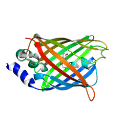
3DQA
DownloadVisualize
BU of 3dqa by Molmil
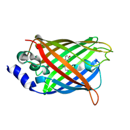
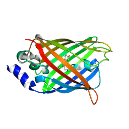
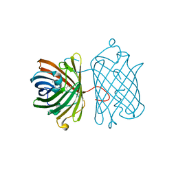
Structure of the Yellow Fluorescent Protein Citrine Frozen at 1250 Atmospheres Number 4: Structure 14 in a Series of 26 High Pressure Structures
Descriptor: Green fluorescent protein
Authors:Barstow, B, Kim, C.U.
Deposit date:2008-07-09
Release date:2008-09-23
Last modified:2023-11-15
Method:X-RAY DIFFRACTION (1.44 Å)
Cite:Alteration of citrine structure by hydrostatic pressure explains the accompanying spectral shift.
Proc.Natl.Acad.Sci.Usa, 105, 2008
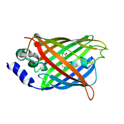
3DQC
DownloadVisualize
BU of 3dqc by Molmil
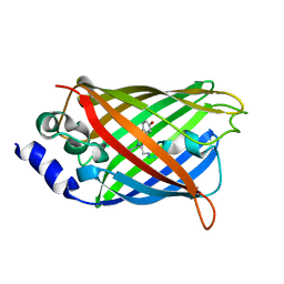
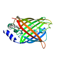
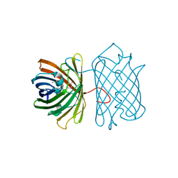
Structure of the Yellow Fluorescent Protein Citrine Frozen at 1250 Atmospheres Number 3: Structure 13 in a Series of 26 High Pressure Structures
Descriptor: Green fluorescent protein
Authors:Barstow, B, Kim, C.U.
Deposit date:2008-07-09
Release date:2008-09-23
Last modified:2023-11-15
Method:X-RAY DIFFRACTION (1.49 Å)
Cite:Alteration of citrine structure by hydrostatic pressure explains the accompanying spectral shift.
Proc.Natl.Acad.Sci.Usa, 105, 2008
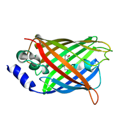
3DQD
DownloadVisualize
BU of 3dqd by Molmil
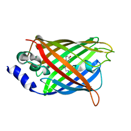
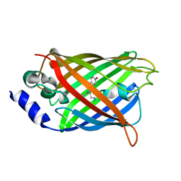
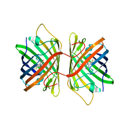
Structure of the Yellow Fluorescent Protein Citrine Frozen at 1250 Atmospheres Number 2: Structure 12 in a Series of 26 High Pressure Structures
Descriptor: Green fluorescent protein
Authors:Barstow, B, Kim, C.U.
Deposit date:2008-07-09
Release date:2008-09-23
Last modified:2023-11-15
Method:X-RAY DIFFRACTION (1.4 Å)
Cite:Alteration of citrine structure by hydrostatic pressure explains the accompanying spectral shift.
Proc.Natl.Acad.Sci.Usa, 105, 2008
3DQE
DownloadVisualize
BU of 3dqe by Molmil
Structure of the Yellow Fluorescent Protein Citrine Frozen at 1250 Atmospheres Number 1: Structure 11 in a Series of 26 High Pressure Structures
Descriptor: Green fluorescent protein
Authors:Barstow, B, Kim, C.U.
Deposit date:2008-07-09
Release date:2008-09-23
Last modified:2023-11-15
Method:X-RAY DIFFRACTION (1.43 Å)
Cite:Alteration of citrine structure by hydrostatic pressure explains the accompanying spectral shift.
Proc.Natl.Acad.Sci.Usa, 105, 2008
3DQF
DownloadVisualize
BU of 3dqf by Molmil
Structure of the Yellow Fluorescent Protein Citrine Frozen at 1000 Atmospheres Number 6: Structure 10 in a Series of 26 High Pressure Structures
Descriptor: Green fluorescent protein
Authors:Barstow, B, Kim, C.U.
Deposit date:2008-07-09
Release date:2008-09-23
Last modified:2023-11-15
Method:X-RAY DIFFRACTION (1.46 Å)
Cite:Alteration of citrine structure by hydrostatic pressure explains the accompanying spectral shift.
Proc.Natl.Acad.Sci.Usa, 105, 2008
3DQH
DownloadVisualize
BU of 3dqh by Molmil
Structure of the Yellow Fluorescent Protein Citrine Frozen at 1000 Atmospheres Number 5: Structure 9 in a Series of 26 High Pressure Structures
Descriptor: Green fluorescent protein
Authors:Barstow, B, Kim, C.U.
Deposit date:2008-07-09
Release date:2008-09-23
Last modified:2023-11-15
Method:X-RAY DIFFRACTION (1.45 Å)
Cite:Alteration of citrine structure by hydrostatic pressure explains the accompanying spectral shift.
Proc.Natl.Acad.Sci.Usa, 105, 2008
3DQI
DownloadVisualize
BU of 3dqi by Molmil
Structure of the Yellow Fluorescent Protein Citrine Frozen at 1000 Atmospheres Number 4: Structure 8 in a Series of 26 High Pressure Structures
Descriptor: Green fluorescent protein
Authors:Barstow, B, Kim, C.U.
Deposit date:2008-07-09
Release date:2008-09-23
Last modified:2023-11-15
Method:X-RAY DIFFRACTION (1.42 Å)
Cite:Alteration of citrine structure by hydrostatic pressure explains the accompanying spectral shift.
Proc.Natl.Acad.Sci.Usa, 105, 2008
3DQJ
DownloadVisualize
BU of 3dqj by Molmil
Structure of the Yellow Fluorescent Protein Citrine Frozen at 1000 Atmospheres Number 3: Structure 7 in a Series of 26 High Pressure Structures
Descriptor: Green fluorescent protein
Authors:Barstow, B, Kim, C.U.
Deposit date:2008-07-09
Release date:2008-09-23
Last modified:2023-11-15
Method:X-RAY DIFFRACTION (1.51 Å)
Cite:Alteration of citrine structure by hydrostatic pressure explains the accompanying spectral shift.
Proc.Natl.Acad.Sci.Usa, 105, 2008
3DQK
DownloadVisualize
BU of 3dqk by Molmil
Structure of the Yellow Fluorescent Protein Citrine Frozen at 1000 Atmospheres Number 2: Structure 6 in a Series of 26 High Pressure Structures
Descriptor: Green fluorescent protein
Authors:Barstow, B, Kim, C.U.
Deposit date:2008-07-09
Release date:2008-09-23
Last modified:2023-11-15
Method:X-RAY DIFFRACTION (1.4 Å)
Cite:Alteration of citrine structure by hydrostatic pressure explains the accompanying spectral shift.
Proc.Natl.Acad.Sci.Usa, 105, 2008
3DQL
DownloadVisualize
BU of 3dql by Molmil
Structure of the Yellow Fluorescent Protein Citrine Frozen at 1000 Atmospheres Number 1: Structure 5 in a Series of 26 High Pressure Structures
Descriptor: Green fluorescent protein
Authors:Barstow, B, Kim, C.U.
Deposit date:2008-07-09
Release date:2008-09-23
Last modified:2023-11-15
Method:X-RAY DIFFRACTION (1.47 Å)
Cite:Alteration of citrine structure by hydrostatic pressure explains the accompanying spectral shift.
Proc.Natl.Acad.Sci.Usa, 105, 2008
3DQM
DownloadVisualize
BU of 3dqm by Molmil
Structure of the Yellow Fluorescent Protein Citrine Frozen at 750 Atmospheres: Structure 4 in a Series of 26 High Pressure Structures
Descriptor: Green fluorescent protein
Authors:Barstow, B, Kim, C.U.
Deposit date:2008-07-09
Release date:2008-09-23
Last modified:2023-11-15
Method:X-RAY DIFFRACTION (1.44 Å)
Cite:Alteration of citrine structure by hydrostatic pressure explains the accompanying spectral shift.
Proc.Natl.Acad.Sci.Usa, 105, 2008
3DQN
DownloadVisualize
BU of 3dqn by Molmil
Structure of the Yellow Fluorescent Protein Citrine Frozen at 500 Atmospheres: Structure 3 in a Series of 26 High Pressure Structures
Descriptor: Green fluorescent protein
Authors:Barstow, B, Kim, C.U.
Deposit date:2008-07-09
Release date:2008-09-23
Last modified:2023-11-15
Method:X-RAY DIFFRACTION (1.44 Å)
Cite:Alteration of citrine structure by hydrostatic pressure explains the accompanying spectral shift.
Proc.Natl.Acad.Sci.Usa, 105, 2008
3DQO
DownloadVisualize
BU of 3dqo by Molmil
Structure of the Yellow Fluorescent Protein Citrine Frozen at 1 Atmosphere Number 2: Structure 2 in a Series of 26 High Pressure Structures
Descriptor: Green fluorescent protein
Authors:Barstow, B, Kim, C.U.
Deposit date:2008-07-09
Release date:2008-09-23
Last modified:2023-11-15
Method:X-RAY DIFFRACTION (1.5 Å)
Cite:Alteration of citrine structure by hydrostatic pressure explains the accompanying spectral shift.
Proc.Natl.Acad.Sci.Usa, 105, 2008
3DQU
DownloadVisualize
BU of 3dqu by Molmil
Structure of the Yellow Fluorescent Protein Citrine Frozen at 2000 Atmospheres Number 1: Structure 20 in a Series of 26 High Pressure Structures
Descriptor: Green fluorescent protein
Authors:Barstow, B, Kim, C.U.
Deposit date:2008-07-09
Release date:2008-09-23
Last modified:2023-11-15
Method:X-RAY DIFFRACTION (1.42 Å)
Cite:Alteration of citrine structure by hydrostatic pressure explains the accompanying spectral shift.
Proc.Natl.Acad.Sci.Usa, 105, 2008
3E5T
DownloadVisualize
BU of 3e5t by Molmil
Crystal Structure Analysis of FP611
Descriptor: Red fluorescent protein eqFP611
Authors:Nar, H, Nienhaus, K, Nienhaus, U, Wiedenmann, J.
Deposit date:2008-08-14
Release date:2008-09-23
Last modified:2023-11-15
Method:X-RAY DIFFRACTION (1.1 Å)
Cite:Trans-cis isomerization is responsible for the red-shifted fluorescence in variants of the red fluorescent protein eqFP611.
J.Am.Chem.Soc., 130, 2008
3E5V
DownloadVisualize
BU of 3e5v by Molmil
Crystal Structure Analysis of eqFP611 Double Mutant T122R, N143S
Descriptor: Red fluorescent protein eqFP611
Authors:Nar, H, Nienhaus, K, Nienhaus, U, Wiedenmann, J.
Deposit date:2008-08-14
Release date:2008-09-23
Last modified:2023-11-15
Method:X-RAY DIFFRACTION (2.1 Å)
Cite:Trans-cis isomerization is responsible for the red-shifted fluorescence in variants of the red fluorescent protein eqFP611.
J.Am.Chem.Soc., 130, 2008
3E5W
DownloadVisualize
BU of 3e5w by Molmil
Crystal Structure Analysis of FP611
Descriptor: Red fluorescent protein eqFP611
Authors:Nienhaus, K, Nar, H, Heilker, R, Wiedenmann, J, Nienhaus, G.U.
Deposit date:2008-08-14
Release date:2008-09-23
Last modified:2023-11-15
Method:X-RAY DIFFRACTION (1.71 Å)
Cite:Trans-cis isomerization is responsible for the red-shifted fluorescence in variants of the red fluorescent protein eqFP611.
J.Am.Chem.Soc., 130, 2008
3EEO
DownloadVisualize
BU of 3eeo by Molmil
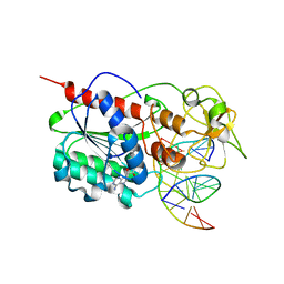
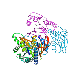
M. HhaI co-crystallized with synthetic dsDNA containing a propane diol in place of the deoxycytidine residue targeted for methylation.
Descriptor: 5'-D(P*DCP*DCP*DAP*DTP*DGP*DCP*DGP*DCP*DTP*DGP*DAP*DC)-3', 5'-D(P*DGP*DTP*DCP*DAP*DGP*(PDI)P*DGP*DCP*DAP*DTP*DGP*DG)-3', Modification methylase HhaI, ...
Authors:Porta, J.C, Christman, J.K, Borgstahl, G.E.O.
Deposit date:2008-09-05
Release date:2010-03-02
Last modified:2024-02-21
Method:X-RAY DIFFRACTION (1.94 Å)
Cite:M. HhaI co-crystallized with synthetic dsDNA containing a propane diol in place of the deoxycytidine residue targeted for methylation.
To be Published
3EK4
DownloadVisualize
BU of 3ek4 by Molmil
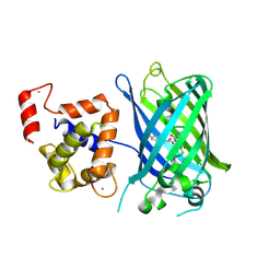
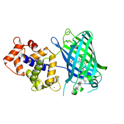
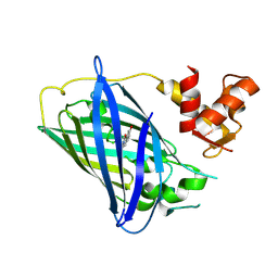
Calcium-saturated GCaMP2 Monomer
Descriptor: CALCIUM ION, Myosin light chain kinase, Green fluorescent protein, ...
Authors:Akerboom, J, Velez Rivera, J.D, Looger, L.L, Schreiter, E.R.
Deposit date:2008-09-18
Release date:2008-12-16
Last modified:2023-11-15
Method:X-RAY DIFFRACTION (2.65 Å)
Cite:Crystal Structures of the GCaMP Calcium Sensor Reveal the Mechanism of Fluorescence Signal Change and Aid Rational Design
J.Biol.Chem., 284, 2009
3EK7
DownloadVisualize
BU of 3ek7 by Molmil
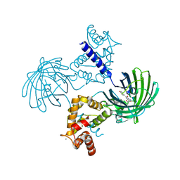
Calcium-saturated GCaMP2 dimer
Descriptor: CALCIUM ION, Myosin light chain kinase, Green fluorescent protein, ...
Authors:Akerboom, J, Velez Rivera, J.D, Looger, L.L, Schreiter, E.R.
Deposit date:2008-09-18
Release date:2008-12-16
Last modified:2023-11-15
Method:X-RAY DIFFRACTION (1.85 Å)
Cite:Crystal Structures of the GCaMP Calcium Sensor Reveal the Mechanism of Fluorescence Signal Change and Aid Rational Design
J.Biol.Chem., 284, 2009
3EK8
DownloadVisualize
BU of 3ek8 by Molmil
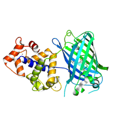
Calcium-saturated GCaMP2 T116V/G87R mutant monomer
Descriptor: CALCIUM ION, Myosin light chain kinase, Green fluorescent protein, ...
Authors:Akerboom, J, Velez Rivera, J.D, Looger, L.L, Schreiter, E.R.
Deposit date:2008-09-19
Release date:2008-12-16
Last modified:2023-11-15
Method:X-RAY DIFFRACTION (2.8 Å)
Cite:Crystal Structures of the GCaMP Calcium Sensor Reveal the Mechanism of Fluorescence Signal Change and Aid Rational Design
J.Biol.Chem., 284, 2009
3EKH
DownloadVisualize
BU of 3ekh by Molmil
Calcium-saturated GCaMP2 T116V/K378W mutant monomer
Descriptor: CALCIUM ION, GLYCEROL, Myosin light chain kinase, ...
Authors:Akerboom, J, Velez Rivera, J.D, Looger, L.L, Schreiter, E.R.
Deposit date:2008-09-19
Release date:2008-12-16
Last modified:2023-11-15
Method:X-RAY DIFFRACTION (2 Å)
Cite:Crystal Structures of the GCaMP Calcium Sensor Reveal the Mechanism of Fluorescence Signal Change and Aid Rational Design
J.Biol.Chem., 284, 2009
3EKJ
DownloadVisualize
BU of 3ekj by Molmil
Calcium-free GCaMP2 (calcium binding deficient mutant)
Descriptor: Myosin light chain kinase, Green fluorescent protein, Calmodulin chimera
Authors:Akerboom, J, Velez Rivera, J.D, Looger, L.L, Schreiter, E.R.
Deposit date:2008-09-19
Release date:2008-12-16
Last modified:2023-11-15
Method:X-RAY DIFFRACTION (2.8 Å)
Cite:Crystal Structures of the GCaMP Calcium Sensor Reveal the Mechanism of Fluorescence Signal Change and Aid Rational Design
J.Biol.Chem., 284, 2009
3EOZ
DownloadVisualize
BU of 3eoz by Molmil
Crystal Structure of Phosphoglycerate Mutase from Plasmodium Falciparum, PFD0660w
Descriptor: GLYCEROL, PHOSPHATE ION, putative Phosphoglycerate mutase
Authors:Wernimont, A.K, Tempel, W, Lam, A, Zhao, Y, Lew, J, Lin, Y.H, Wasney, G, Vedadi, M, Kozieradzki, I, Cossar, D, Schapira, M, Weigelt, J, Arrowsmith, C.H, Bochkarev, A, Edwards, A.M, Hui, R, Pizarro, J, Hills, T, Structural Genomics Consortium (SGC)
Deposit date:2008-09-29
Release date:2008-11-25
Last modified:2017-10-25
Method:X-RAY DIFFRACTION (2.4 Å)
Cite:Characterization of a new phosphatase from Plasmodium.
Mol.Biochem.Parasitol., 179, 2011

219140

数据于2024-05-01公开中

PDB statisticsPDBj update infoContact PDBjnumon