Loading
PDBj
MenuPDBj@FacebookPDBj@TwitterPDBj@YouTubewwPDB FoundationwwPDB
RCSB PDBPDBeBMRBAdv. SearchSearch help

3H8U
DownloadVisualize
BU of 3h8u by Molmil
Crystal structure of uncharacterized conserved protein with double-stranded beta-helix domain (YP_001338853.1) from Klebsiella pneumoniae subsp. pneumoniae MGH 78578 at 1.80 A resolution
Descriptor: GLYCEROL, NONAETHYLENE GLYCOL, SULFATE ION, ...
Authors:Joint Center for Structural Genomics (JCSG)
Deposit date:2009-04-29
Release date:2009-05-12
Last modified:2023-02-01
Method:X-RAY DIFFRACTION (1.8 Å)
Cite:Crystal structure of uncharacterized conserved protein with double-stranded beta-helix domain (YP_001338853.1) from Klebsiella pneumoniae subsp. pneumoniae MGH 78578 at 1.80 A resolution
To be published
3GYD
DownloadVisualize
BU of 3gyd by Molmil
Crystal structure of a cyclic nucleotide-binding domain (mfla_1926) from methylobacillus flagellatus kt at 1.79 A resolution
Descriptor: ADENOSINE-3',5'-CYCLIC-MONOPHOSPHATE, Cyclic nucleotide-binding domain, GLYCEROL
Authors:Joint Center for Structural Genomics (JCSG)
Deposit date:2009-04-03
Release date:2009-04-21
Last modified:2023-02-01
Method:X-RAY DIFFRACTION (1.79 Å)
Cite:Crystal structure of cyclic nucleotide-binding domain (YP_546034.1) from Methylobacillus flagellatus KT at 1.79 A resolution
To be published
3H3Z
DownloadVisualize
BU of 3h3z by Molmil
Crystal structure of a putative cyclic nucleotide binding protein (spoa0323) from ruegeria pomeroyi dss-3 at 2.35 A resolution
Descriptor: CHLORIDE ION, Cyclic nucleotide-binding protein
Authors:Joint Center for Structural Genomics (JCSG)
Deposit date:2009-04-17
Release date:2009-05-05
Last modified:2023-02-01
Method:X-RAY DIFFRACTION (2.35 Å)
Cite:Crystal structure of PUTATIVE CAMP-BINDING REGULATORY PROTEIN (YP_165150.1) from SILICIBACTER POMEROYI DSS-3 at 2.35 A resolution
To be published
3H4O
DownloadVisualize
BU of 3h4o by Molmil
Crystal structure of a nitroreductase family protein (cd3355) from clostridium difficile 630 at 1.50 A resolution
Descriptor: (4R)-2-METHYLPENTANE-2,4-DIOL, (4S)-2-METHYL-2,4-PENTANEDIOL, FLAVIN MONONUCLEOTIDE, ...
Authors:Joint Center for Structural Genomics (JCSG)
Deposit date:2009-04-20
Release date:2009-05-19
Last modified:2023-02-01
Method:X-RAY DIFFRACTION (1.5 Å)
Cite:Crystal structure of NITROREDUCTASE FAMILY PROTEIN (YP_001089872.1) from CLOSTRIDIUM DIFFICILE 630 at 1.50 A resolution
To be published
4DGU
DownloadVisualize
BU of 4dgu by Molmil
Crystal structure of a putative cell adhesion protein (BT0320) from Bacteroides thetaiotaomicron VPI-5482 at 2.37 A resolution
Descriptor: CHLORIDE ION, ZINC ION, putative cell adhesion protein
Authors:Joint Center for Structural Genomics (JCSG)
Deposit date:2012-01-26
Release date:2012-02-15
Last modified:2023-02-01
Method:X-RAY DIFFRACTION (2.37 Å)
Cite:A Distinct Type of Pilus from the Human Microbiome.
Cell, 165, 2016
5HD1
DownloadVisualize
BU of 5hd1 by Molmil
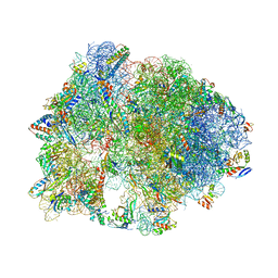
Crystal structure of antimicrobial peptide Pyrrhocoricin bound to the Thermus thermophilus 70S ribosome
Descriptor: 16S Ribosomal RNA, 23S Ribosomal RNA, 30S ribosomal protein S10, ...
Authors:Gagnon, M.G, Roy, R.N, Lomakin, I.B, Florin, T, Mankin, A.S, Steitz, T.A.
Deposit date:2016-01-04
Release date:2016-04-06
Last modified:2024-03-06
Method:X-RAY DIFFRACTION (2.7 Å)
Cite:Structures of proline-rich peptides bound to the ribosome reveal a common mechanism of protein synthesis inhibition.
Nucleic Acids Res., 44, 2016
5HCP
DownloadVisualize
BU of 5hcp by Molmil
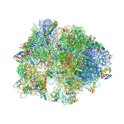
Crystal structure of antimicrobial peptide Metalnikowin bound to the Thermus thermophilus 70S ribosome
Descriptor: 16S Ribosomal RNA, 23S Ribosomal RNA, 30S ribosomal protein S10, ...
Authors:Gagnon, M.G, Roy, R.N, Lomakin, I.B, Florin, T, Mankin, A.S, Steitz, T.A.
Deposit date:2016-01-04
Release date:2016-04-06
Last modified:2024-03-06
Method:X-RAY DIFFRACTION (2.894 Å)
Cite:Structures of proline-rich peptides bound to the ribosome reveal a common mechanism of protein synthesis inhibition.
Nucleic Acids Res., 44, 2016
4DF9
DownloadVisualize
BU of 4df9 by Molmil
Crystal structure of a putative peptidase (BF3526) from Bacteroides fragilis NCTC 9343 at 2.17 A resolution
Descriptor: CHLORIDE ION, GLYCEROL, NONAETHYLENE GLYCOL, ...
Authors:Joint Center for Structural Genomics (JCSG)
Deposit date:2012-01-23
Release date:2012-02-15
Last modified:2023-02-01
Method:X-RAY DIFFRACTION (2.17 Å)
Cite:Crystal structure of a putative peptidase (BF3526) from Bacteroides fragilis NCTC 9343 at 2.17 A resolution
To be published
3HWU
DownloadVisualize
BU of 3hwu by Molmil
Crystal structure of Putative DNA-binding protein (YP_299413.1) from Ralstonia eutrophA JMP134 at 1.30 A resolution
Descriptor: Putative DNA-binding protein
Authors:Joint Center for Structural Genomics (JCSG)
Deposit date:2009-06-18
Release date:2009-06-30
Last modified:2023-02-01
Method:X-RAY DIFFRACTION (1.3 Å)
Cite:Crystal structure of Putative DNA-binding protein (YP_299413.1) from Ralstonia eutropha JMP134 at 1.30 A resolution
To be published
3I09
DownloadVisualize
BU of 3i09 by Molmil
CRYSTAL STRUCTURE OF A PERIPLASMIC BINDING PROTEIN (BMA2936) FROM BURKHOLDERIA MALLEI AT 1.80 A RESOLUTION
Descriptor: 1,2-ETHANEDIOL, ACETOACETIC ACID, CITRIC ACID, ...
Authors:Joint Center for Structural Genomics (JCSG)
Deposit date:2009-06-24
Release date:2009-07-21
Last modified:2023-02-01
Method:X-RAY DIFFRACTION (1.8 Å)
Cite:Crystal structure of Periplasmic Binding Protein BMA293 (YP_104442.1) from Burkholderia mallei ATCC 23344 at 1.80 A resolution
To be published
4DN6
DownloadVisualize
BU of 4dn6 by Molmil
Crystal structure of a putative pilus assembly protein (cpaE) from Burkholderia thailandensis E264 at 2.80 A resolution
Descriptor: Putative pilus assembly protein CpaE
Authors:Joint Center for Structural Genomics (JCSG), Shapiro, L.
Deposit date:2012-02-08
Release date:2012-02-29
Last modified:2023-02-01
Method:X-RAY DIFFRACTION (2.8 Å)
Cite:Crystal structure of a putative pilus assembly protein (cpaE) from Burkholderia thailandensis E264 at 2.80 A resolution
To be published
3HTV
DownloadVisualize
BU of 3htv by Molmil
Crystal structure of D-allose kinase (NP_418508.1) from ESCHERICHIA COLI K12 at 1.95 A resolution
Descriptor: D-allose kinase
Authors:Joint Center for Structural Genomics (JCSG)
Deposit date:2009-06-12
Release date:2009-06-23
Last modified:2023-02-01
Method:X-RAY DIFFRACTION (1.95 Å)
Cite:Crystal structure of D-allose kinase (NP_418508.1) from ESCHERICHIA COLI K12 at 1.95 A resolution
To be published
3HOI
DownloadVisualize
BU of 3hoi by Molmil
Crystal structure of FMN-dependent nitroreductase BF3017 from Bacteroides fragilis NCTC 9343 (YP_212631.1) from Bacteroides fragilis NCTC 9343 at 1.55 A resolution
Descriptor: CITRATE ANION, DI(HYDROXYETHYL)ETHER, FLAVIN MONONUCLEOTIDE, ...
Authors:Joint Center for Structural Genomics (JCSG)
Deposit date:2009-06-02
Release date:2009-06-23
Last modified:2023-02-01
Method:X-RAY DIFFRACTION (1.55 Å)
Cite:Crystal structure of FMN-dependent nitroreductase BF3017 from Bacteroides fragilis NCTC 9343 (YP_212631.1) from Bacteroides fragilis NCTC 9343 at 1.55 A resolution
To be published
3HVY
DownloadVisualize
BU of 3hvy by Molmil
Crystal structure of putative cystathionine beta-lyase involved in aluminum resistance (NP_348457.1) from Clostridium acetobutylicum at 2.00 A resolution
Descriptor: 1,2-ETHANEDIOL, Cystathionine beta-lyase family protein, YNBB B.subtilis ortholog, ...
Authors:Joint Center for Structural Genomics (JCSG)
Deposit date:2009-06-16
Release date:2009-06-30
Last modified:2023-02-01
Method:X-RAY DIFFRACTION (2 Å)
Cite:Crystal structure of putative cystathionine beta-lyase involved in aluminum resistance (NP_348457.1) from Clostridium acetobutylicum at 2.00 A resolution
To be published
3HX8
DownloadVisualize
BU of 3hx8 by Molmil
Crystal structure of putative ketosteroid isomerase (NP_103587.1) from Mesorhizobium loti at 1.45 A resolution
Descriptor: IMIDAZOLE, TETRAETHYLENE GLYCOL, UNKNOWN LIGAND, ...
Authors:Joint Center for Structural Genomics (JCSG)
Deposit date:2009-06-19
Release date:2009-07-07
Last modified:2023-02-01
Method:X-RAY DIFFRACTION (1.45 Å)
Cite:Crystal structure of putative ketosteroid isomerase (NP_103587.1) from MESORHIZOBIUM LOTI at 1.45 A resolution
To be published
4FXT
DownloadVisualize
BU of 4fxt by Molmil
Crystal structure of a DUF3823 family protein (BACOVA_02663) from Bacteroides ovatus ATCC 8483 at 2.77 A resolution
Descriptor: 1,2-ETHANEDIOL, Uncharacterized protein
Authors:Joint Center for Structural Genomics (JCSG)
Deposit date:2012-07-03
Release date:2012-07-25
Last modified:2023-02-01
Method:X-RAY DIFFRACTION (2.77 Å)
Cite:Crystal structure of a hypothetical protein (BACOVA_02663) from Bacteroides ovatus ATCC 8483 at 2.77 A resolution
To be published
3FSD
DownloadVisualize
BU of 3fsd by Molmil
Crystal structure of NTF2-like protein of unknown function in nutrient uptake (YP_427473.1) from RHODOSPIRILLUM RUBRUM ATCC 11170 at 1.70 A resolution
Descriptor: 1,2-ETHANEDIOL, NTF2-like protein of unknown function in nutrient uptake, UNKNOWN LIGAND
Authors:Joint Center for Structural Genomics (JCSG)
Deposit date:2009-01-09
Release date:2009-02-10
Last modified:2023-02-01
Method:X-RAY DIFFRACTION (1.7 Å)
Cite:Crystal structure of NTF2-like protein of unknown function in nutrient uptake (YP_427473.1) from RHODOSPIRILLUM RUBRUM ATCC 11170 at 1.70 A resolution
To be published
3G7Q
DownloadVisualize
BU of 3g7q by Molmil
Crystal structure of valine-pyruvate aminotransferase AvtA (NP_462565.1) from Salmonella typhimurium LT2 at 1.80 A resolution
Descriptor: 1,2-ETHANEDIOL, CHLORIDE ION, Valine-pyruvate aminotransferase
Authors:Joint Center for Structural Genomics (JCSG)
Deposit date:2009-02-10
Release date:2009-03-03
Last modified:2023-02-01
Method:X-RAY DIFFRACTION (1.8 Å)
Cite:Crystal structure of valine-pyruvate aminotransferase AvtA (NP_462565.1) from Salmonella typhimurium LT2 at 1.80 A resolution
To be published
3HTY
DownloadVisualize
BU of 3hty by Molmil
Crystal structure of a lipocalin-like protein (BT_0869) from Bacteroides thetaiotaomicron VPI-5482 at 1.95 A resolution
Descriptor: GLYCEROL, SULFATE ION, hypothetical protein BT_0869
Authors:Joint Center for Structural Genomics, Joint Center for Structural Genomics (JCSG)
Deposit date:2009-06-12
Release date:2009-07-07
Last modified:2023-02-01
Method:X-RAY DIFFRACTION (1.95 Å)
Cite:Crystal structure of hypothetical protein BT_0869 from Bacteroides thetaiotaomicron VPI-5482 (NP_809782.1) at 1.95 A resolution
To be Published
4F53
DownloadVisualize
BU of 4f53 by Molmil
Crystal structure of a susd homolog (BACOVA_04803) from Bacteroides ovatus ATCC 8483 at 2.25 A resolution
Descriptor: ACETATE ION, CALCIUM ION, CHLORIDE ION, ...
Authors:Joint Center for Structural Genomics (JCSG)
Deposit date:2012-05-11
Release date:2012-07-11
Last modified:2023-02-01
Method:X-RAY DIFFRACTION (2.25 Å)
Cite:Crystal structure of a susd homolog (BACOVA_04803) from Bacteroides ovatus ATCC 8483 at 2.25 A resolution
To be published
3I4G
DownloadVisualize
BU of 3i4g by Molmil
CRYSTAL STRUCTURE OF A SUSD-LIKE CARBOHYDRATE BINDING PROTEIN (BF0978) FROM BACTEROIDES FRAGILIS NCTC 9343 AT 1.35 A RESOLUTION
Descriptor: 1,2-ETHANEDIOL, 4-(2-HYDROXYETHYL)-1-PIPERAZINE ETHANESULFONIC ACID, SusD-like carbohydrate binding protein BF1063
Authors:Joint Center for Structural Genomics (JCSG)
Deposit date:2009-07-01
Release date:2009-07-21
Last modified:2023-02-01
Method:X-RAY DIFFRACTION (1.35 Å)
Cite:Crystal structure of SusD-like carbohydrate binding protein BF1063 from Bacteroides fragilis YCH46 (YP_210668.1) from Bacteroides fragilis NCTC 9343 at 1.35 A resolution
To be published
3HN0
DownloadVisualize
BU of 3hn0 by Molmil
CRYSTAL STRUCTURE OF AN ABC TRANSPORTER (BDI_1369) FROM PARABACTEROIDES DISTASONIS AT 1.75 A RESOLUTION
Descriptor: 1,2-ETHANEDIOL, DI(HYDROXYETHYL)ETHER, Nitrate transport protein, ...
Authors:Joint Center for Structural Genomics (JCSG)
Deposit date:2009-05-29
Release date:2009-07-07
Last modified:2023-02-01
Method:X-RAY DIFFRACTION (1.75 Å)
Cite:Crystal structure of Nitrate transport protein (YP_001302749.1) from Parabacteroides distasonis ATCC 8503 at 1.75 A resolution
To be published
3HOS
DownloadVisualize
BU of 3hos by Molmil
Crystal structure of the mariner Mos1 paired end complex with Mg
Descriptor: MAGNESIUM ION, Mos1 NTS inverted repeat DNA, Mos1 TS inverted repeat DNA, ...
Authors:Richardson, J.M, Walkinshaw, M.D.
Deposit date:2009-06-03
Release date:2009-09-22
Last modified:2023-11-01
Method:X-RAY DIFFRACTION (3.5 Å)
Cite:Molecular architecture of the Mos1 paired-end complex: the structural basis of DNA transposition in a eukaryote
Cell(Cambridge,Mass.), 138, 2009
4F0J
DownloadVisualize
BU of 4f0j by Molmil
Crystal structure of a probable hydrolytic enzyme (PA3053) from Pseudomonas aeruginosa PAO1 at 1.50 A resolution
Descriptor: 1,2-ETHANEDIOL, CHLORIDE ION, NONAETHYLENE GLYCOL, ...
Authors:Joint Center for Structural Genomics (JCSG)
Deposit date:2012-05-04
Release date:2012-07-04
Last modified:2023-02-01
Method:X-RAY DIFFRACTION (1.5 Å)
Cite:Crystal structure of a probable hydrolytic enzyme (PA3053) from Pseudomonas aeruginosa PAO1 at 1.50 A resolution
To be published
3GR3
DownloadVisualize
BU of 3gr3 by Molmil
Crystal structure of a nitroreductase-like family protein (pnba, bh06130) from bartonella henselae str. houston-1 at 1.45 A resolution
Descriptor: 1,2-ETHANEDIOL, ACETATE ION, CHLORIDE ION, ...
Authors:Joint Center for Structural Genomics (JCSG)
Deposit date:2009-03-24
Release date:2009-04-07
Last modified:2023-02-01
Method:X-RAY DIFFRACTION (1.45 Å)
Cite:Crystal structure of nitroreductase-like family protein (YP_033442.1) from BARTONELLA HENSELAE HOUSTON-1 at 1.45 A resolution
To be published

224931

PDB entries from 2024-09-11

PDB statisticsPDBj update infoContact PDBjnumon