9AZS
 
 | |
9BNA
 | |
7RYE
 | |
2VMK
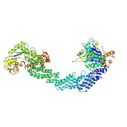
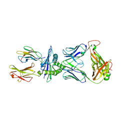
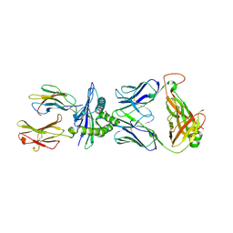
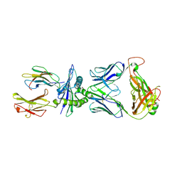
 | | Crystal Structure of E. coli RNase E Apoprotein - Catalytic Domain | | Descriptor: | RIBONUCLEASE E, SULFATE ION, ZINC ION | | Authors: | Koslover, D.J, Callaghan, A.J, Marcaida, M.J, Martick, M, Scott, W.G, Luisi, B.F. | | Deposit date: | 2008-01-28 | | Release date: | 2008-07-22 | | Last modified: | 2023-12-13 | | Method: | X-RAY DIFFRACTION (3.3 Å) | | Cite: | The Crystal Structure of the Escherichia Coli Rnase E Apoprotein and a Mechanism for RNA Degradation.
Structure, 16, 2008
|
|
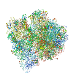
4V65
 | | Structure of the E. coli ribosome in the Pre-accommodation state | | Descriptor: | 16S rRNA, 23S rRNA, 30S ribosomal protein S10, ... | | Authors: | Devkota, B, Caulfield, T.R, Tan, R.-Z, Harvey, S.C. | | Deposit date: | 2008-08-03 | | Release date: | 2014-07-09 | | Last modified: | 2024-02-28 | | Method: | ELECTRON MICROSCOPY (9 Å) | | Cite: | The Structure of the E. coli Ribosome Before and After Accommodation: Implications for Proofreading
To be Published
|
|
4V66
 | | Structure of the E. coli ribosome and the tRNAs in Post-accommodation state | | Descriptor: | 16S rRNA, 23S rRNA, 30S ribosomal protein S10, ... | | Authors: | Devkota, B, Caulfield, T.R, Tan, R.-Z, Harvey, S.C. | | Deposit date: | 2008-08-03 | | Release date: | 2014-07-09 | | Last modified: | 2024-02-28 | | Method: | ELECTRON MICROSCOPY (9 Å) | | Cite: | The Structure of the E. coli Ribosome Before and After Accommodation: Implications for Proofreading
To be Published
|
|
5K06
 | | Recombinant bovine beta-lactoglobulin with uncleaved N-terminal methionine (rBlgB) | | Descriptor: | Beta-lactoglobulin, GLYCEROL, MYRISTIC ACID, ... | | Authors: | Loch, J.I, Bonarek, P, Tworzydlo, M, Polit, A, Hawro, B, Lach, A, Ludwin, E, Lewinski, K. | | Deposit date: | 2016-05-17 | | Release date: | 2016-07-20 | | Last modified: | 2024-11-13 | | Method: | X-RAY DIFFRACTION (2.5 Å) | | Cite: | Engineered beta-Lactoglobulin Produced in E. coli: Purification, Biophysical and Structural Characterisation.
Mol Biotechnol., 58, 2016
|
|
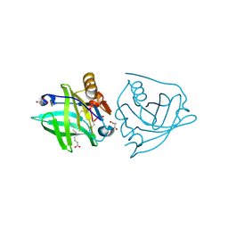
5JOQ
 | | Crystal Structure of an ABC Transporter Substrate-Binding Protein from Listeria monocytogenes EGD-e | | Descriptor: | CHLORIDE ION, CITRIC ACID, Lmo2184 protein | | Authors: | Brunzelle, J.S, Wawrzak, Z, Kudritska, M, Savchenko, A, Anderson, W.F, Center for Structural Genomics of Infectious Diseases (CSGID) | | Deposit date: | 2016-05-02 | | Release date: | 2016-07-27 | | Last modified: | 2023-09-27 | | Method: | X-RAY DIFFRACTION (1.99 Å) | | Cite: | Crystal Structure of an ABC Transporter Substrate-Binding Protein from Listeria monocytogenes EGD-e
To Be Published
|
|
9K6P
 | | Cryo-EM Structure of hAGO2D669A-siRNA-target (12-nt) | | Descriptor: | Protein argonaute-2, RNA (5'-R(P*GP*GP*CP*UP*CP*UP*UP*GP*U)-3'), RNA (5'-R(P*UP*AP*CP*AP*AP*GP*AP*GP*CP*C)-3') | | Authors: | Li, Z.Z, Xu, Q.K, Wu, J.P, Shen, E.Z. | | Deposit date: | 2024-10-22 | | Release date: | 2025-06-25 | | Method: | ELECTRON MICROSCOPY (3.2 Å) | | Cite: | Mechanistic insights into RNA cleavage by human Argonaute2-siRNA complex.
Cell Res., 35, 2025
|
|
9K6R
 | | Cryo-EM Structure of hAGO2D669A-siRNA-target (14-nt, uni-lobed) | | Descriptor: | MAGNESIUM ION, Protein argonaute-2, RNA (5'-R(P*GP*AP*AP*AP*GP*GP*CP*UP*CP*UP*UP*GP*UP*U)-3'), ... | | Authors: | Li, Z.Z, Xu, Q.K, Wu, J.P, Shen, E.Z. | | Deposit date: | 2024-10-22 | | Release date: | 2025-06-25 | | Method: | ELECTRON MICROSCOPY (2.7 Å) | | Cite: | Mechanistic insights into RNA cleavage by human Argonaute2-siRNA complex.
Cell Res., 35, 2025
|
|
9K6T
 | | Cryo-EM Structure of hAGO2D669A-siRNA-target (21-nt) | | Descriptor: | MAGNESIUM ION, Protein argonaute-2, RNA (5'-R(P*AP*AP*CP*AP*AP*CP*AP*GP*AP*AP*AP*GP*GP*CP*UP*CP*UP*UP*GP*UP*U)-3'), ... | | Authors: | Li, Z.Z, Xu, Q.K, Wu, J.P, Shen, E.Z. | | Deposit date: | 2024-10-22 | | Release date: | 2025-06-25 | | Method: | ELECTRON MICROSCOPY (2.8 Å) | | Cite: | Mechanistic insights into RNA cleavage by human Argonaute2-siRNA complex.
Cell Res., 35, 2025
|
|
9K6S
 | | Cryo-EM Structure of hAGO2D669A-siRNA-target (19-nt) | | Descriptor: | MAGNESIUM ION, Protein argonaute-2, RNA (5'-R(P*CP*AP*AP*CP*AP*GP*AP*AP*AP*GP*GP*CP*UP*CP*UP*UP*GP*UP*U)-3'), ... | | Authors: | Li, Z.Z, Xu, Q.K, Wu, J.P, Shen, E.Z. | | Deposit date: | 2024-10-22 | | Release date: | 2025-06-25 | | Method: | ELECTRON MICROSCOPY (2.8 Å) | | Cite: | Mechanistic insights into RNA cleavage by human Argonaute2-siRNA complex.
Cell Res., 35, 2025
|
|
9K6Q
 | | "Cryo-EM Structure of hAGO2D669A-siRNA-target (14-nt, sesqui-lobed) | | Descriptor: | MAGNESIUM ION, Protein argonaute-2, RNA (5'-R(P*GP*AP*AP*AP*GP*GP*CP*UP*CP*UP*UP*GP*U)-3'), ... | | Authors: | Li, Z.Z, Xu, Q.K, Wu, J.P, Shen, E.Z. | | Deposit date: | 2024-10-22 | | Release date: | 2025-06-25 | | Method: | ELECTRON MICROSCOPY (2.7 Å) | | Cite: | Mechanistic insights into RNA cleavage by human Argonaute2-siRNA complex.
Cell Res., 35, 2025
|
|
9B0O
 | |
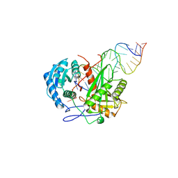
4UMA
 | | Structural analysis of substrate-mimicking inhibitors in complex with Neisseria meningitidis 3 deoxy D arabino heptulosonate 7 phosphate synthase the importance of accommodating the active site water | | Descriptor: | (E)-2-METHYL-3-PHOSPHONOACRYLATE, MANGANESE (II) ION, PHOSPHO-2-DEHYDRO-3-DEOXYHEPTONATE ALDOLASE | | Authors: | Heyes, L.C, Reichau, S, Cross, P.J, Parker, E.J. | | Deposit date: | 2014-05-16 | | Release date: | 2014-10-08 | | Last modified: | 2024-01-10 | | Method: | X-RAY DIFFRACTION (1.76 Å) | | Cite: | Structural Analysis of Substrate-Mimicking Inhibitors in Complex with Neisseria Meningitidis 3-Deoxy-D-Arabino-Heptulosonate 7-Phosphate Synthase - the Importance of Accommodating the Active Site Water.
Bioorg.Chem., 57, 2014
|
|
9OMA
 | | Cryo-EM structure of PCMTD1-ELOBC-CUL5-RBX2 (CRL5-PCMTD1) | | Descriptor: | Cullin-5, Elongin-B, Elongin-C, ... | | Authors: | Pang, E.Z, Zhao, B, Flowers, C, Oroudjeva, E, Winters, J.B, Pandey, V, Sawaya, M.R, Wohlschlegel, W, Loo, J.A, Rodriguez, J.A, Clarke, S.G. | | Deposit date: | 2025-05-13 | | Release date: | 2025-06-04 | | Last modified: | 2025-06-18 | | Method: | ELECTRON MICROSCOPY (4.14 Å) | | Cite: | Structural basis for L-isoaspartyl-containing protein recognition by the PCMTD1 cullin-RING E3 ubiquitin ligase.
Biorxiv, 2025
|
|
9OMF
 | | Cryo-EM structure of neddylated PCMTD1-ELOBC-CUL5-RBX2 (N8-CRL5-PCMTD1) | | Descriptor: | Cullin-5, Elongin-B, Elongin-C, ... | | Authors: | Pang, E.Z, Zhao, B, Flowers, C, Oroudjeva, E, Winters, J.B, Pandey, V, Sawaya, M.R, Wohlschlegel, W, Loo, J.A, Rodriguez, J.A, Clarke, S.G. | | Deposit date: | 2025-05-13 | | Release date: | 2025-06-04 | | Last modified: | 2025-06-18 | | Method: | ELECTRON MICROSCOPY (9.72 Å) | | Cite: | Structural basis for L-isoaspartyl-containing protein recognition by the PCMTD1 cullin-RING E3 ubiquitin ligase.
Biorxiv, 2025
|
|
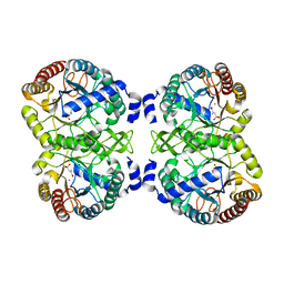
4U7Y
 | | Structure of the complex of VPS4B MIT and IST1 MIM | | Descriptor: | IST1 homolog, Vacuolar protein sorting-associated protein 4B | | Authors: | Guo, E.Z, Xu, Z. | | Deposit date: | 2014-07-31 | | Release date: | 2015-02-11 | | Last modified: | 2023-09-27 | | Method: | X-RAY DIFFRACTION (2.502 Å) | | Cite: | Distinct Mechanisms of Recognizing Endosomal Sorting Complex Required for Transport III (ESCRT-III) Protein IST1 by Different Microtubule Interacting and Trafficking (MIT) Domains.
J.Biol.Chem., 290, 2015
|
|
4U7I
 | | Structure of the complex of Spartin MIT and IST1 MIM | | Descriptor: | IST1 homolog, Spartin | | Authors: | Guo, E.Z, Xu, Z. | | Deposit date: | 2014-07-30 | | Release date: | 2015-02-11 | | Last modified: | 2023-09-27 | | Method: | X-RAY DIFFRACTION (1.794 Å) | | Cite: | Distinct Mechanisms of Recognizing Endosomal Sorting Complex Required for Transport III (ESCRT-III) Protein IST1 by Different Microtubule Interacting and Trafficking (MIT) Domains.
J.Biol.Chem., 290, 2015
|
|
6FK0
 | |
8RLU
 | |
8RLT
 | |
8RLV
 | |
8BYX
 | |
7PH4
 | | AMP-PNP bound nanodisc reconstituted MsbA with nanobodies, spin-labeled at position T68C | | Descriptor: | (1~{R},4~{R},11~{S},14~{S},19~{Z})-19-[2-[2,5-bis(oxidanylidene)pyrrolidin-1-yl]ethylimino]-7,8,17,18-tetraoxa-1,4,11,14-tetrazatricyclo[12.6.2.2^{4,11}]tetracosane-6,9,16-trione, ATP-dependent lipid A-core flippase, DODECYL-BETA-D-MALTOSIDE, ... | | Authors: | Parey, K, Januliene, D, Galazzo, L, Meier, G, Vecchis, D, Striednig, B, Hilbi, H, Schaefer, L.V, Kuprov, I, Bordignon, E, Seeger, M.A, Moeller, A. | | Deposit date: | 2021-08-16 | | Release date: | 2022-08-24 | | Last modified: | 2025-07-02 | | Method: | ELECTRON MICROSCOPY (2.8 Å) | | Cite: | The ABC transporter MsbA adopts the wide inward-open conformation in E. coli cells.
Sci Adv, 8, 2022
|
|


