Loading
PDBj
MenuPDBj@FacebookPDBj@TwitterPDBj@YouTubewwPDB FoundationwwPDB
RCSB PDBPDBeBMRBAdv. SearchSearch help
PDB: 522 results

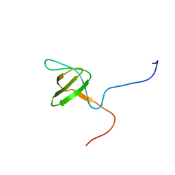
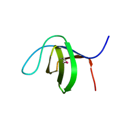
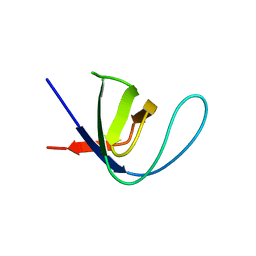
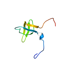
2YUP
DownloadVisualize
BU of 2yup by Molmil
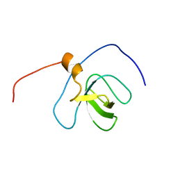
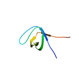
Solution structure of the second SH3 domain of human Vinexin
Descriptor: Vinexin
Authors:Abe, H, Tochio, N, Miyamoto, K, Saito, K, Sasagawa, A, Koshiba, S, Inoue, M, Kigawa, T, Yokoyama, S, RIKEN Structural Genomics/Proteomics Initiative (RSGI)
Deposit date:2007-04-06
Release date:2007-10-09
Last modified:2022-03-16
Method:SOLUTION NMR
Cite:Solution structure of the second SH3 domain of human Vinexin
To be Published
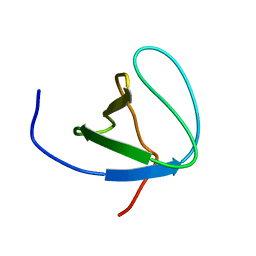
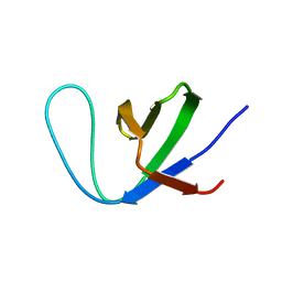
2YT6
DownloadVisualize
BU of 2yt6 by Molmil
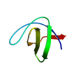
Solution structure of the SH3_1 domain of Yamaguchi sarcoma viral (v-yes) oncogene homolog 1
Descriptor: Adult male urinary bladder cDNA, RIKEN full-length enriched library, clone:9530076O17 product:Yamaguchi sarcoma viral (v-yes) oncogene homolog
Authors:Dang, W, Muto, Y, Inoue, M, Kigawa, T, Shirouzu, M, Tarada, T, Yokoyama, S, RIKEN Structural Genomics/Proteomics Initiative (RSGI)
Deposit date:2007-04-05
Release date:2008-04-08
Last modified:2022-03-16
Method:SOLUTION NMR
Cite:Solution structure of the SH3_1 domain of Yamaguchi sarcoma viral (v-yes) oncogene homolog 1
To be Published
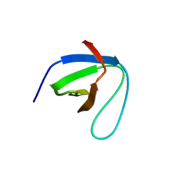
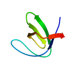
6RO9
DownloadVisualize
BU of 6ro9 by Molmil
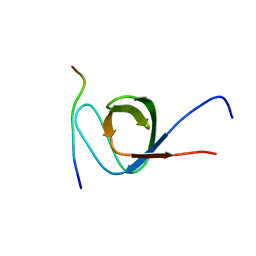
Human spectrin SH3 domain D48G, E7V, K60V
Descriptor: Spectrin alpha, non-erythrocytic 1
Authors:Reverter, D, Navarro, S, Ventura, S.
Deposit date:2019-05-10
Release date:2020-06-03
Last modified:2024-01-24
Method:X-RAY DIFFRACTION (1.814 Å)
Cite:Human spectrin SH3 domain D48G, E7V, K60V
To Be Published
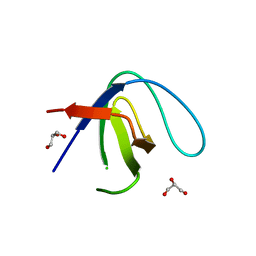
7TZK
DownloadVisualize
BU of 7tzk by Molmil
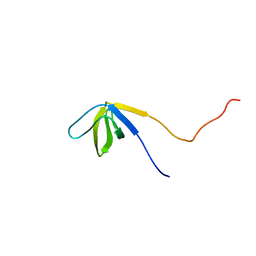
EPS8 SH3 Domain with NleH1 PxxDY Motif
Descriptor: Epidermal growth factor receptor kinase substrate 8, T3SS secreted effector NleH homolog
Authors:Grishin, A.M, Cygler, M.
Deposit date:2022-02-15
Release date:2022-07-06
Last modified:2023-10-18
Method:X-RAY DIFFRACTION (1.43 Å)
Cite:Targeting of microvillus protein Eps8 by the NleH effector kinases from enteropathogenic E. coli
Proc.Natl.Acad.Sci.USA, 119, 2022
2YUO
DownloadVisualize
BU of 2yuo by Molmil
Solution structure of the SH3 domain of mouse RUN and TBC1 domain containing 3
Descriptor: RUN and TBC1 domain containing 3
Authors:Abe, H, Tochio, N, Miyamoto, K, Saito, K, Sasagawa, A, Koshiba, S, Inoue, M, Kigawa, T, Yokoyama, S, RIKEN Structural Genomics/Proteomics Initiative (RSGI)
Deposit date:2007-04-06
Release date:2007-10-09
Last modified:2022-03-16
Method:SOLUTION NMR
Cite:Solution structure of the SH3 domain of mouse RUN and TBC1 domain containing 3
To be Published
2YUQ
DownloadVisualize
BU of 2yuq by Molmil
Solution structure of the SH3 domain of human Tyrosine-protein kinase ITK/TSK
Descriptor: Tyrosine-protein kinase ITK/TSK
Authors:Abe, H, Miyamoto, K, Tochio, N, Sato, M, Koshiba, S, Harada, T, Watanabe, S, Kigawa, T, Yokoyama, S, RIKEN Structural Genomics/Proteomics Initiative (RSGI)
Deposit date:2007-04-06
Release date:2007-10-09
Last modified:2022-03-16
Method:SOLUTION NMR
Cite:Solution structure of the SH3 domain of human Tyrosine-protein kinase ITK/TSK
To be Published
6SCW
DownloadVisualize
BU of 6scw by Molmil
SH3-subunit of chicken alpha spectrin solved by NMR
Descriptor: Spectrin alpha chain, non-erythrocytic 1 isoform X11
Authors:Grohe, K, Hebrank, C, Linser, R.
Deposit date:2019-07-25
Release date:2020-08-12
Last modified:2024-01-31
Method:SOLUTION NMR
Cite:Protein Motional Details Revealed by Complementary Structural Biology Techniques.
Structure, 28, 2020
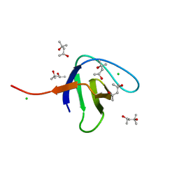
6SDF
DownloadVisualize
BU of 6sdf by Molmil
N-terminal SH3 domain of Grb2 protein
Descriptor: (4S)-2-METHYL-2,4-PENTANEDIOL, CHLORIDE ION, Growth factor receptor-bound protein 2
Authors:Bolgov, A.A, Korban, S.A, Luzik, D.A, Rogacheva, O.N, Zhemkov, V.A, Kim, M, Skrynnikov, N.R, Bezprozvanny, I.B.
Deposit date:2019-07-26
Release date:2020-01-29
Last modified:2024-01-24
Method:X-RAY DIFFRACTION (2.5 Å)
Cite:Crystal structure of the SH3 domain of growth factor receptor-bound protein 2.
Acta Crystallogr.,Sect.F, 76, 2020
7CFZ
DownloadVisualize
BU of 7cfz by Molmil
SH3 domain of NADPH oxidase activator 1
Descriptor: NADPH oxidase activator 1
Authors:Kim, M, Park, J.H, Attri, P, Lee, W.
Deposit date:2020-06-29
Release date:2021-07-07
Last modified:2023-11-29
Method:X-RAY DIFFRACTION (1.89 Å)
Cite:Structural modification of NADPH oxidase activator (Noxa 1) by oxidative stress: An experimental and computational study.
Int.J.Biol.Macromol., 163, 2020
3C0C
DownloadVisualize
BU of 3c0c by Molmil
X-ray Crystal Structure of the Rat Endophilin A2 SH3 Domain
Descriptor: Endophilin-A2
Authors:Loll, P.J, Swain, E.
Deposit date:2008-01-19
Release date:2008-03-18
Last modified:2011-07-13
Method:X-RAY DIFFRACTION (1.7 Å)
Cite:Crystal structure of the SH3 domain of rat endophilin A2
To be Published
7Y4N
DownloadVisualize
BU of 7y4n by Molmil
Insight into the C-terminal SH3 domain mediated binding of Drosophila Drk to Sos and Dos
Descriptor: Growth factor receptor-bound protein 2
Authors:Pooppadi, M.S, Ikeya, T, Sugasawa, H, Watanabe, R, Mishima, M, Inomata, K, Ito, Y.
Deposit date:2022-06-15
Release date:2022-08-31
Last modified:2024-05-15
Method:SOLUTION NMR
Cite:Insight into the C-terminal SH3 domain mediated binding of Drosophila Drk to Sos and Dos.
Biochem.Biophys.Res.Commun., 625, 2022
3CQT
DownloadVisualize
BU of 3cqt by Molmil
N53I V55L MUTANT of FYN SH3 DOMAIN
Descriptor: Proto-oncogene tyrosine-protein kinase Fyn
Authors:Neculai, A.M, Zarrine-Afsar, A, Howell, P.L, Davidson, A, Chan, H.S.
Deposit date:2008-04-03
Release date:2008-07-01
Last modified:2023-08-30
Method:X-RAY DIFFRACTION (1.6 Å)
Cite:Theoretical and experimental demonstration of the importance of specific nonnative interactions in protein folding.
Proc.Natl.Acad.Sci.Usa, 105, 2008
6XVM
DownloadVisualize
BU of 6xvm by Molmil
Crystal structure of c-Src SH3 domain without ATCUN motif: monomer 2
Descriptor: GLYCEROL, Proto-oncogene tyrosine-protein kinase Src
Authors:Camara-Artigas, A, Plaza-Garrido, M.
Deposit date:2020-01-22
Release date:2020-04-22
Last modified:2024-01-24
Method:X-RAY DIFFRACTION (0.9 Å)
Cite:The effect of an engineered ATCUN motif on the structure and biophysical properties of the SH3 domain of c-Src tyrosine kinase.
J.Biol.Inorg.Chem., 25, 2020
6XVN
DownloadVisualize
BU of 6xvn by Molmil
Crystal structure of c-Src SH3 domain without ATCUN motif: monomer 1
Descriptor: Proto-oncogene tyrosine-protein kinase Src
Authors:Camara-Artigas, A, Plaza-Garrido, M.
Deposit date:2020-01-22
Release date:2020-04-22
Last modified:2024-01-24
Method:X-RAY DIFFRACTION (1.7 Å)
Cite:The effect of an engineered ATCUN motif on the structure and biophysical properties of the SH3 domain of c-Src tyrosine kinase.
J.Biol.Inorg.Chem., 25, 2020
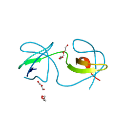
6XVO
DownloadVisualize
BU of 6xvo by Molmil
Crystal structure of the intertwined dimer of the c-Src SH3 domain without ATCUN motif
Descriptor: DI(HYDROXYETHYL)ETHER, Proto-oncogene tyrosine-protein kinase Src, TRIETHYLENE GLYCOL
Authors:Camara-Artigas, A, Plaza-Garrido, M.
Deposit date:2020-01-22
Release date:2020-04-22
Last modified:2024-01-24
Method:X-RAY DIFFRACTION (1.7 Å)
Cite:The effect of an engineered ATCUN motif on the structure and biophysical properties of the SH3 domain of c-Src tyrosine kinase.
J.Biol.Inorg.Chem., 25, 2020
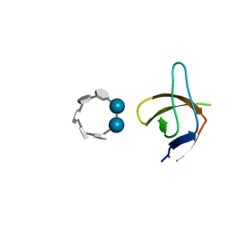
6XX3
DownloadVisualize
BU of 6xx3 by Molmil
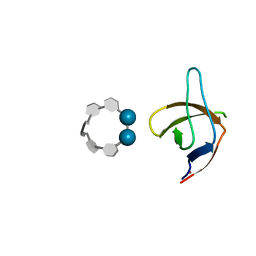
Crystal structure of the c-Src SH3 domain H122R-Q128E mutant in complex with Cu(II) at pH 6.5 co-crystallized with methyl beta-cyclodextrin
Descriptor: COPPER (II) ION, Cyclic 2,3-di-O-methyl-alpha-D-glucopyranose-(1-4)-2-O-methyl-alpha-D-glucopyranose-(1-4)-2,6-di-O-methyl-alpha-D-glucopyranose-(1-4)-2-O-methyl-alpha-D-glucopyranose-(1-4)-alpha-D-glucopyranose-(1-4)-alpha-D-glucopyranose-(1-4)-3-O-methyl-alpha-D-glucopyranose, Proto-oncogene tyrosine-protein kinase Src
Authors:Camara-Artigas, A.
Deposit date:2020-01-26
Release date:2020-04-22
Last modified:2024-01-24
Method:X-RAY DIFFRACTION (1.36 Å)
Cite:The effect of an engineered ATCUN motif on the structure and biophysical properties of the SH3 domain of c-Src tyrosine kinase.
J.Biol.Inorg.Chem., 25, 2020
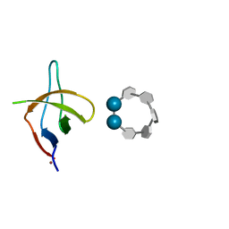
6XX4
DownloadVisualize
BU of 6xx4 by Molmil
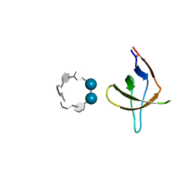
Crystal structure of the c-Src SH3 domain H122R-Q128E mutant in complex with Ni(II) at pH 7.5 co-crystallized with methyl beta-cyclodextrin
Descriptor: Cyclic 2,3-di-O-methyl-alpha-D-glucopyranose-(1-4)-2-O-methyl-alpha-D-glucopyranose-(1-4)-2,6-di-O-methyl-alpha-D-glucopyranose-(1-4)-2-O-methyl-alpha-D-glucopyranose-(1-4)-alpha-D-glucopyranose-(1-4)-alpha-D-glucopyranose-(1-4)-3-O-methyl-alpha-D-glucopyranose, NICKEL (II) ION, Proto-oncogene tyrosine-protein kinase Src
Authors:Camara-Artigas, A.
Deposit date:2020-01-26
Release date:2020-04-22
Last modified:2024-01-24
Method:X-RAY DIFFRACTION (1.05 Å)
Cite:The effect of an engineered ATCUN motif on the structure and biophysical properties of the SH3 domain of c-Src tyrosine kinase.
J.Biol.Inorg.Chem., 25, 2020
6XX5
DownloadVisualize
BU of 6xx5 by Molmil
Crystal structure of the c-Src SH3 domain H122R-Q128K mutant in complex with Ni(II) at pH 7.5 co-crystallized with methyl beta-cyclodextrin
Descriptor: Cyclic 2,3-di-O-methyl-alpha-D-glucopyranose-(1-4)-2-O-methyl-alpha-D-glucopyranose-(1-4)-2,6-di-O-methyl-alpha-D-glucopyranose-(1-4)-2-O-methyl-alpha-D-glucopyranose-(1-4)-alpha-D-glucopyranose-(1-4)-alpha-D-glucopyranose-(1-4)-3-O-methyl-alpha-D-glucopyranose, NICKEL (II) ION, Proto-oncogene tyrosine-protein kinase Src
Authors:Camara-Artigas, A.
Deposit date:2020-01-26
Release date:2020-05-27
Last modified:2024-01-24
Method:X-RAY DIFFRACTION (1.3 Å)
Cite:The effect of an engineered ATCUN motif on the structure and biophysical properties of the SH3 domain of c-Src tyrosine kinase.
J.Biol.Inorg.Chem., 25, 2020
6XX2
DownloadVisualize
BU of 6xx2 by Molmil
Crystal structure of the c-Src SH3 domain H122R-Q128K mutant in complex with Cu(II) at pH 7.5 co-crystallized with methyl beta-cyclodextrin
Descriptor: COPPER (II) ION, Cyclic 2,3-di-O-methyl-alpha-D-glucopyranose-(1-4)-2-O-methyl-alpha-D-glucopyranose-(1-4)-2,6-di-O-methyl-alpha-D-glucopyranose-(1-4)-2-O-methyl-alpha-D-glucopyranose-(1-4)-alpha-D-glucopyranose-(1-4)-alpha-D-glucopyranose-(1-4)-3-O-methyl-alpha-D-glucopyranose, Proto-oncogene tyrosine-protein kinase Src
Authors:Camara-Artigas, A.
Deposit date:2020-01-26
Release date:2020-04-22
Last modified:2024-01-24
Method:X-RAY DIFFRACTION (1.25 Å)
Cite:The effect of an engineered ATCUN motif on the structure and biophysical properties of the SH3 domain of c-Src tyrosine kinase.
J.Biol.Inorg.Chem., 25, 2020
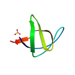
5XGG
DownloadVisualize
BU of 5xgg by Molmil
Crystal Structure C-terminal SH3 domain of Myosin IB from Entamoeba histolytica
Descriptor: SULFATE ION, Unconventional myosin IB
Authors:Gautam, G, Gourinath, S.
Deposit date:2017-04-13
Release date:2017-08-16
Last modified:2023-11-22
Method:X-RAY DIFFRACTION (1.72 Å)
Cite:Crystal structure of the PEG-bound SH3 domain of myosin IB from Entamoeba histolytica reveals its mode of ligand recognition
Acta Crystallogr D Struct Biol, 73, 2017
5XG9
DownloadVisualize
BU of 5xg9 by Molmil
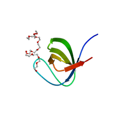
Crystal Structure of PEG-bound SH3 domain of Myosin IB from Entamoeba histolytica
Descriptor: 1-(2-METHOXY-ETHOXY)-2-{2-[2-(2-METHOXY-ETHOXY]-ETHOXY}-ETHANE, 2,5,8,11,14,17,20,23,26,29,32,35,38,41,44,47,50,53,56,59,62,65,68,71,74,77,80-HEPTACOSAOXADOOCTACONTAN-82-OL, PENTAETHYLENE GLYCOL, ...
Authors:Gautam, G, Gourinath, S.
Deposit date:2017-04-13
Release date:2017-08-16
Last modified:2023-11-22
Method:X-RAY DIFFRACTION (1.78 Å)
Cite:Crystal structure of the PEG-bound SH3 domain of myosin IB from Entamoeba histolytica reveals its mode of ligand recognition
Acta Crystallogr D Struct Biol, 73, 2017
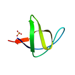
3M0P
DownloadVisualize
BU of 3m0p by Molmil
Crystal Structure of the R21D mutant of alpha-spectrin SH3 domain. Crystal obtained in ammonium sulphate at pH 4.
Descriptor: SULFATE ION, Spectrin alpha chain, brain
Authors:Camara-Artigas, A, Gavira, J.A.
Deposit date:2010-03-03
Release date:2011-01-19
Last modified:2023-09-06
Method:X-RAY DIFFRACTION (2.6 Å)
Cite:Understanding the polymorphic behaviour of a mutant of the alpha-spectrin SH3 domain by means of two 1.1 A structures
Acta Crystallogr.,Sect.D, 2011
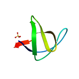
3M0S
DownloadVisualize
BU of 3m0s by Molmil
Crystal Structure of the R21D mutant of alpha-spectrin SH3 domain. Crystal obtained in ammonium sulphate at pH 7
Descriptor: SULFATE ION, Spectrin alpha chain, brain
Authors:Camara-Artigas, A, Gavira, J.A.
Deposit date:2010-03-03
Release date:2011-01-19
Last modified:2023-09-06
Method:X-RAY DIFFRACTION (1.6 Å)
Cite:Understanding the polymorphic behaviour of a mutant of the alpha-spectrin SH3 domain by means of two 1.1 A structures
Acta Crystallogr.,Sect.D, 2011
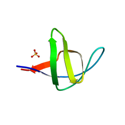
3M0R
DownloadVisualize
BU of 3m0r by Molmil
Crystal Structure of the R21D mutant of alpha-spectrin SH3 domain. Crystal obtained in ammonium sulphate at pH 6.
Descriptor: SULFATE ION, Spectrin alpha chain, brain
Authors:Gavira, J.A, Camara-Artigas, A.
Deposit date:2010-03-03
Release date:2011-01-19
Last modified:2023-09-06
Method:X-RAY DIFFRACTION (1.1 Å)
Cite:Understanding the polymorphic behaviour of a mutant of the alpha-spectrin SH3 domain by means of two 1.1 A structures
Acta Crystallogr.,Sect.D, 2011
3M0T
DownloadVisualize
BU of 3m0t by Molmil
Crystal Structure of the R21D mutant of alpha-spectrin SH3 domain. Crystal obtained in ammonium sulphate at pH 9.
Descriptor: SULFATE ION, Spectrin alpha chain, brain
Authors:Camara-Artigas, A, Gavira, J.A.
Deposit date:2010-03-03
Release date:2011-01-19
Last modified:2024-02-21
Method:X-RAY DIFFRACTION (1.7 Å)
Cite:Understanding the polymorphic behaviour of a mutant of the alpha-spectrin SH3 domain by means of two 1.1 A structures
Acta Crystallogr.,Sect.D, 2011

220113

PDB entries from 2024-05-22

PDB statisticsPDBj update infoContact PDBjnumon