Loading
PDBj
MenuPDBj@FacebookPDBj@TwitterPDBj@YouTubewwPDB FoundationwwPDB
RCSB PDBPDBeBMRBAdv. SearchSearch help
PDB: 12 results

6QTS
DownloadVisualize
BU of 6qts by Molmil
Crystal structure of a mutant Arabidopsis WD40 domain in complex with a photoreceptor
Descriptor: E3 ubiquitin-protein ligase COP1, GLYCEROL, SULFATE ION, ...
Authors:Hothorn, M, Lau, K.
Deposit date:2019-02-25
Release date:2019-07-10
Last modified:2024-01-24
Method:X-RAY DIFFRACTION (1.11 Å)
Cite:Plant photoreceptors and their signaling components compete for COP1 binding via VP peptide motifs.
Embo J., 38, 2019
6QTO
DownloadVisualize
BU of 6qto by Molmil
Crystal structure of an Arabidopsis WD40 domain in complex with a transcription factor
Descriptor: E3 ubiquitin-protein ligase COP1, GLYCEROL, SULFATE ION, ...
Authors:Hothorn, M, Lau, K.
Deposit date:2019-02-25
Release date:2019-07-10
Last modified:2024-01-24
Method:X-RAY DIFFRACTION (1.27 Å)
Cite:Plant photoreceptors and their signaling components compete for COP1 binding via VP peptide motifs.
Embo J., 38, 2019
6QTQ
DownloadVisualize
BU of 6qtq by Molmil
Crystal structure of an Arabidopsis WD40 domain in complex with photoreceptor
Descriptor: E3 ubiquitin-protein ligase COP1, GLYCEROL, SULFATE ION, ...
Authors:Hothorn, M, Lau, K.
Deposit date:2019-02-25
Release date:2019-07-10
Last modified:2024-01-24
Method:X-RAY DIFFRACTION (1.3 Å)
Cite:Plant photoreceptors and their signaling components compete for COP1 binding via VP peptide motifs.
Embo J., 38, 2019
6QTU
DownloadVisualize
BU of 6qtu by Molmil
Crystal structure of Arabidopsis WD40 domain in complex with a BBX transcription factor
Descriptor: B-box zinc finger protein 24, E3 ubiquitin-protein ligase COP1, GLYCEROL, ...
Authors:Hothorn, M, Lau, K.
Deposit date:2019-02-25
Release date:2019-07-10
Last modified:2024-01-24
Method:X-RAY DIFFRACTION (1.3 Å)
Cite:Plant photoreceptors and their signaling components compete for COP1 binding via VP peptide motifs.
Embo J., 38, 2019
6QTV
DownloadVisualize
BU of 6qtv by Molmil
Crystal structure of an Arabidopsis WD40 domain in complex with an atypical bHLH transcription factor
Descriptor: E3 ubiquitin-protein ligase COP1, GLYCEROL, MALONATE ION, ...
Authors:Hothorn, M, Lau, K.
Deposit date:2019-02-25
Release date:2019-07-10
Last modified:2024-01-24
Method:X-RAY DIFFRACTION (1.31 Å)
Cite:Plant photoreceptors and their signaling components compete for COP1 binding via VP peptide motifs.
Embo J., 38, 2019
6QTR
DownloadVisualize
BU of 6qtr by Molmil
Crystal structure of a mutant Arabidopsis WD40 domain in complex with a transcription factor
Descriptor: E3 ubiquitin-protein ligase COP1, GLYCEROL, SULFATE ION, ...
Authors:Hothorn, M, Lau, K.
Deposit date:2019-02-25
Release date:2019-07-10
Last modified:2024-01-24
Method:X-RAY DIFFRACTION (1.37 Å)
Cite:Plant photoreceptors and their signaling components compete for COP1 binding via VP peptide motifs.
Embo J., 38, 2019
6QTW
DownloadVisualize
BU of 6qtw by Molmil
Crystal structure of an Arabidopsis WD40 domain in complex with a blue light photoreceptor
Descriptor: Cryptochrome-1, E3 ubiquitin-protein ligase COP1, GLYCEROL, ...
Authors:Hothorn, M, Lau, K.
Deposit date:2019-02-25
Release date:2019-07-10
Last modified:2024-01-24
Method:X-RAY DIFFRACTION (1.39 Å)
Cite:Plant photoreceptors and their signaling components compete for COP1 binding via VP peptide motifs.
Embo J., 38, 2019
5KWN
DownloadVisualize
BU of 5kwn by Molmil
The WD domain ofArabidopsis thaliana E3 Ubiquitin Ligase COP1 in complex with peptide from HY5
Descriptor: E3 ubiquitin-protein ligase COP1, peptide 16-mer
Authors:Uljon, S, Blacklow, S.
Deposit date:2016-07-18
Release date:2017-07-26
Last modified:2023-10-04
Method:X-RAY DIFFRACTION (1.42 Å)
Cite:The COP1 WD domain in complex with HY5 peptide
To Be Published
6QTT
DownloadVisualize
BU of 6qtt by Molmil
Crystal structure of an Arabidopsis WD40 domain in complex with a transcription factor homolog
Descriptor: E3 ubiquitin-protein ligase COP1, GLYCEROL, MALONATE ION, ...
Authors:Hothorn, M, Lau, K.
Deposit date:2019-02-25
Release date:2019-07-10
Last modified:2024-01-24
Method:X-RAY DIFFRACTION (1.51 Å)
Cite:Plant photoreceptors and their signaling components compete for COP1 binding via VP peptide motifs.
Embo J., 38, 2019
5IGO
DownloadVisualize
BU of 5igo by Molmil
WD40 domain of Arabidopsis thaliana E3 Ubiquitin Ligase COP1 in complex with peptide from Trib1
Descriptor: E3 ubiquitin-protein ligase COP1, Tribbles homolog 1
Authors:Uljon, S, Blacklow, S.C.
Deposit date:2016-02-28
Release date:2016-04-20
Last modified:2023-09-27
Method:X-RAY DIFFRACTION (1.6 Å)
Cite:Structural Basis for Substrate Selectivity of the E3 Ligase COP1.
Structure, 24, 2016
6QTX
DownloadVisualize
BU of 6qtx by Molmil
Crystal structure of an Arabidopsis WD40 domain in complex with a flowering transcription factor homolog
Descriptor: E3 ubiquitin-protein ligase COP1, GLYCEROL, SULFATE ION, ...
Authors:Hothorn, M, Lau, K.
Deposit date:2019-02-25
Release date:2019-07-10
Last modified:2019-10-02
Method:X-RAY DIFFRACTION (1.95 Å)
Cite:Plant photoreceptors and their signaling components compete for COP1 binding via VP peptide motifs.
Embo J., 38, 2019
7VGG
DownloadVisualize
BU of 7vgg by Molmil
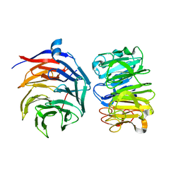
Cryo-EM structure of Ultraviolet-B activated UVR8 in complex with COP1
Descriptor: E3 ubiquitin-protein ligase COP1, Ultraviolet-B receptor UVR8
Authors:Wang, Y.D, Wang, L.X, Guan, Z.Y, Yin, P.
Deposit date:2021-09-16
Release date:2022-05-04
Last modified:2024-06-19
Method:ELECTRON MICROSCOPY (3.1 Å)
Cite:Structural insight into UV-B-activated UVR8 bound to COP1.
Sci Adv, 8, 2022

223532

PDB entries from 2024-08-07

PDB statisticsPDBj update infoContact PDBjnumon