Loading
PDBj
MenuPDBj@FacebookPDBj@TwitterPDBj@YouTubewwPDB FoundationwwPDB
RCSB PDBPDBeBMRBAdv. SearchSearch help
PDB: 2 results

1A7G
DownloadVisualize
BU of 1a7g by Molmil
THE CRYSTAL STRUCTURE OF THE E2 DNA-BINDING DOMAIN FROM HUMAN PAPILLOMAVIRUS AT 2.4 ANGSTROMS
Descriptor: REGULATORY PROTEIN E2, SULFATE ION
Authors:Bussiere, D.E, Giranda, V.L.
Deposit date:1998-03-13
Release date:1999-04-27
Last modified:2024-04-03
Method:X-RAY DIFFRACTION (2.4 Å)
Cite:Structure of the E2 DNA-binding domain from human papillomavirus serotype 31 at 2.4 A.
Acta Crystallogr.,Sect.D, 54, 1998
1DHM
DownloadVisualize
BU of 1dhm by Molmil
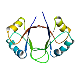
DNA-BINDING DOMAIN OF E2 FROM HUMAN PAPILLOMAVIRUS-31, NMR, MINIMIZED AVERAGE STRUCTURE
Descriptor: E2 PROTEIN
Authors:Liang, H, Petros, A.P, Meadows, R.P, Yoon, H.S, Egan, D.A, Walter, K, Holzman, T.F, Robins, T, Fesik, S.W.
Deposit date:1995-08-15
Release date:1996-12-07
Last modified:2024-04-10
Method:SOLUTION NMR
Cite:Solution structure of the DNA-binding domain of a human papillomavirus E2 protein: evidence for flexible DNA-binding regions.
Biochemistry, 35, 1996

224201

PDB entries from 2024-08-28

PDB statisticsPDBj update infoContact PDBjnumon