Loading
PDBj
MenuPDBj@FacebookPDBj@X(formerly Twitter)PDBj@BlueSkyPDBj@YouTubewwPDB FoundationwwPDBDonate
RCSB PDBPDBeBMRBAdv. SearchSearch help
PDB: 3 results

1GHL
DownloadVisualize
BU of 1ghl by Molmil
THE THREE-DIMENSIONAL STRUCTURE OF PHEASANT AND GUINEA-FOWL EGG LYSOZYMES
Descriptor: PHEASANT EGG WHITE LYSOZYME
Authors:Alzari, P.M, Lescar, J.
Deposit date:1993-05-04
Release date:1993-10-31
Last modified:2024-11-06
Method:X-RAY DIFFRACTION (2.1 Å)
Cite:Crystal structures of pheasant and guinea fowl egg-white lysozymes
Protein Sci., 3, 1994
1HHL
DownloadVisualize
BU of 1hhl by Molmil
THE THREE-DIMENSIONAL STRUCTURE OF PHEASANT AND GUINEA-FOWL EGG LYSOZYMES
Descriptor: GUINEA FOWL LYSOZYME
Authors:Alzari, P.M, Lescar, J.
Deposit date:1993-05-04
Release date:1993-10-31
Last modified:2024-10-30
Method:X-RAY DIFFRACTION (1.9 Å)
Cite:Crystal structures of pheasant and guinea fowl egg-white lysozymes
Protein Sci., 3, 1994
2GV0
DownloadVisualize
BU of 2gv0 by Molmil
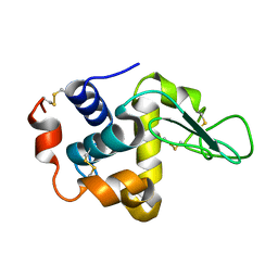
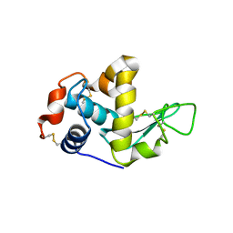
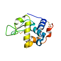
The structure of the orthorhombic form of soft-shelled turtle lysozyme at 1.9 angstroms resolution
Descriptor: Lysozyme C, SULFATE ION
Authors:Siritapetawee, J, Thammasirirak, S, Yuvaniyama, J, Robinson, R.C.
Deposit date:2006-05-02
Release date:2007-05-08
Last modified:2025-01-29
Method:X-RAY DIFFRACTION (1.9 Å)
Cite:The 1.9 A X-ray structure of egg-white lysozyme from Taiwanese soft-shelled turtle (Trionyx Sinensis Wiegmann) exhibits structural differences from the standard chicken-type lysozyme.
J.Biochem., 145, 2009

231029

PDB entries from 2025-02-05

PDB statisticsPDBj update infoContact PDBjnumon