3SPE
 
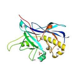 | | Crystal structure of the tail sheath protein protease resistant fragment from bacteriophage phiKZ | | Descriptor: | GLYCEROL, PHIKZ029, PHOSPHATE ION | | Authors: | Aksyuk, A.A, Kurochkina, L.P, Fokine, A, Mesyanzhinov, V.V, Rossmann, M.G. | | Deposit date: | 2011-07-01 | | Release date: | 2011-12-14 | | Last modified: | 2017-11-08 | | Method: | X-RAY DIFFRACTION (2.3996 Å) | | Cite: | Structural conservation of the myoviridae phage tail sheath protein fold.
Structure, 19, 2011
|
|
3J0I
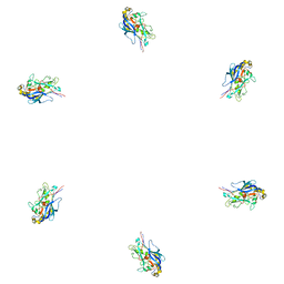 | | Fitting of the phiKZ gp29PR structure into the cryo-EM density map of the phiKZ polysheath | | Descriptor: | PHIKZ029 | | Authors: | Aksyuk, A.A, Fokine, A, Kurochkina, L.P, Mesyanzhinov, V.V, Rossmann, M.G. | | Deposit date: | 2011-08-16 | | Release date: | 2011-12-14 | | Last modified: | 2024-02-21 | | Method: | ELECTRON MICROSCOPY (19 Å) | | Cite: | Structural conservation of the myoviridae phage tail sheath protein fold.
Structure, 19, 2011
|
|
3J0H
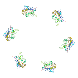 | | Fitting of the bacteriophage phiKZ gp29PR structure into the cryo-EM density map of the phiKZ extended tail sheath | | Descriptor: | PHIKZ029 | | Authors: | Aksyuk, A.A, Fokine, A, Kurochkina, L.P, Mesyanzhinov, V.V, Rossmann, M.G. | | Deposit date: | 2011-08-16 | | Release date: | 2011-12-14 | | Last modified: | 2024-02-21 | | Method: | ELECTRON MICROSCOPY (18 Å) | | Cite: | Structural conservation of the myoviridae phage tail sheath protein fold.
Structure, 19, 2011
|
|


