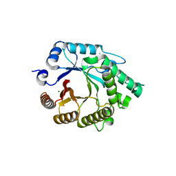
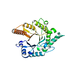
Loading
PDBj
MenuPDBj@FacebookPDBj@TwitterPDBj@YouTubewwPDB FoundationwwPDB
RCSB PDBPDBeBMRBAdv. SearchSearch help
PDB: 3 results

4E4P
DownloadVisualize
BU of 4e4p by Molmil
Second native structure of Xylanase A1 from Paenibacillus sp. JDR-2
Descriptor: CHLORIDE ION, Endo-1,4-beta-xylanase A, MAGNESIUM ION
Authors:Pozharski, E, St John, F.J.
Deposit date:2012-03-13
Release date:2012-04-18
Last modified:2024-02-28
Method:X-RAY DIFFRACTION (1.92 Å)
Cite:Novel structural features of xylanase A1 from Paenibacillus sp. JDR-2.
J.Struct.Biol., 180, 2012
3RDK
DownloadVisualize
BU of 3rdk by Molmil
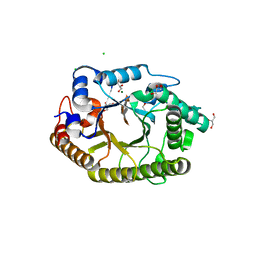
Protein crystal structure of xylanase A1 of Paenibacillus sp. JDR-2
Descriptor: 4-O-methyl-alpha-D-glucopyranuronic acid-(1-2)-beta-D-xylopyranose-(1-4)-beta-D-xylopyranose-(1-4)-alpha-D-xylopyranose, CHLORIDE ION, Endo-1,4-beta-xylanase, ...
Authors:Pozharski, E, St John, F.J.
Deposit date:2011-04-01
Release date:2012-04-04
Last modified:2024-02-21
Method:X-RAY DIFFRACTION (1.49 Å)
Cite:Novel structural features of xylanase A1 from Paenibacillus sp. JDR-2.
J.Struct.Biol., 180, 2012
3RO8
DownloadVisualize
BU of 3ro8 by Molmil
Crystal structure of the catalytic domain of XynA1 from Paenibacillus sp. JDR-2
Descriptor: CHLORIDE ION, Endo-1,4-beta-xylanase, MAGNESIUM ION
Authors:Pozharski, E, St John, F.J.
Deposit date:2011-04-25
Release date:2012-05-16
Last modified:2024-02-28
Method:X-RAY DIFFRACTION (1.34 Å)
Cite:Novel structural features of xylanase A1 from Paenibacillus sp. JDR-2.
J.Struct.Biol., 180, 2012

222415

PDB entries from 2024-07-10

PDB statisticsPDBj update infoContact PDBjnumon