Loading
PDBj
MenuPDBj@FacebookPDBj@TwitterPDBj@YouTubewwPDB FoundationwwPDB
RCSB PDBPDBeBMRBAdv. SearchSearch help

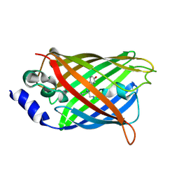
3DQF
DownloadVisualize
BU of 3dqf by Molmil
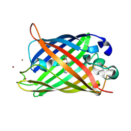
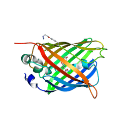
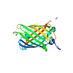
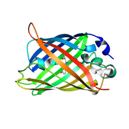
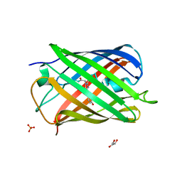
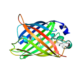
Structure of the Yellow Fluorescent Protein Citrine Frozen at 1000 Atmospheres Number 6: Structure 10 in a Series of 26 High Pressure Structures
Descriptor: Green fluorescent protein
Authors:Barstow, B, Kim, C.U.
Deposit date:2008-07-09
Release date:2008-09-23
Last modified:2023-11-15
Method:X-RAY DIFFRACTION (1.46 Å)
Cite:Alteration of citrine structure by hydrostatic pressure explains the accompanying spectral shift.
Proc.Natl.Acad.Sci.Usa, 105, 2008
2OTE
DownloadVisualize
BU of 2ote by Molmil
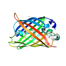
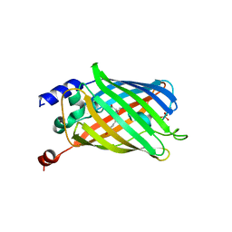
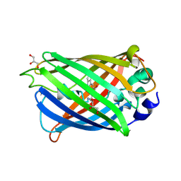
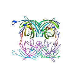
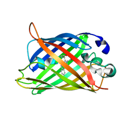
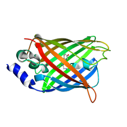
Crystal structure of a monomeric cyan fluorescent protein in the photobleached state
Descriptor: ACETATE ION, GFP-like fluorescent chromoprotein cFP484
Authors:Henderson, J.N, Ai, H, Campbell, R.E, Remington, S.J.
Deposit date:2007-02-07
Release date:2007-04-03
Last modified:2023-11-15
Method:X-RAY DIFFRACTION (1.47 Å)
Cite:Structural basis for reversible photobleaching of a green fluorescent protein homologue.
Proc.Natl.Acad.Sci.Usa, 104, 2007
3DQL
DownloadVisualize
BU of 3dql by Molmil
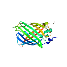
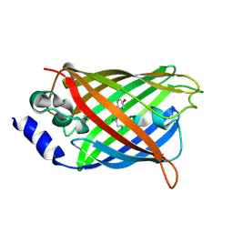
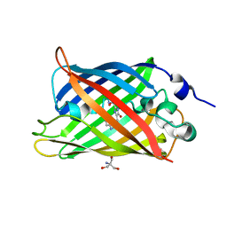
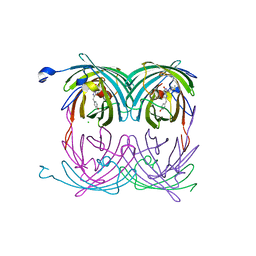
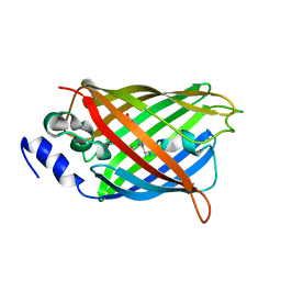
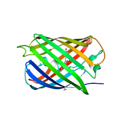
Structure of the Yellow Fluorescent Protein Citrine Frozen at 1000 Atmospheres Number 1: Structure 5 in a Series of 26 High Pressure Structures
Descriptor: Green fluorescent protein
Authors:Barstow, B, Kim, C.U.
Deposit date:2008-07-09
Release date:2008-09-23
Last modified:2023-11-15
Method:X-RAY DIFFRACTION (1.47 Å)
Cite:Alteration of citrine structure by hydrostatic pressure explains the accompanying spectral shift.
Proc.Natl.Acad.Sci.Usa, 105, 2008
2YE0
DownloadVisualize
BU of 2ye0 by Molmil
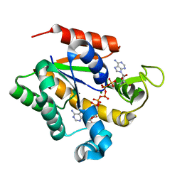
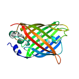
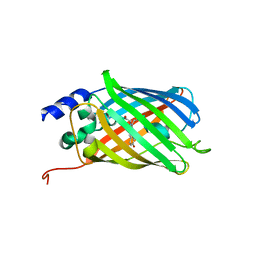
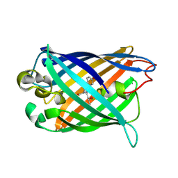
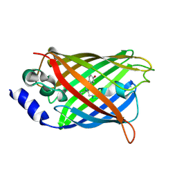
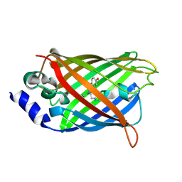
X-ray structure of the cyan fluorescent protein mTurquoise (K206A mutant)
Descriptor: GREEN FLUORESCENT PROTEIN
Authors:von Stetten, D, Goedhart, J, Noirclerc-Savoye, M, Lelimousin, M, Joosen, L, Hink, M.A, van Weeren, L, Gadella, T.W.J, Royant, A.
Deposit date:2011-03-25
Release date:2012-03-21
Last modified:2023-12-20
Method:X-RAY DIFFRACTION (1.47 Å)
Cite:Structure-Guided Evolution of Cyan Fluorescent Proteins Towards a Quantum Yield of 93%
Nat.Commun, 3, 2012
3DQ4
DownloadVisualize
BU of 3dq4 by Molmil
Structure of the Yellow Fluorescent Protein Citrine Frozen at 2000 Atmospheres Number 2: Structure 20 in a Series of 26 High Pressure Structures
Descriptor: Green fluorescent protein
Authors:Barstow, B, Kim, C.U.
Deposit date:2008-07-09
Release date:2008-09-23
Last modified:2023-11-15
Method:X-RAY DIFFRACTION (1.47 Å)
Cite:Alteration of citrine structure by hydrostatic pressure explains the accompanying spectral shift.
Proc.Natl.Acad.Sci.Usa, 105, 2008
2H6V
DownloadVisualize
BU of 2h6v by Molmil
Spectroscopic and structural study of the heterotropic linkage between halide and proton ion binding to GFP proteins- E2(GFP) APO FORM
Descriptor: Green fluorescent protein, SODIUM ION
Authors:Garau, G.
Deposit date:2006-06-01
Release date:2007-05-15
Last modified:2021-10-20
Method:X-RAY DIFFRACTION (1.47 Å)
Cite:Spectroscopic and Structural Study of Proton and Halide Ion Cooperative Binding to GFP
Biophys.J., 93, 2007
7A7P
DownloadVisualize
BU of 7a7p by Molmil
rsGreen0.7-K206A partially in the green-off state
Descriptor: 1,2-ETHANEDIOL, Green fluorescent protein, NITRATE ION
Authors:De Zitter, E, Dedecker, P, Van Meervelt, L.
Deposit date:2020-08-30
Release date:2021-02-17
Last modified:2021-05-05
Method:X-RAY DIFFRACTION (1.48 Å)
Cite:Structure-Function Dataset Reveals Environment Effects within a Fluorescent Protein Model System*.
Angew.Chem.Int.Ed.Engl., 60, 2021
1Q4B
DownloadVisualize
BU of 1q4b by Molmil
S65T Q80R Green Fluorescent Protein (GFP) pH 5.5
Descriptor: Green Fluorescent Protein
Authors:Jain, R.K, Ranganathan, R.
Deposit date:2003-08-02
Release date:2004-02-03
Last modified:2023-11-15
Method:X-RAY DIFFRACTION (1.48 Å)
Cite:Local complexity of amino acid interactions in a protein core.
Proc.Natl.Acad.Sci.USA, 101, 2004
6OFM
DownloadVisualize
BU of 6ofm by Molmil
Crystal structure of green fluorescent protein (GFP); S65T, Y66(3-CH3Y); ih circular permutant (50-51)
Descriptor: Green fluorescent protein (GFP); S65T, Y66(3-CH3Y); ih circular permutant (50-51)
Authors:Lin, C.-Y, Romei, M.G, Mathews, I.I, Boxer, S.G.
Deposit date:2019-03-31
Release date:2019-07-10
Last modified:2023-11-15
Method:X-RAY DIFFRACTION (1.48 Å)
Cite:Unified Model for Photophysical and Electro-Optical Properties of Green Fluorescent Proteins.
J.Am.Chem.Soc., 141, 2019
3CBE
DownloadVisualize
BU of 3cbe by Molmil
Development of a family of redox-sensitive green fluorescent protein indicators for use in relatively oxidizing subcellular environments
Descriptor: Green fluorescent protein, MAGNESIUM ION
Authors:Lohman, J.R.
Deposit date:2008-02-21
Release date:2008-08-19
Last modified:2021-10-20
Method:X-RAY DIFFRACTION (1.488 Å)
Cite:Development of a family of redox-sensitive green fluorescent protein indicators for use in relatively oxidizing subcellular environments
Biochemistry, 47, 2008
3DQC
DownloadVisualize
BU of 3dqc by Molmil
Structure of the Yellow Fluorescent Protein Citrine Frozen at 1250 Atmospheres Number 3: Structure 13 in a Series of 26 High Pressure Structures
Descriptor: Green fluorescent protein
Authors:Barstow, B, Kim, C.U.
Deposit date:2008-07-09
Release date:2008-09-23
Last modified:2023-11-15
Method:X-RAY DIFFRACTION (1.49 Å)
Cite:Alteration of citrine structure by hydrostatic pressure explains the accompanying spectral shift.
Proc.Natl.Acad.Sci.Usa, 105, 2008
3SVE
DownloadVisualize
BU of 3sve by Molmil
Engineered low-affinity halide-binding protein derived from YFP: bromide complex
Descriptor: BROMIDE ION, Green fluorescent protein
Authors:Wang, W, Grimley, J.S, Beese, L.S, Hellinga, H.W.
Deposit date:2011-07-12
Release date:2012-07-18
Last modified:2023-11-15
Method:X-RAY DIFFRACTION (1.492 Å)
Cite:Determination of engineered chloride-binding site structures in fluorescent proteins reveals principles of halide recognition
To be Published
3SS0
DownloadVisualize
BU of 3ss0 by Molmil
Engineered high-affinity halide-binding protein derived from YFP: fluoride complex
Descriptor: Green fluorescent protein
Authors:Wang, W, Grimley, J.S, Beese, L.S, Hellinga, H.W.
Deposit date:2011-07-07
Release date:2012-07-11
Last modified:2023-11-15
Method:X-RAY DIFFRACTION (1.492 Å)
Cite:Determination of engineered chloride-binding site structures in fluorescent proteins reveals principles of halide recognition
To be Published
4XGY
DownloadVisualize
BU of 4xgy by Molmil
GFP based antibody (fluorobody)
Descriptor: 3,6,9,12,15,18,21,24-OCTAOXAHEXACOSAN-1-OL, Green fluorescent protein, mAb LCDR3
Authors:Shi, N, Chen, Y.G, Wang, S.H.
Deposit date:2015-01-03
Release date:2015-07-01
Last modified:2023-11-15
Method:X-RAY DIFFRACTION (1.494 Å)
Cite:The structure of a GFP-based antibody (fluorobody) to TLH, a toxin from Vibrio parahaemolyticus.
Acta Crystallogr.,Sect.F, 71, 2015
6UN4
DownloadVisualize
BU of 6un4 by Molmil
Crystal structure of rsEGFP2, Y67(3-ClY), Y107(3-ClY)
Descriptor: Green fluorescent protein, SULFATE ION
Authors:Lin, C.-Y, Romei, M.G, Boxer, S.G, Chang, J.
Deposit date:2019-10-10
Release date:2020-07-15
Last modified:2023-11-15
Method:X-RAY DIFFRACTION (1.499 Å)
Cite:Structural and spectroscopic characterization of photoactive yellow protein and photoswitchable fluorescent protein constructs containing heavy atoms.
J Photochem Photobiol A Chem, 401, 2020
6UN6
DownloadVisualize
BU of 6un6 by Molmil
Crystal structure of green fluorescent protein (GFP); S65T, Y66(3-NO2Y); ih circular permutant (50-51)
Descriptor: Green fluorescent protein
Authors:Lin, C.-Y, Boxer, S.G.
Deposit date:2019-10-10
Release date:2020-10-14
Last modified:2023-11-15
Method:X-RAY DIFFRACTION (1.5 Å)
Cite:Unusual Spectroscopic and Electric Field Sensitivity of Chromophores with Short Hydrogen Bonds: GFP and PYP as Model Systems.
J.Phys.Chem.B, 124, 2020
2RH7
DownloadVisualize
BU of 2rh7 by Molmil
Crystal Structures of the Luciferase and Green Fluorescent Protein from Renilla Reniformis
Descriptor: Green fluorescent protein
Authors:Loening, A.M, Fenn, T.D, Gambhir, S.S.
Deposit date:2007-10-06
Release date:2007-10-23
Last modified:2023-11-15
Method:X-RAY DIFFRACTION (1.5 Å)
Cite:Crystal Structures of the Luciferase and Green Fluorescent Protein from Renilla reniformis.
J.Mol.Biol., 374, 2007
2DUF
DownloadVisualize
BU of 2duf by Molmil
crystal structure of a green fluorescent protein variant S65T/H148D at pH 5.6
Descriptor: Green fluorescent protein
Authors:Shu, X, Remington, S.J.
Deposit date:2006-07-23
Release date:2007-07-10
Last modified:2021-11-10
Method:X-RAY DIFFRACTION (1.5 Å)
Cite:Ultrafast excited-state dynamics in the green fluorescent protein variant S65T/H148D. 1. Mutagenesis and structural studies.
Biochemistry, 46, 2007
7QGK
DownloadVisualize
BU of 7qgk by Molmil
The mRubyFT protein, Genetically Encoded Blue-to-Red Fluorescent Timer in its red state
Descriptor: MAGNESIUM ION, The red form of the mRubyFT protein, Genetically Encoded Blue-to-Red Fluorescent Timer
Authors:Boyko, K.M, Nikolaeva, A.Y, Gaivoronskii, F.A, Vlaskina, A.V, Subach, O.M, Popov, V.O, Subach, F.V.
Deposit date:2021-12-08
Release date:2022-03-23
Last modified:2024-02-07
Method:X-RAY DIFFRACTION (1.5 Å)
Cite:The mRubyFT Protein, Genetically Encoded Blue-to-Red Fluorescent Timer.
Int J Mol Sci, 23, 2022
3LVA
DownloadVisualize
BU of 3lva by Molmil
Crystal structure of colorless GFP-like protein from Aequorea coerulescens
Descriptor: GLYCEROL, Green fluorescent protein, SULFATE ION
Authors:Pletneva, N.V, Pletnev, V.Z, Pletnev, S.V.
Deposit date:2010-02-19
Release date:2010-03-09
Last modified:2023-11-15
Method:X-RAY DIFFRACTION (1.5 Å)
Cite:Structural evidence for a dehydrated intermediate in green fluorescent protein chromophore biosynthesis.
J.Biol.Chem., 285, 2010
3W1D
DownloadVisualize
BU of 3w1d by Molmil
Structure of a pressure sensitive YFP variant YFP-G3
Descriptor: Green Fluorescent protein
Authors:Imada, K, Yoshizawa, K, Kinoshita, M, Watanabe, T.M.
Deposit date:2012-11-14
Release date:2013-10-02
Last modified:2023-12-06
Method:X-RAY DIFFRACTION (1.5 Å)
Cite:Glycine insertion makes yellow fluorescent protein sensitive to hydrostatic pressure.
Plos One, 8, 2013
7QLN
DownloadVisualize
BU of 7qln by Molmil
rsKiiro pump probe structure by TR-SFX
Descriptor: rsKiiro
Authors:van Thor, J.J.
Deposit date:2021-12-20
Release date:2023-10-18
Last modified:2023-11-15
Method:X-RAY DIFFRACTION (1.5 Å)
Cite:Optical control of ultrafast structural dynamics in a fluorescent protein.
Nat.Chem., 15, 2023
5DTY
DownloadVisualize
BU of 5dty by Molmil
Crystal structure of rsEGFP2 in the non-fluorescent off-state
Descriptor: Green fluorescent protein
Authors:Adam, V, Dos-Santos Martins, A.
Deposit date:2015-09-18
Release date:2016-01-20
Method:X-RAY DIFFRACTION (1.5 Å)
Cite:Rational design of ultrastable and reversibly photoswitchable fluorescent proteins for super-resolution imaging of the bacterial periplasm.
Sci Rep, 6, 2016
1H6R
DownloadVisualize
BU of 1h6r by Molmil
The oxidized state of a redox sensitive variant of green fluorescent protein
Descriptor: CHLORIDE ION, GREEN FLUORESCENT PROTEIN
Authors:Ostergaard, H, Henriksen, A, Hansen, F.G, Winther, J.R.
Deposit date:2001-06-22
Release date:2001-11-15
Last modified:2023-12-13
Method:X-RAY DIFFRACTION (1.5 Å)
Cite:Shedding Light on Disulfide Bond Formation: Engineering a Redox Switch in Green Fluorescent Protein
Embo J., 20, 2001
2Y0G
DownloadVisualize
BU of 2y0g by Molmil
X-ray structure of Enhanced Green Fluorescent Protein (EGFP)
Descriptor: GREEN FLUORESCENT PROTEIN
Authors:Royant, A, Noirclerc-Savoye, M.
Deposit date:2010-12-02
Release date:2011-04-27
Last modified:2023-12-20
Method:X-RAY DIFFRACTION (1.5 Å)
Cite:Stabilizing Role of Glutamic Acid 222 in the Structure of Enhanced Green Fluorescent Protein.
J.Struct.Biol., 174, 2011

221051

數據於2024-06-12公開中

PDB statisticsPDBj update infoContact PDBjnumon