Loading
PDBj
MenuPDBj@FacebookPDBj@TwitterPDBj@YouTubewwPDB FoundationwwPDB
RCSB PDBPDBeBMRBAdv. SearchSearch help

1MUA
DownloadVisualize
BU of 1mua by Molmil
STRUCTURE AND ENERGETICS OF A NON-PROLINE CIS-PEPTIDYL LINKAGE IN AN ENGINEERED PROTEIN
Descriptor: CARBONIC ANHYDRASE II, MERCURY (II) ION, ZINC ION
Authors:Nair, S.K, Christianson, D.W.
Deposit date:1993-06-18
Release date:1993-10-31
Last modified:2024-02-14
Method:X-RAY DIFFRACTION (1.7 Å)
Cite:Structure and energetics of a non-proline cis-peptidyl linkage in a proline-202-->alanine carbonic anhydrase II variant.
Biochemistry, 32, 1993
1BYH
DownloadVisualize
BU of 1byh by Molmil
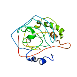
MOLECULAR AND ACTIVE-SITE STRUCTURE OF A BACILLUS (1-3,1-4)-BETA-GLUCANASE
Descriptor: CALCIUM ION, HYBRID, N-BUTANE, ...
Authors:Keitel, T, Heinemann, U.
Deposit date:1992-12-31
Release date:1993-10-31
Last modified:2020-07-29
Method:X-RAY DIFFRACTION (2.8 Å)
Cite:Molecular and active-site structure of a Bacillus 1,3-1,4-beta-glucanase.
Proc.Natl.Acad.Sci.USA, 90, 1993
1CVB
DownloadVisualize
BU of 1cvb by Molmil
STRUCTURAL AND FUNCTIONAL IMPORTANCE OF A CONSERVED HYDROGEN BOND NETWORK IN HUMAN CARBONIC ANHYDRASE II
Descriptor: CARBONIC ANHYDRASE II, SULFATE ION, ZINC ION
Authors:Ippolito, J.A, Christianson, D.W.
Deposit date:1993-02-04
Release date:1993-10-31
Last modified:2024-02-07
Method:X-RAY DIFFRACTION (2.4 Å)
Cite:Structural and functional importance of a conserved hydrogen bond network in human carbonic anhydrase II.
J.Biol.Chem., 268, 1993
1BW4
DownloadVisualize
BU of 1bw4 by Molmil
THREE-DIMENSIONAL STRUCTURE IN SOLUTION OF BARWIN, A PROTEIN FROM BARLEY SEED
Descriptor: BARWIN, BASIC BARLEY SEED PROTEIN
Authors:Poulsen, F.M.
Deposit date:1992-07-06
Release date:1993-10-31
Last modified:2017-11-29
Method:SOLUTION NMR
Cite:Three-dimensional structure in solution of barwin, a protein from barley seed.
Biochemistry, 31, 1992
1CPS
DownloadVisualize
BU of 1cps by Molmil
STRUCTURAL COMPARISON OF SULFODIIMINE AND SULFONAMIDE INHIBITORS IN THEIR COMPLEXES WITH ZINC ENZYMES
Descriptor: CARBOXYPEPTIDASE A, S-(2-CARBOXY-3-PHENYLPROPYL)THIODIIMINE-S-METHANE, ZINC ION
Authors:Cappalonga, A.M, Alexander, R.S, Christianson, D.W.
Deposit date:1992-02-18
Release date:1993-10-31
Last modified:2024-06-05
Method:X-RAY DIFFRACTION (2.25 Å)
Cite:Structural comparison of sulfodiimine and sulfonamide inhibitors in their complexes with zinc enzymes.
J.Biol.Chem., 267, 1992
2BPP
DownloadVisualize
BU of 2bpp by Molmil
PHOSPHOLIPASE A2 ENGINEERING. X-RAY STRUCTURAL AND FUNCTIONAL EVIDENCE FOR THE INTERACTION OF LYSINE-56 WITH SUBSTRATES
Descriptor: CALCIUM ION, PHOSPHOLIPASE A2
Authors:Sundaralingam, M.
Deposit date:1992-01-17
Release date:1993-10-31
Last modified:2017-11-29
Method:X-RAY DIFFRACTION (1.8 Å)
Cite:Phospholipase A2 engineering. X-ray structural and functional evidence for the interaction of lysine-56 with substrates.
Biochemistry, 30, 1991
1CLM
DownloadVisualize
BU of 1clm by Molmil
STRUCTURE OF PARAMECIUM TETRAURELIA CALMODULIN AT 1.8 ANGSTROMS RESOLUTION
Descriptor: CALCIUM ION, CALMODULIN
Authors:Sundaralingam, M.
Deposit date:1993-01-23
Release date:1993-10-31
Last modified:2024-02-07
Method:X-RAY DIFFRACTION (1.8 Å)
Cite:Structure of Paramecium tetraurelia calmodulin at 1.8 A resolution.
Protein Sci., 2, 1993
1FBF
DownloadVisualize
BU of 1fbf by Molmil
CRYSTALLOGRAPHIC STUDIES OF THE CATALYTIC MECHANISM OF THE NEUTRAL FORM OF FRUCTOSE-1,6-BISPHOSPHATASE
Descriptor: 2,5-anhydro-1,6-di-O-phosphono-D-mannitol, FRUCTOSE 1,6-BISPHOSPHATASE, MAGNESIUM ION
Authors:Zhang, Y, Liang, J.-Y, Huang, S, Ke, H, Lipscomb, W.N.
Deposit date:1992-10-16
Release date:1993-10-31
Last modified:2024-02-07
Method:X-RAY DIFFRACTION (2.7 Å)
Cite:Crystallographic studies of the catalytic mechanism of the neutral form of fructose-1,6-bisphosphatase.
Biochemistry, 32, 1993
1FAN
DownloadVisualize
BU of 1fan by Molmil
CREVICE-FORMING MUTANTS IN THE RIGID CORE OF BOVINE PANCREATIC TRYPSIN INHIBITOR: CRYSTAL STRUCTURES OF F22A, Y23A, N43G, AND F45A
Descriptor: BOVINE PANCREATIC TRYPSIN INHIBITOR
Authors:Danishefsky, A.T, Wlodawer, A, Kim, K.-S, Tao, F, Woodward, C.
Deposit date:1992-08-21
Release date:1993-10-31
Last modified:2019-08-14
Method:X-RAY DIFFRACTION (2 Å)
Cite:Crevice-forming mutants in the rigid core of bovine pancreatic trypsin inhibitor: crystal structures of F22A, Y23A, N43G, and F45A.
Protein Sci., 2, 1993
1BPQ
DownloadVisualize
BU of 1bpq by Molmil
PHOSPHOLIPASE A2 ENGINEERING. X-RAY STRUCTURAL AND FUNCTIONAL EVIDENCE FOR THE INTERACTION OF LYSINE-56 WITH SUBSTRATES
Descriptor: CALCIUM ION, PHOSPHOLIPASE A2
Authors:Sundaralingam, M.
Deposit date:1991-10-28
Release date:1993-10-31
Last modified:2017-11-29
Method:X-RAY DIFFRACTION (1.8 Å)
Cite:Phospholipase A2 engineering. X-ray structural and functional evidence for the interaction of lysine-56 with substrates.
Biochemistry, 30, 1991
1RBG
DownloadVisualize
BU of 1rbg by Molmil
CRYSTALLOGRAPHIC STRUCTURES OF RIBONUCLEASE S VARIANTS WITH NONPOLAR SUBSTITUTION AT POSITION 13: PACKING AND CAVITIES
Descriptor: RIBONUCLEASE S (S-PEPTIDE), RIBONUCLEASE S (S-PROTEIN), SULFATE ION
Authors:Varadarajan, R, Richards, F.M.
Deposit date:1992-06-12
Release date:1993-10-31
Last modified:2017-11-29
Method:X-RAY DIFFRACTION (1.8 Å)
Cite:Crystallographic structures of ribonuclease S variants with nonpolar substitution at position 13: packing and cavities.
Biochemistry, 31, 1992
7AAT
DownloadVisualize
BU of 7aat by Molmil
X-RAY STRUCTURE REFINEMENT AND COMPARISON OF THREE FORMS OF MITOCHONDRIAL ASPARTATE AMINOTRANSFERASE
Descriptor: ASPARTATE AMINOTRANSFERASE, PYRIDOXAL-5'-PHOSPHATE
Authors:Mcphalen, C.A, Vincent, M.G, Jansonius, J.N.
Deposit date:1991-12-02
Release date:1993-10-31
Last modified:2017-11-29
Method:X-RAY DIFFRACTION (1.9 Å)
Cite:X-ray structure refinement and comparison of three forms of mitochondrial aspartate aminotransferase.
J.Mol.Biol., 225, 1992
1RDD
DownloadVisualize
BU of 1rdd by Molmil
CRYSTAL STRUCTURE OF ESCHERICHIA COLI RNASE HI IN COMPLEX WITH MG2+ AT 2.8 ANGSTROMS RESOLUTION: PROOF FOR A SINGLE MG2+ SITE
Descriptor: MAGNESIUM ION, RIBONUCLEASE H
Authors:Katayanagi, K, Morikawa, K.
Deposit date:1993-06-23
Release date:1993-10-31
Last modified:2024-02-14
Method:X-RAY DIFFRACTION (2.8 Å)
Cite:Crystal structure of Escherichia coli RNase HI in complex with Mg2+ at 2.8 A resolution: proof for a single Mg(2+)-binding site.
Proteins, 17, 1993
1RNE
DownloadVisualize
BU of 1rne by Molmil
THE CRYSTAL STRUCTURE OF RECOMBINANT GLYCOSYLATED HUMAN RENIN ALONE AND IN COMPLEX WITH A TRANSITION STATE ANALOG INHIBITOR
Descriptor: 2-acetamido-2-deoxy-beta-D-glucopyranose, RENIN, [[[3-(2-METHYL-PROPANE-2-SULFONYL)-1-BENZENYL]-2-PROPYL]-CARBONYL-HISTIDYL]-AMINO-[CYCLOHEXYLMETHYL]-[2-HYDROXY-4-ISOPROPYL]-PENTAN-5-OIC ACID BUTYLAMIDE
Authors:Gruetter, M.G, Rahuel, J, Priestle, J.P.
Deposit date:1991-12-12
Release date:1993-10-31
Last modified:2020-10-21
Method:X-RAY DIFFRACTION (2.4 Å)
Cite:The crystal structures of recombinant glycosylated human renin alone and in complex with a transition state analog inhibitor.
J.Struct.Biol., 107, 1991
1FCS
DownloadVisualize
BU of 1fcs by Molmil
CRYSTAL STRUCTURE OF A DISTAL SITE DOUBLE MUTANT OF SPERM WHALE MYOGLOBIN AT 1.6 ANGSTROMS RESOLUTION
Descriptor: MYOGLOBIN, PROTOPORPHYRIN IX CONTAINING FE, SULFATE ION
Authors:Rizzi, M, Bolognesi, M, Coda, A, Cutruzzola, F, Travaglini Allocatelli, C, Brancaccio, A, Brunori, M.
Deposit date:1993-04-20
Release date:1993-10-31
Last modified:2024-02-07
Method:X-RAY DIFFRACTION (1.6 Å)
Cite:Crystal structure of a distal site double mutant of sperm whale myoglobin at 1.6 A resolution.
FEBS Lett., 320, 1993
1RCM
DownloadVisualize
BU of 1rcm by Molmil
CRYSTAL STRUCTURE OF A UBIQUITIN-DEPENDENT DEGRADATION SUBSTRATE: A THREE-DISULFIDE FORM OF LYSOZYME
Descriptor: ACETIC ACID, HEN EGG WHITE LYSOZYME
Authors:Hill, C.P, Johnston, N.L, Cohen, R.E.
Deposit date:1993-01-10
Release date:1993-10-31
Last modified:2020-01-15
Method:X-RAY DIFFRACTION (1.9 Å)
Cite:Crystal structure of a ubiquitin-dependent degradation substrate: a three-disulfide form of lysozyme.
Proc.Natl.Acad.Sci.USA, 90, 1993
1REA
DownloadVisualize
BU of 1rea by Molmil
STRUCTURE OF THE RECA PROTEIN-ADP COMPLEX
Descriptor: ADENOSINE-5'-DIPHOSPHATE, REC A
Authors:Story, R.M, Steitz, T.A.
Deposit date:1991-12-19
Release date:1993-10-31
Last modified:2024-02-14
Method:X-RAY DIFFRACTION (2.7 Å)
Cite:Structure of the recA protein-ADP complex.
Nature, 355, 1992
1RTC
DownloadVisualize
BU of 1rtc by Molmil
THE STRUCTURE OF RECOMBINANT RICIN A CHAIN AT 2.3 ANGSTROMS
Descriptor: RICIN
Authors:Mlsna, D, Monzingo, A.F, Katzin, B.J, Ernst, S, Robertus, J.D.
Deposit date:1992-10-29
Release date:1993-10-31
Last modified:2024-02-14
Method:X-RAY DIFFRACTION (2.3 Å)
Cite:Structure of recombinant ricin A chain at 2.3 A.
Protein Sci., 2, 1993
2HHM
DownloadVisualize
BU of 2hhm by Molmil
STRUCTURE OF INOSITOL MONOPHOSPHATASE, THE PUTATIVE TARGET OF LITHIUM THERAPY
Descriptor: GADOLINIUM ATOM, INOSITOL MONOPHOSPHATASE, SULFATE ION
Authors:Bone, R.
Deposit date:1992-10-20
Release date:1993-10-31
Last modified:2024-02-14
Method:X-RAY DIFFRACTION (2.1 Å)
Cite:Structure of inositol monophosphatase, the putative target of lithium therapy.
Proc.Natl.Acad.Sci.USA, 89, 1992
1RIN
DownloadVisualize
BU of 1rin by Molmil
X-RAY CRYSTAL STRUCTURE OF A PEA LECTIN-TRIMANNOSIDE COMPLEX AT 2.6 ANGSTROMS RESOLUTION
Descriptor: CALCIUM ION, MANGANESE (II) ION, PEA LECTIN, ...
Authors:Rini, J.M, Hardman, K.D, Einspahr, H, Suddath, F.L, Carver, J.P.
Deposit date:1993-01-27
Release date:1993-10-31
Last modified:2024-02-14
Method:X-RAY DIFFRACTION (2.6 Å)
Cite:X-ray crystal structure of a pea lectin-trimannoside complex at 2.6 A resolution.
J.Biol.Chem., 268, 1993
1RBD
DownloadVisualize
BU of 1rbd by Molmil
CRYSTALLOGRAPHIC STRUCTURES OF RIBONUCLEASE S VARIANTS WITH NONPOLAR SUBSTITUTION AT POSITION 13: PACKING AND CAVITIES
Descriptor: RIBONUCLEASE S (S-PEPTIDE), RIBONUCLEASE S (S-PROTEIN), SULFATE ION
Authors:Varadarajan, R, Richards, F.M.
Deposit date:1992-06-12
Release date:1993-10-31
Last modified:2017-11-29
Method:X-RAY DIFFRACTION (1.7 Å)
Cite:Crystallographic structures of ribonuclease S variants with nonpolar substitution at position 13: packing and cavities.
Biochemistry, 31, 1992
1CVA
DownloadVisualize
BU of 1cva by Molmil
STRUCTURAL AND FUNCTIONAL IMPORTANCE OF A CONSERVED HYDROGEN BOND NETWORK IN HUMAN CARBONIC ANHYDRASE II
Descriptor: AZIDE ION, CARBONIC ANHYDRASE II, ZINC ION
Authors:Ippolito, J.A, Christianson, D.W.
Deposit date:1993-02-04
Release date:1993-10-31
Last modified:2024-02-07
Method:X-RAY DIFFRACTION (2.25 Å)
Cite:Structural and functional importance of a conserved hydrogen bond network in human carbonic anhydrase II.
J.Biol.Chem., 268, 1993
1CMA
DownloadVisualize
BU of 1cma by Molmil
MET REPRESSOR/DNA COMPLEX + S-ADENOSYL-METHIONINE
Descriptor: DNA (5'-D(*AP*GP*AP*CP*GP*TP*CP*TP*A)-3'), DNA (5'-D(*TP*TP*AP*GP*AP*CP*GP*TP*CP*T)-3'), PROTEIN (MET REPRESSOR), ...
Authors:Somers, W.S, Phillips, S.E.V.
Deposit date:1992-08-24
Release date:1993-10-31
Last modified:2024-02-07
Method:X-RAY DIFFRACTION (2.8 Å)
Cite:Crystal structure of the met repressor-operator complex at 2.8 A resolution reveals DNA recognition by beta-strands.
Nature, 359, 1992
1COB
DownloadVisualize
BU of 1cob by Molmil
CRYSTAL STRUCTURE SOLUTION AND REFINEMENT OF THE SEMISYNTHETIC COBALT SUBSTITUTED BOVINE ERYTHROCYTE ENZYME SUPEROXIDE DISMUTASE AT 2.0 ANGSTROMS RESOLUTION
Descriptor: COBALT (II) ION, COPPER (II) ION, SUPEROXIDE DISMUTASE
Authors:Djinovic, K, Coda, A, Antolini, L, Pelosi, G, Desideri, A, Falconi, M, Rotilio, G, Bolognesi, M.
Deposit date:1992-02-19
Release date:1993-10-31
Last modified:2011-07-13
Method:X-RAY DIFFRACTION (2 Å)
Cite:Crystal structure solution and refinement of the semisynthetic cobalt-substituted bovine erythrocyte superoxide dismutase at 2.0 A resolution.
J.Mol.Biol., 226, 1992
1RBH
DownloadVisualize
BU of 1rbh by Molmil
CRYSTALLOGRAPHIC STRUCTURES OF RIBONUCLEASE S VARIANTS WITH NONPOLAR SUBSTITUTION AT POSITION 13: PACKING AND CAVITIES
Descriptor: RIBONUCLEASE S (S-PEPTIDE), RIBONUCLEASE S (S-PROTEIN), SULFATE ION
Authors:Varadarajan, R, Richards, F.M.
Deposit date:1992-06-12
Release date:1993-10-31
Last modified:2017-11-29
Method:X-RAY DIFFRACTION (1.7 Å)
Cite:Crystallographic structures of ribonuclease S variants with nonpolar substitution at position 13: packing and cavities.
Biochemistry, 31, 1992

225158

數據於2024-09-18公開中

PDB statisticsPDBj update infoContact PDBjnumon