Loading
PDBj
MenuPDBj@FacebookPDBj@TwitterPDBj@YouTubewwPDB FoundationwwPDB
RCSB PDBPDBeBMRBAdv. SearchSearch help

5YEV
DownloadVisualize
BU of 5yev by Molmil
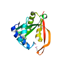
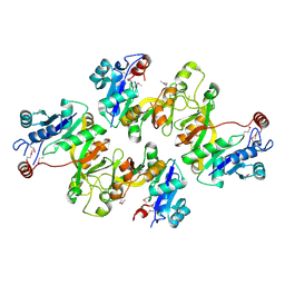
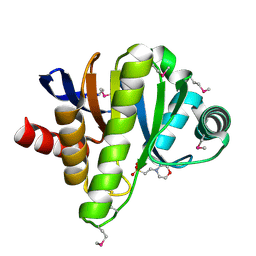
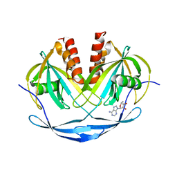
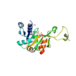
Murine DR3 death domain
Descriptor: SULFATE ION, TNFRSF25 death domain
Authors:Yin, X, Jin, T.
Deposit date:2017-09-19
Release date:2018-09-19
Last modified:2023-11-22
Method:X-RAY DIFFRACTION (2.5 Å)
Cite:Crystal structure and activation mechanism of DR3 death domain.
Febs J., 286, 2019
5O2F
DownloadVisualize
BU of 5o2f by Molmil
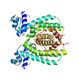
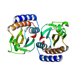
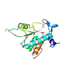
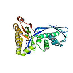
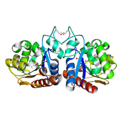
Crystal structure of NDM-1 in complex with hydrolyzed ampicillin - new refinement
Descriptor: (2R,4S)-2-[(R)-{[(2R)-2-amino-2-phenylacetyl]amino}(carboxy)methyl]-5,5-dimethyl-1,3-thiazolidine-4-carboxylic acid, 1,2-ETHANEDIOL, CHLORIDE ION, ...
Authors:Raczynska, J.E, Shabalin, I.G, Jaskolski, M, Minor, W, Wlodawer, A.
Deposit date:2017-05-20
Release date:2018-12-26
Last modified:2024-02-07
Method:X-RAY DIFFRACTION (2.01 Å)
Cite:A close look onto structural models and primary ligands of metallo-beta-lactamases.
Drug Resist. Updat., 40, 2018
6IN7
DownloadVisualize
BU of 6in7 by Molmil
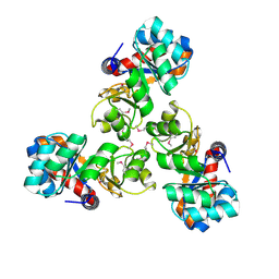
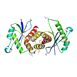
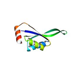
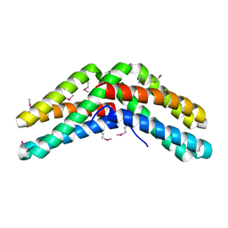
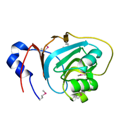
Crystal structure of AlgU in complex with MucA(cyto)
Descriptor: NICOTINAMIDE, RNA polymerase sigma-H factor, Sigma factor AlgU negative regulatory protein
Authors:Li, S, Zhang, Q, Bartlam, M.
Deposit date:2018-10-24
Release date:2019-07-24
Last modified:2023-11-22
Method:X-RAY DIFFRACTION (1.96 Å)
Cite:Structural basis for the recognition of MucA by MucB and AlgU in Pseudomonas aeruginosa.
Febs J., 286, 2019
5O2E
DownloadVisualize
BU of 5o2e by Molmil
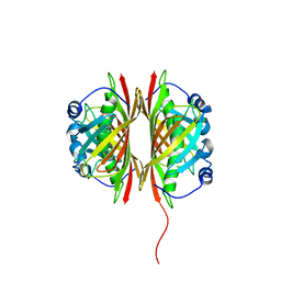
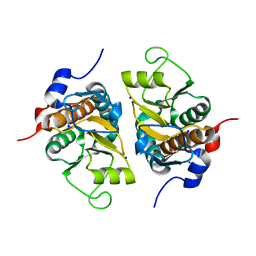
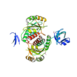
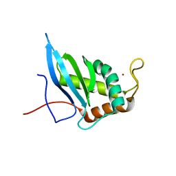
Crystal structure of NDM-1 in complex with hydrolyzed cefuroxime - new refinement
Descriptor: (2R,5S)-5-[(carbamoyloxy)methyl]-2-[(R)-carboxy{[(2Z)-2-(furan-2-yl)-2-(methoxyimino)acetyl]amino}methyl]-5,6-dihydro-2H-1,3-thiazine-4-carboxylic acid, Metallo-beta-lactamase type 2, SULFATE ION, ...
Authors:Raczynska, J.E, Shabalin, I.G, Jaskolski, M, Minor, W, Wlodawer, A.
Deposit date:2017-05-20
Release date:2018-12-26
Last modified:2024-06-19
Method:X-RAY DIFFRACTION (1.3 Å)
Cite:A close look onto structural models and primary ligands of metallo-beta-lactamases.
Drug Resist. Updat., 40, 2018
6JOY
DownloadVisualize
BU of 6joy by Molmil
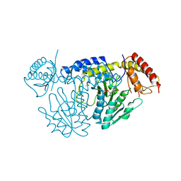
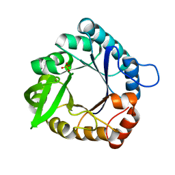
The X-ray Crystallographic Structure of Branching Enzyme from Rhodothermus obamensis STB05
Descriptor: 1,4-alpha-glucan branching enzyme GlgB
Authors:Li, Z.F, Ban, X.F, Jiang, H.M, Wang, Z, Jin, T.C, Li, C.M, Gu, Z.B.
Deposit date:2019-03-25
Release date:2020-03-04
Last modified:2024-03-27
Method:X-RAY DIFFRACTION (2.392 Å)
Cite:Flexible Loop in Carbohydrate-Binding Module 48 Allosterically Modulates Substrate Binding of the 1,4-alpha-Glucan Branching Enzyme.
J.Agric.Food Chem., 69, 2021
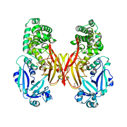
6H34
DownloadVisualize
BU of 6h34 by Molmil
The crystal structure of human carbonic anhydrase II in complex with 4-[(4-fluorophenyl)methyl]-1-piperazinyl]benzenesulfonamide.
Descriptor: 4-[4-[(4-fluorophenyl)methyl]piperazin-1-yl]carbonylbenzenesulfonamide, Carbonic anhydrase 2, GLYCEROL, ...
Authors:Buemi, M.R, Di Fiore, A, De Luca, L, Ferro, S, Mancuso, F, Monti, S.M, Buonanno, M, Angeli, A, Russo, E, De Sarro, G, Supuran, C.T, De Simone, G, Gitto, R.
Deposit date:2018-07-17
Release date:2018-12-19
Last modified:2024-01-17
Method:X-RAY DIFFRACTION (1.55 Å)
Cite:Exploring structural properties of potent human carbonic anhydrase inhibitors bearing a 4-(cycloalkylamino-1-carbonyl)benzenesulfonamide moiety.
Eur J Med Chem, 163, 2018
5OFT
DownloadVisualize
BU of 5oft by Molmil
Structural basis for OXA-48 dimerization
Descriptor: Beta-lactamase
Authors:Lund, B.A, Nesheim, B.H.B, Leiros, H.K.S.
Deposit date:2017-07-11
Release date:2018-08-01
Last modified:2024-01-17
Method:X-RAY DIFFRACTION (3.2 Å)
Cite:The biological assembly of OXA-48 reveals a dimer interface with high charge complementarity and very high affinity.
FEBS J., 285, 2018
6JNA
DownloadVisualize
BU of 6jna by Molmil
Cryo-EM structure of glutamate dehydrogenase from Thermococcus profundus
Descriptor: 2-AMINO-2-HYDROXYMETHYL-PROPANE-1,3-DIOL, Glutamate dehydrogenase
Authors:Oide, M, Kato, T, Oroguchi, T, Nakasako, M.
Deposit date:2019-03-14
Release date:2020-02-12
Last modified:2024-03-27
Method:ELECTRON MICROSCOPY (3.8 Å)
Cite:Energy landscape of domain motion in glutamate dehydrogenase deduced from cryo-electron microscopy.
Febs J., 287, 2020
6JDP
DownloadVisualize
BU of 6jdp by Molmil
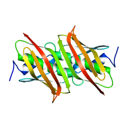
the imm52 family protein TsiT (PA3908) from Pseudomonas aeruginosa
Descriptor: Imm52 family protein, PHOSPHATE ION, SODIUM ION
Authors:She, Z.
Deposit date:2019-02-02
Release date:2019-03-20
Last modified:2024-03-27
Method:X-RAY DIFFRACTION (1.601 Å)
Cite:Structural characterization of the Imm52 family protein TsiT in Pseudomonas aeruginosa.
Protein Sci., 28, 2019
6JN9
DownloadVisualize
BU of 6jn9 by Molmil
Cryo-EM structure of glutamate dehydrogenase from Thermococcus profundus
Descriptor: 2-AMINO-2-HYDROXYMETHYL-PROPANE-1,3-DIOL, Glutamate dehydrogenase
Authors:Oide, M, Kato, T, Oroguchi, T, Nakasako, M.
Deposit date:2019-03-14
Release date:2020-02-12
Last modified:2024-03-27
Method:ELECTRON MICROSCOPY (3.8 Å)
Cite:Energy landscape of domain motion in glutamate dehydrogenase deduced from cryo-electron microscopy.
Febs J., 287, 2020
6JNC
DownloadVisualize
BU of 6jnc by Molmil
Cryo-EM structure of glutamate dehydrogenase from Thermococcus profundus
Descriptor: 2-AMINO-2-HYDROXYMETHYL-PROPANE-1,3-DIOL, Glutamate dehydrogenase
Authors:Oide, M, Kato, T, Oroguchi, T, Nakasako, M.
Deposit date:2019-03-14
Release date:2020-02-12
Last modified:2024-03-27
Method:ELECTRON MICROSCOPY (3.7 Å)
Cite:Energy landscape of domain motion in glutamate dehydrogenase deduced from cryo-electron microscopy.
Febs J., 287, 2020
6H2Z
DownloadVisualize
BU of 6h2z by Molmil
The crystal structure of human carbonic anhydrase II in complex with 4-(4-phenylpiperidine-1-carbonyl)benzenesulfonamide.
Descriptor: 4-(4-phenylpiperidin-1-yl)carbonylbenzenesulfonamide, Carbonic anhydrase 2, GLYCEROL, ...
Authors:Buemi, M.R, Di Fiore, A, De Luca, L, Ferro, S, Mancuso, F, Monti, S.M, Buonanno, M, Angeli, A, Russo, E, De Sarro, G, Supuran, C.T, De Simone, G, Gitto, R.
Deposit date:2018-07-17
Release date:2018-12-19
Last modified:2024-01-17
Method:X-RAY DIFFRACTION (1.94 Å)
Cite:Exploring structural properties of potent human carbonic anhydrase inhibitors bearing a 4-(cycloalkylamino-1-carbonyl)benzenesulfonamide moiety.
Eur J Med Chem, 163, 2018
6H33
DownloadVisualize
BU of 6h33 by Molmil
The crystal structure of human carbonic anhydrase II in complex with 4-(4-phenyl)-4-hydroxy-1-piperidine-1-carbonyl)benzenesulfonamide.
Descriptor: 4-(4-oxidanyl-4-phenyl-piperidin-1-yl)carbonylbenzenesulfonamide, Carbonic anhydrase 2, GLYCEROL, ...
Authors:Buemi, M.R, Di Fiore, A, De Luca, L, Ferro, S, Mancuso, F, Monti, S.M, Buonanno, M, Angeli, A, Russo, E, De Sarro, G, Supuran, C.T, De Simone, G, Gitto, R.
Deposit date:2018-07-17
Release date:2018-12-19
Last modified:2024-01-17
Method:X-RAY DIFFRACTION (1.58 Å)
Cite:Exploring structural properties of potent human carbonic anhydrase inhibitors bearing a 4-(cycloalkylamino-1-carbonyl)benzenesulfonamide moiety.
Eur J Med Chem, 163, 2018
1IE7
DownloadVisualize
BU of 1ie7 by Molmil
PHOSPHATE INHIBITED BACILLUS PASTEURII UREASE CRYSTAL STRUCTURE
Descriptor: NICKEL (II) ION, PHOSPHATE ION, UREASE ALPHA SUBUNIT, ...
Authors:Benini, S, Rypniewski, W.R, Wilson, K.S, Ciurli, S, Mangani, S.
Deposit date:2001-04-09
Release date:2001-04-25
Last modified:2023-11-15
Method:X-RAY DIFFRACTION (1.85 Å)
Cite:Structure-based rationalization of urease inhibition by phosphate: novel insights into the enzyme mechanism.
J.Biol.Inorg.Chem., 6, 2001
5Z09
DownloadVisualize
BU of 5z09 by Molmil
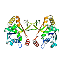
ST0452(Y97N)-UTP binding form
Descriptor: Dual sugar-1-phosphate nucleotidylyltransferase, URIDINE 5'-TRIPHOSPHATE
Authors:Honda, Y, Nakano, S, Ito, S, Dadashipour, M, Zhang, Z, Kawarabayasi, Y.
Deposit date:2017-12-19
Release date:2018-10-31
Last modified:2023-11-22
Method:X-RAY DIFFRACTION (2.91 Å)
Cite:Improvement of ST0452N-Acetylglucosamine-1-Phosphate Uridyltransferase Activity by the Cooperative Effect of Two Single Mutations Identified through Structure-Based Protein Engineering
Appl. Environ. Microbiol., 84, 2018
6JND
DownloadVisualize
BU of 6jnd by Molmil
Cryo-EM structure of glutamate dehydrogenase from Thermococcus profundus
Descriptor: 2-AMINO-2-HYDROXYMETHYL-PROPANE-1,3-DIOL, Glutamate dehydrogenase
Authors:Oide, M, Kato, T, Oroguchi, T, Nakasako, M.
Deposit date:2019-03-14
Release date:2020-02-12
Last modified:2024-03-27
Method:ELECTRON MICROSCOPY (3.9 Å)
Cite:Energy landscape of domain motion in glutamate dehydrogenase deduced from cryo-electron microscopy.
Febs J., 287, 2020
6H38
DownloadVisualize
BU of 6h38 by Molmil
The crystal structure of human carbonic anhydrase VII in complex with 4-[(4-fluorophenyl)methyl]-1-piperazinyl]benzenesulfonamide.
Descriptor: 4-[4-[(4-fluorophenyl)methyl]piperazin-1-yl]carbonylbenzenesulfonamide, Carbonic anhydrase 7, GLYCEROL, ...
Authors:Buemi, M.R, Di Fiore, A, De Luca, L, Ferro, S, Mancuso, F, Monti, S.M, Buonanno, M, Angeli, A, Russo, E, De Sarro, G, Supuran, C.T, De Simone, G, Gitto, R.
Deposit date:2018-07-17
Release date:2018-12-19
Last modified:2024-01-17
Method:X-RAY DIFFRACTION (1.7 Å)
Cite:Exploring structural properties of potent human carbonic anhydrase inhibitors bearing a 4-(cycloalkylamino-1-carbonyl)benzenesulfonamide moiety.
Eur J Med Chem, 163, 2018
6H36
DownloadVisualize
BU of 6h36 by Molmil
The crystal structure of human carbonic anhydrase VII in complex with 4-(4-phenylpiperidine-1-carbonyl)benzenesulfonamide.
Descriptor: 4-(4-phenylpiperidin-1-yl)carbonylbenzenesulfonamide, Carbonic anhydrase 7, ZINC ION
Authors:Buemi, M.R, Di Fiore, A, De Luca, L, Ferro, S, Mancuso, F, Monti, S.M, Buonanno, M, Angeli, A, Russo, E, De Sarro, G, Supuran, C.T, De Simone, G, Gitto, R.
Deposit date:2018-07-17
Release date:2018-12-19
Last modified:2024-01-17
Method:X-RAY DIFFRACTION (1.85 Å)
Cite:Exploring structural properties of potent human carbonic anhydrase inhibitors bearing a 4-(cycloalkylamino-1-carbonyl)benzenesulfonamide moiety.
Eur J Med Chem, 163, 2018
5K59
DownloadVisualize
BU of 5k59 by Molmil
Crystal structure of LukGH from Staphylococcus aureus in complex with a neutralising antibody
Descriptor: CHLORIDE ION, Fab heavy chain, Fab light chain, ...
Authors:Welin, M, Logan, D.T, Badarau, A, Mirkina, I, Zauner, G, Dolezilkova, I, Nagy, E.
Deposit date:2016-05-23
Release date:2016-08-10
Last modified:2018-01-17
Method:X-RAY DIFFRACTION (2.84 Å)
Cite:Context matters: The importance of dimerization-induced conformation of the LukGH leukocidin of Staphylococcus aureus for the generation of neutralizing antibodies.
Mabs, 8, 2016
6H37
DownloadVisualize
BU of 6h37 by Molmil
The crystal structure of human carbonic anhydrase VII in complex with 4-(4-phenyl)-4-hydroxy-1-piperidine-1-carbonyl)benzenesulfonamide
Descriptor: 4-(4-oxidanyl-4-phenyl-piperidin-1-yl)carbonylbenzenesulfonamide, Carbonic anhydrase 7, ZINC ION
Authors:Buemi, M.R, Di Fiore, A, De Luca, L, Ferro, S, Mancuso, F, Monti, S.M, Buonanno, M, Angeli, A, Russo, E, De Sarro, G, Supuran, C.T, De Simone, G, Gitto, R.
Deposit date:2018-07-17
Release date:2018-12-19
Last modified:2024-01-17
Method:X-RAY DIFFRACTION (1.9 Å)
Cite:Exploring structural properties of potent human carbonic anhydrase inhibitors bearing a 4-(cycloalkylamino-1-carbonyl)benzenesulfonamide moiety.
Eur J Med Chem, 163, 2018
1LKA
DownloadVisualize
BU of 1lka by Molmil
Porcine Pancreatic Elastase/Ca-Complex
Descriptor: ACETATE ION, CALCIUM ION, CHLORIDE ION, ...
Authors:Weiss, M.S, Panjikar, S, Nowak, E, Tucker, P.A.
Deposit date:2002-04-24
Release date:2002-08-28
Last modified:2023-08-16
Method:X-RAY DIFFRACTION (1.7 Å)
Cite:Metal binding to porcine pancreatic elastase: calcium or not calcium.
Acta Crystallogr.,Sect.D, 58, 2002
1LKB
DownloadVisualize
BU of 1lkb by Molmil
Porcine Pancreatic Elastase/Na-Complex
Descriptor: CHLORIDE ION, Elastase 1, SODIUM ION, ...
Authors:Weiss, M.S, Panjikar, S, Nowak, E, Tucker, P.A.
Deposit date:2002-04-24
Release date:2002-08-28
Last modified:2023-08-16
Method:X-RAY DIFFRACTION (1.7 Å)
Cite:Metal binding to porcine pancreatic elastase: calcium or not calcium.
Acta Crystallogr.,Sect.D, 58, 2002
6K35
DownloadVisualize
BU of 6k35 by Molmil
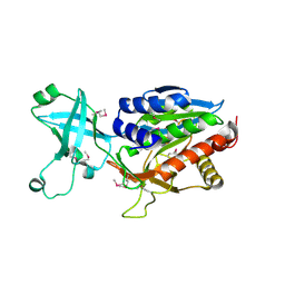
Crystal structure of GH20 exo beta-N-acetylglucosaminidase from Vibrio harveyi in complex with NAG-thiazoline
Descriptor: 3AR,5R,6S,7R,7AR-5-HYDROXYMETHYL-2-METHYL-5,6,7,7A-TETRAHYDRO-3AH-PYRANO[3,2-D]THIAZOLE-6,7-DIOL, Beta-N-acetylglucosaminidase Nag2
Authors:Meekrathok, P, Stubbs, K.A, Bulmer, D.M, van den Berg, B, Suginta, W.
Deposit date:2019-05-16
Release date:2020-05-13
Last modified:2023-11-22
Method:X-RAY DIFFRACTION (2.36 Å)
Cite:NAG-thiazoline is a potent inhibitor of the Vibrio campbellii GH20 beta-N-Acetylglucosaminidase.
Febs J., 287, 2020
5OQ1
DownloadVisualize
BU of 5oq1 by Molmil
Crystal structure of Serratia marcescens ChiX (used as MR model for superior PDB 5OPZ)
Descriptor: CHLORIDE ION, ChiX, ZINC ION
Authors:Owen, R.A, Fyfe, P.K, Lodge, A, Biboy, J, Vollmer, W, Hunter, W.N, Sargent, F.
Deposit date:2017-08-10
Release date:2018-01-17
Last modified:2024-05-08
Method:X-RAY DIFFRACTION (1.34 Å)
Cite:Structure and activity of ChiX: a peptidoglycan hydrolase required for chitinase secretion by Serratia marcescens.
Biochem. J., 475, 2018
2KQO
DownloadVisualize
BU of 2kqo by Molmil
A 3D-structural model of unsulphated chondroitin from high-field NMR: 4-sulphation has little effect on backbone conformation
Descriptor: beta-D-glucopyranuronic acid-(1-3)-2-acetamido-2-deoxy-beta-D-galactopyranose-(1-4)-beta-D-glucopyranuronic acid-(1-3)-2-acetamido-2-deoxy-beta-D-galactopyranose-(1-4)-beta-D-glucopyranuronic acid-(1-3)-2-acetamido-2-deoxy-beta-D-galactopyranose
Authors:Sattelle, B.M, Shakeri, J, Roberts, I.S, Almond, A.
Deposit date:2009-11-12
Release date:2009-12-01
Last modified:2024-05-01
Method:SOLUTION NMR
Cite:A 3D-structural model of unsulfated chondroitin from high-field NMR: 4-sulfation has little effect on backbone conformation.
Carbohydr.Res., 345, 2010

222415

數據於2024-07-10公開中

PDB statisticsPDBj update infoContact PDBjnumon