Loading
PDBj
MenuPDBj@FacebookPDBj@TwitterPDBj@YouTubewwPDB FoundationwwPDB
RCSB PDBPDBeBMRBAdv. SearchSearch help

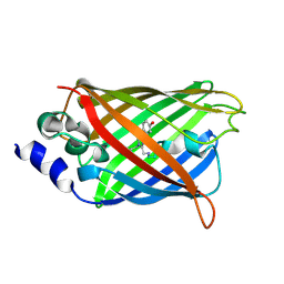
3SSY
DownloadVisualize
BU of 3ssy by Molmil
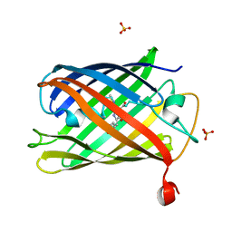
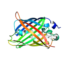
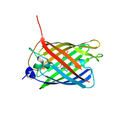
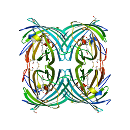
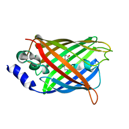
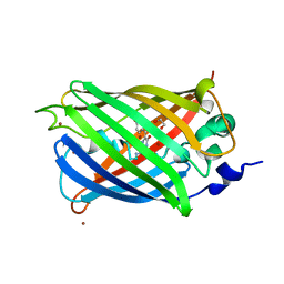
Engineered low-affinity halide-binding protein derived from YFP: iodide complex
Descriptor: Green fluorescent protein, IODIDE ION
Authors:Wang, W, Grimley, J.S, Beese, L.S, Hellinga, H.W.
Deposit date:2011-07-08
Release date:2012-07-11
Last modified:2023-11-15
Method:X-RAY DIFFRACTION (1.769 Å)
Cite:Determination of engineered chloride-binding site structures in fluorescent proteins reveals principles of halide recognition
To be Published
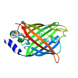
3ST0
DownloadVisualize
BU of 3st0 by Molmil
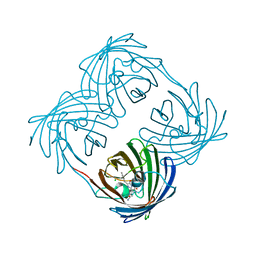
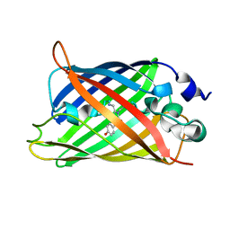
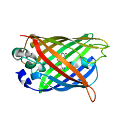
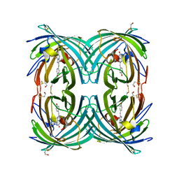
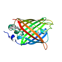
Engineered medium-affinity halide-binding protein derived from YFP: halide-free
Descriptor: 1,2-ETHANEDIOL, FORMIC ACID, Green fluorescent protein
Authors:Wang, W, Grimley, J.S, Beese, L.S, Hellinga, H.W.
Deposit date:2011-07-08
Release date:2012-07-11
Last modified:2024-04-03
Method:X-RAY DIFFRACTION (1.19 Å)
Cite:Visualization of Synaptic Inhibition with an Optogenetic Sensor Developed by Cell-Free Protein Engineering Automation.
J.Neurosci., 33, 2013
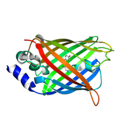
3SSP
DownloadVisualize
BU of 3ssp by Molmil
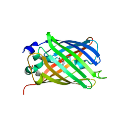
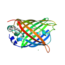
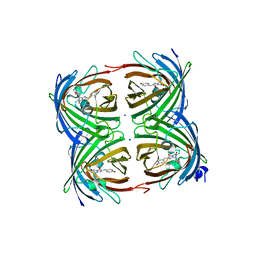
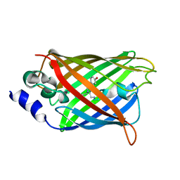
Engineered low-affinity halide-binding protein derived from YFP: halide-free
Descriptor: Green fluorescent protein
Authors:Wang, W, Grimley, J.S, Beese, L.S, Hellinga, H.W.
Deposit date:2011-07-08
Release date:2012-07-11
Last modified:2023-11-15
Method:X-RAY DIFFRACTION (1.628 Å)
Cite:Determination of engineered chloride-binding site structures in fluorescent proteins reveals principles of halide recognition
To be Published
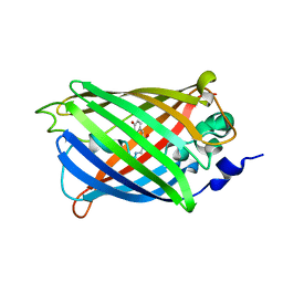
3SSV
DownloadVisualize
BU of 3ssv by Molmil
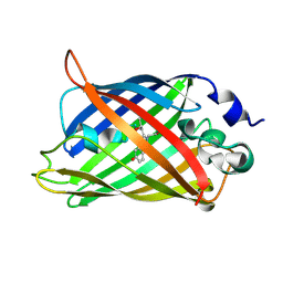
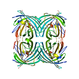
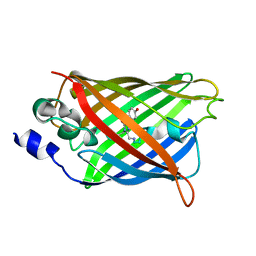
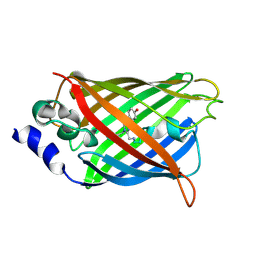
Engineered low-affinity halide-binding protein derived from YFP: fluoride complex
Descriptor: FLUORIDE ION, Green fluorescent protein
Authors:Wang, W, Grimley, J.S, Beese, L.S, Hellinga, H.W.
Deposit date:2011-07-08
Release date:2012-07-11
Last modified:2023-11-15
Method:X-RAY DIFFRACTION (1.861 Å)
Cite:Determination of engineered chloride-binding site structures in fluorescent proteins reveals principles of halide recognition
To be Published
3SVD
DownloadVisualize
BU of 3svd by Molmil
Engineered medium-affinity halide-binding protein derived from YFP: bromide complex
Descriptor: 1,2-ETHANEDIOL, BROMIDE ION, Green fluorescent protein
Authors:Wang, W, Grimley, J.S, Beese, L.S, Hellinga, H.W.
Deposit date:2011-07-12
Release date:2012-07-18
Last modified:2023-11-15
Method:X-RAY DIFFRACTION (1.78 Å)
Cite:Determination of engineered chloride-binding site structures in fluorescent proteins reveals principles of halide recognition
To be Published
3U0N
DownloadVisualize
BU of 3u0n by Molmil
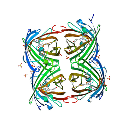
Crystal structure of the engineered fluorescent protein mRuby, crystal form 2
Descriptor: SULFATE ION, mRuby
Authors:Akerboom, J, Looger, L.L, Schreiter, E.R.
Deposit date:2011-09-28
Release date:2012-10-03
Last modified:2023-12-06
Method:X-RAY DIFFRACTION (1.6 Å)
Cite:Genetically encoded calcium indicators for multi-color neural activity imaging and combination with optogenetics.
Front Mol Neurosci, 6, 2013
3TMT
DownloadVisualize
BU of 3tmt by Molmil
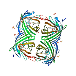
IrisFP, distorted chromophore
Descriptor: Green to red photoconvertible GPF-like protein EosFP, SULFATE ION
Authors:Adam, V, Carpentier, P, Roy, A, Field, M, Bourgeois, D.
Deposit date:2011-08-31
Release date:2011-11-16
Last modified:2023-11-15
Method:X-RAY DIFFRACTION (2 Å)
Cite:The nature of transient dark States in a photoactivatable fluorescent protein.
J.Am.Chem.Soc., 133, 2011
3E5V
DownloadVisualize
BU of 3e5v by Molmil
Crystal Structure Analysis of eqFP611 Double Mutant T122R, N143S
Descriptor: Red fluorescent protein eqFP611
Authors:Nar, H, Nienhaus, K, Nienhaus, U, Wiedenmann, J.
Deposit date:2008-08-14
Release date:2008-09-23
Last modified:2023-11-15
Method:X-RAY DIFFRACTION (2.1 Å)
Cite:Trans-cis isomerization is responsible for the red-shifted fluorescence in variants of the red fluorescent protein eqFP611.
J.Am.Chem.Soc., 130, 2008
3E5T
DownloadVisualize
BU of 3e5t by Molmil
Crystal Structure Analysis of FP611
Descriptor: Red fluorescent protein eqFP611
Authors:Nar, H, Nienhaus, K, Nienhaus, U, Wiedenmann, J.
Deposit date:2008-08-14
Release date:2008-09-23
Last modified:2023-11-15
Method:X-RAY DIFFRACTION (1.1 Å)
Cite:Trans-cis isomerization is responsible for the red-shifted fluorescence in variants of the red fluorescent protein eqFP611.
J.Am.Chem.Soc., 130, 2008
3E5W
DownloadVisualize
BU of 3e5w by Molmil
Crystal Structure Analysis of FP611
Descriptor: Red fluorescent protein eqFP611
Authors:Nienhaus, K, Nar, H, Heilker, R, Wiedenmann, J, Nienhaus, G.U.
Deposit date:2008-08-14
Release date:2008-09-23
Last modified:2023-11-15
Method:X-RAY DIFFRACTION (1.71 Å)
Cite:Trans-cis isomerization is responsible for the red-shifted fluorescence in variants of the red fluorescent protein eqFP611.
J.Am.Chem.Soc., 130, 2008
3ED8
DownloadVisualize
BU of 3ed8 by Molmil
Application of the superfolder YFP bimolecular fluorescence complementation for studying protein-protein interactions in vitro
Descriptor: yellow fluorescence protein
Authors:Ottmann, C, Weyand, M.
Deposit date:2008-09-02
Release date:2009-01-20
Last modified:2022-12-21
Method:X-RAY DIFFRACTION (2.7 Å)
Cite:Applicability of superfolder YFP bimolecular fluorescence complementation in vitro.
Biol.Chem., 390, 2009
3EVP
DownloadVisualize
BU of 3evp by Molmil
crystal structure of circular-permutated EGFP
Descriptor: Green fluorescent protein,Green fluorescent protein
Authors:Wang, Q, Shui, B, Kotlikoff, M.I, Sondermann, H.
Deposit date:2008-10-13
Release date:2008-12-09
Last modified:2023-12-27
Method:X-RAY DIFFRACTION (1.453 Å)
Cite:Structural Basis for Calcium Sensing by GCaMP2.
Structure, 16, 2008
3DQ1
DownloadVisualize
BU of 3dq1 by Molmil
Structure of the Yellow Fluorescent Protein Citrine Frozen at 4000 Atmospheres Number 2: Structure 24 in a Series of 26 High Pressure Structures
Descriptor: Green fluorescent protein
Authors:Barstow, B, Kim, C.U.
Deposit date:2008-07-09
Release date:2008-09-23
Last modified:2023-11-15
Method:X-RAY DIFFRACTION (1.7 Å)
Cite:Alteration of citrine structure by hydrostatic pressure explains the accompanying spectral shift.
Proc.Natl.Acad.Sci.Usa, 105, 2008
3DQA
DownloadVisualize
BU of 3dqa by Molmil
Structure of the Yellow Fluorescent Protein Citrine Frozen at 1250 Atmospheres Number 4: Structure 14 in a Series of 26 High Pressure Structures
Descriptor: Green fluorescent protein
Authors:Barstow, B, Kim, C.U.
Deposit date:2008-07-09
Release date:2008-09-23
Last modified:2023-11-15
Method:X-RAY DIFFRACTION (1.44 Å)
Cite:Alteration of citrine structure by hydrostatic pressure explains the accompanying spectral shift.
Proc.Natl.Acad.Sci.Usa, 105, 2008
3CFH
DownloadVisualize
BU of 3cfh by Molmil
Photoswitchable red fluorescent protein psRFP, off-state
Descriptor: GFP-like photoswitchable fluorescent protein
Authors:Kachalova, G.S, Gundel, S, Bartunik, H.D, Wiedenmann, J.
Deposit date:2008-03-03
Release date:2009-03-10
Last modified:2023-11-15
Method:X-RAY DIFFRACTION (1.75 Å)
Cite:Structure of red fluorescent protein asRFP
To be Published
3CFA
DownloadVisualize
BU of 3cfa by Molmil
Anemonia sulcata red fluorescent protein asRFP
Descriptor: GFP-like fluorescent protein
Authors:Kachalova, G.S, Gundel, S, Wiedenmann, J, Bartunik, H.D.
Deposit date:2008-03-03
Release date:2009-03-10
Last modified:2023-11-15
Method:X-RAY DIFFRACTION (1.75 Å)
Cite:Structure of Anemonia sulcata red fluorescent protein asRFP
To be Published
3DQ6
DownloadVisualize
BU of 3dq6 by Molmil
Structure of the Yellow Fluorescent Protein Citrine Frozen at 1920 Atmospheres Number 2: Structure 18 in a Series of 26 High Pressure Structures
Descriptor: Green fluorescent protein
Authors:Barstow, B, Kim, C.U.
Deposit date:2008-07-09
Release date:2008-09-23
Last modified:2023-11-15
Method:X-RAY DIFFRACTION (1.6 Å)
Cite:Alteration of citrine structure by hydrostatic pressure explains the accompanying spectral shift.
Proc.Natl.Acad.Sci.Usa, 105, 2008
3DQL
DownloadVisualize
BU of 3dql by Molmil
Structure of the Yellow Fluorescent Protein Citrine Frozen at 1000 Atmospheres Number 1: Structure 5 in a Series of 26 High Pressure Structures
Descriptor: Green fluorescent protein
Authors:Barstow, B, Kim, C.U.
Deposit date:2008-07-09
Release date:2008-09-23
Last modified:2023-11-15
Method:X-RAY DIFFRACTION (1.47 Å)
Cite:Alteration of citrine structure by hydrostatic pressure explains the accompanying spectral shift.
Proc.Natl.Acad.Sci.Usa, 105, 2008
3DQ3
DownloadVisualize
BU of 3dq3 by Molmil
Structure of the Yellow Fluorescent Protein Citrine Frozen at 2500 Atmospheres: Structure 22 in a Series of 26 High Pressure Structures
Descriptor: Green fluorescent protein
Authors:Barstow, B, Kim, C.U.
Deposit date:2008-07-09
Release date:2008-09-23
Last modified:2023-11-15
Method:X-RAY DIFFRACTION (1.7 Å)
Cite:Alteration of citrine structure by hydrostatic pressure explains the accompanying spectral shift.
Proc.Natl.Acad.Sci.Usa, 105, 2008
3DQD
DownloadVisualize
BU of 3dqd by Molmil
Structure of the Yellow Fluorescent Protein Citrine Frozen at 1250 Atmospheres Number 2: Structure 12 in a Series of 26 High Pressure Structures
Descriptor: Green fluorescent protein
Authors:Barstow, B, Kim, C.U.
Deposit date:2008-07-09
Release date:2008-09-23
Last modified:2023-11-15
Method:X-RAY DIFFRACTION (1.4 Å)
Cite:Alteration of citrine structure by hydrostatic pressure explains the accompanying spectral shift.
Proc.Natl.Acad.Sci.Usa, 105, 2008
3DQN
DownloadVisualize
BU of 3dqn by Molmil
Structure of the Yellow Fluorescent Protein Citrine Frozen at 500 Atmospheres: Structure 3 in a Series of 26 High Pressure Structures
Descriptor: Green fluorescent protein
Authors:Barstow, B, Kim, C.U.
Deposit date:2008-07-09
Release date:2008-09-23
Last modified:2023-11-15
Method:X-RAY DIFFRACTION (1.44 Å)
Cite:Alteration of citrine structure by hydrostatic pressure explains the accompanying spectral shift.
Proc.Natl.Acad.Sci.Usa, 105, 2008
3CGL
DownloadVisualize
BU of 3cgl by Molmil
Crystal Structure and Raman Studies of dsFP483, a Cyan Fluorescent Protein from Discosoma striata
Descriptor: GFP-like fluorescent chromoprotein dsFP483, SODIUM ION
Authors:Malo, G.D.
Deposit date:2008-03-05
Release date:2008-07-01
Last modified:2023-12-27
Method:X-RAY DIFFRACTION (2.09 Å)
Cite:Crystal structure and Raman studies of dsFP483, a cyan fluorescent protein from Discosoma striata.
J.Mol.Biol., 378, 2008
3DPW
DownloadVisualize
BU of 3dpw by Molmil
Structure of the Yellow Fluorescent Protein Citrine Frozen at 1 Atmosphere Number 1: Structure 1 in a Series of 26 High Pressure Structures
Descriptor: Green fluorescent protein
Authors:Barstow, B, Kim, C.U.
Deposit date:2008-07-09
Release date:2008-09-23
Last modified:2023-11-15
Method:X-RAY DIFFRACTION (1.59 Å)
Cite:Alteration of citrine structure by hydrostatic pressure explains the accompanying spectral shift.
Proc.Natl.Acad.Sci.Usa, 105, 2008
3DQ4
DownloadVisualize
BU of 3dq4 by Molmil
Structure of the Yellow Fluorescent Protein Citrine Frozen at 2000 Atmospheres Number 2: Structure 20 in a Series of 26 High Pressure Structures
Descriptor: Green fluorescent protein
Authors:Barstow, B, Kim, C.U.
Deposit date:2008-07-09
Release date:2008-09-23
Last modified:2023-11-15
Method:X-RAY DIFFRACTION (1.47 Å)
Cite:Alteration of citrine structure by hydrostatic pressure explains the accompanying spectral shift.
Proc.Natl.Acad.Sci.Usa, 105, 2008
3DQF
DownloadVisualize
BU of 3dqf by Molmil
Structure of the Yellow Fluorescent Protein Citrine Frozen at 1000 Atmospheres Number 6: Structure 10 in a Series of 26 High Pressure Structures
Descriptor: Green fluorescent protein
Authors:Barstow, B, Kim, C.U.
Deposit date:2008-07-09
Release date:2008-09-23
Last modified:2023-11-15
Method:X-RAY DIFFRACTION (1.46 Å)
Cite:Alteration of citrine structure by hydrostatic pressure explains the accompanying spectral shift.
Proc.Natl.Acad.Sci.Usa, 105, 2008

221051

數據於2024-06-12公開中

PDB statisticsPDBj update infoContact PDBjnumon