Loading
PDBj
MenuPDBj@FacebookPDBj@TwitterPDBj@YouTubewwPDB FoundationwwPDB
RCSB PDBPDBeBMRBAdv. SearchSearch help

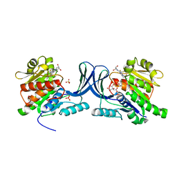
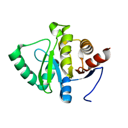
7FOJ
DownloadVisualize
BU of 7foj by Molmil
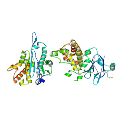
PanDDA analysis group deposition -- Aar2/RNaseH in complex with fragment P08B08 from the F2X-Universal Library
Descriptor: (2S)-N-(3-chlorophenyl)-2-[(2-hydroxyethyl)sulfanyl]propanamide, A1 cistron-splicing factor AAR2, Pre-mRNA-splicing factor 8
Authors:Barthel, T, Wollenhaupt, J, Lima, G.M.A, Wahl, M.C, Weiss, M.S.
Deposit date:2022-08-26
Release date:2022-11-02
Last modified:2024-05-22
Method:X-RAY DIFFRACTION (1.65 Å)
Cite:Large-Scale Crystallographic Fragment Screening Expedites Compound Optimization and Identifies Putative Protein-Protein Interaction Sites.
J.Med.Chem., 65, 2022
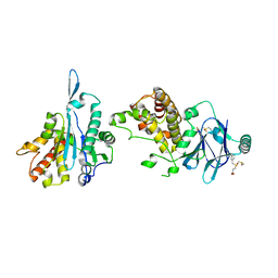
5WBZ
DownloadVisualize
BU of 5wbz by Molmil
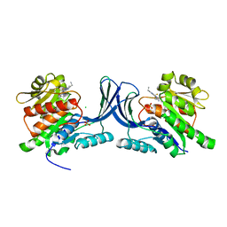
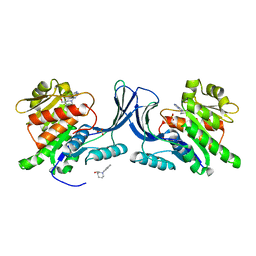
Structure of human Ketohexokinase complexed with hits from fragment screening
Descriptor: 6-[(3S,4S)-3,4-dihydroxypyrrolidin-1-yl]-2-[(3S)-3-hydroxy-3-methylpyrrolidin-1-yl]-4-(trifluoromethyl)pyridine-3-carbonitrile, CITRIC ACID, Ketohexokinase, ...
Authors:Pandit, J.
Deposit date:2017-06-29
Release date:2017-09-13
Last modified:2023-10-04
Method:X-RAY DIFFRACTION (2.4 Å)
Cite:Discovery of Fragment-Derived Small Molecules for in Vivo Inhibition of Ketohexokinase (KHK).
J. Med. Chem., 60, 2017
7FO2
DownloadVisualize
BU of 7fo2 by Molmil
PanDDA analysis group deposition -- Aar2/RNaseH in complex with fragment P07G04 from the F2X-Universal Library
Descriptor: 3-[(3-bromophenyl)methanesulfonyl]propanoic acid, A1 cistron-splicing factor AAR2, DIMETHYL SULFOXIDE, ...
Authors:Barthel, T, Wollenhaupt, J, Lima, G.M.A, Wahl, M.C, Weiss, M.S.
Deposit date:2022-08-26
Release date:2022-11-02
Last modified:2024-05-22
Method:X-RAY DIFFRACTION (2.01 Å)
Cite:Large-Scale Crystallographic Fragment Screening Expedites Compound Optimization and Identifies Putative Protein-Protein Interaction Sites.
J.Med.Chem., 65, 2022
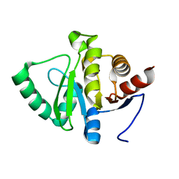
7FRA
DownloadVisualize
BU of 7fra by Molmil
PanDDA analysis group deposition -- Crystal structure of SARS-CoV-2 NSP3 macrodomain in complex with Z1343520564
Descriptor: 6-(azepane-1-sulfonyl)pyrido[2,3-d]pyrimidine-2,4(1H,3H)-dione, Non-structural protein 3
Authors:Correy, G.J, Fraser, J.S.
Deposit date:2022-10-20
Release date:2022-11-09
Last modified:2024-05-22
Method:X-RAY DIFFRACTION (1 Å)
Cite:Ligand screen against SARS-CoV-2 NSP3 macrodomain
To be published
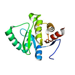
7FR1
DownloadVisualize
BU of 7fr1 by Molmil
PanDDA analysis group deposition -- Crystal structure of SARS-CoV-2 NSP3 macrodomain in complex with Z1423250928
Descriptor: 1-methyl-N'-(7H-purin-6-yl)cyclopropane-1-carbohydrazide, Non-structural protein 3
Authors:Correy, G.J, Fraser, J.S.
Deposit date:2022-10-20
Release date:2022-11-09
Last modified:2024-05-22
Method:X-RAY DIFFRACTION (1.15 Å)
Cite:Ligand screen against SARS-CoV-2 NSP3 macrodomain
To be published
7FR7
DownloadVisualize
BU of 7fr7 by Molmil
PanDDA analysis group deposition -- Crystal structure of SARS-CoV-2 NSP3 macrodomain in complex with Z431872694
Descriptor: 6-(azepane-1-carbonyl)pyrido[2,3-d]pyrimidine-2,4(1H,3H)-dione, Non-structural protein 3
Authors:Correy, G.J, Fraser, J.S.
Deposit date:2022-10-20
Release date:2022-11-09
Last modified:2024-05-22
Method:X-RAY DIFFRACTION (1 Å)
Cite:Ligand screen against SARS-CoV-2 NSP3 macrodomain
To be published
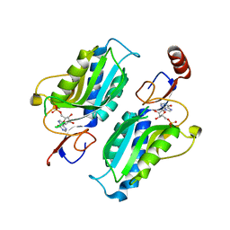
1P19
DownloadVisualize
BU of 1p19 by Molmil
Hypoxanthine Phosphoribosyltransferase from Trypanosoma cruzi, in complex with the product IMP
Descriptor: INOSINIC ACID, hypoxanthine phosphoribosyltransferase
Authors:Medrano, F.J, Eakin, A.E, Craig III, S.P.
Deposit date:2003-04-11
Release date:2004-05-18
Last modified:2024-02-14
Method:X-RAY DIFFRACTION (2.3 Å)
Cite:Interactions at the dimer interface influence the relative efficiencies for purine nucleotide synthesis and pyrophosphorolysis in a phosphoribosyltransferase.
J.Mol.Biol., 335, 2004
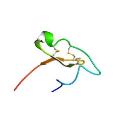
7M1D
DownloadVisualize
BU of 7m1d by Molmil
Structural and functional studies about scorpine showed the presence of blocking channel and cytolytic activities as well as two different structural domains
Descriptor: Scorpine
Authors:del Rio-Portilla, F, Lopez-Giraldo, A.E.
Deposit date:2021-03-12
Release date:2022-01-19
Last modified:2023-06-14
Method:SOLUTION NMR
Cite:Structural and functional studies of scorpine: A channel blocker and cytolytic peptide.
Toxicon, 222, 2022
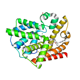
3QPP
DownloadVisualize
BU of 3qpp by Molmil
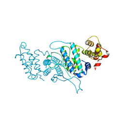
Structure of PDE10-inhibitor complex
Descriptor: 7-methoxy-4-[(3S)-3-phenylpiperidin-1-yl]-6-[2-(quinolin-2-yl)ethoxy]quinazoline, DIMETHYL SULFOXIDE, MAGNESIUM ION, ...
Authors:Pandit, J, Marr, E.S.
Deposit date:2011-02-14
Release date:2011-06-15
Last modified:2024-02-21
Method:X-RAY DIFFRACTION (1.8 Å)
Cite:Use of Structure-Based Design to Discover a Potent, Selective, In Vivo Active Phosphodiesterase 10A Inhibitor Lead Series for the Treatment of Schizophrenia.
J.Med.Chem., 54, 2011
2AIM
DownloadVisualize
BU of 2aim by Molmil
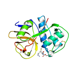
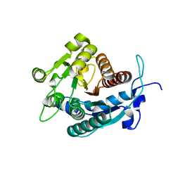
CRUZAIN INHIBITED WITH BENZOYL-ARGININE-ALANINE-FLUOROMETHYLKETONE
Descriptor: BENZOYL-ARGININE-ALANINE-FLUORO-METHYL KETONE, CRUZAIN
Authors:Gillmor, S.A, Fletterick, R.J.
Deposit date:1997-04-21
Release date:1997-10-22
Last modified:2024-04-03
Method:X-RAY DIFFRACTION (2.2 Å)
Cite:Structural determinants of specificity in the cysteine protease cruzain.
Protein Sci., 6, 1997
1YZV
DownloadVisualize
BU of 1yzv by Molmil
HYPOTHETICAL PROTEIN FROM TRYPANOSOMA CRUZI
Descriptor: HYPOTHETICAL PROTEIN, SULFATE ION
Authors:Caruthers, J, Merritt, E.A, Structural Genomics of Pathogenic Protozoa Consortium (SGPP)
Deposit date:2005-02-28
Release date:2005-03-08
Last modified:2024-02-14
Method:X-RAY DIFFRACTION (2.001 Å)
Cite:Crystal structures and proposed structural/functional classification of three protozoan proteins from the isochorismatase superfamily.
Protein Sci., 14, 2005
4DK2
DownloadVisualize
BU of 4dk2 by Molmil
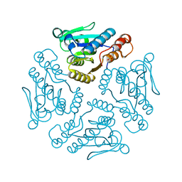
Crystal Structure of Open Trypanosoma brucei dUTPase
Descriptor: Deoxyuridine triphosphatase
Authors:Hemsworth, G.R, Gonzalez-Pacanowska, D, Wilson, K.S.
Deposit date:2012-02-03
Release date:2013-08-07
Last modified:2023-09-13
Method:X-RAY DIFFRACTION (1.47 Å)
Cite:On the catalytic mechanism of dimeric dUTPases.
Biochem.J., 456, 2013
5WBQ
DownloadVisualize
BU of 5wbq by Molmil
Structure of human Ketohexokinase complexed with hits from fragment screening
Descriptor: 2-ethyl-7-[(3S)-3-hydroxy-3-methylpyrrolidin-1-yl]-5-(trifluoromethyl)-1H-pyrrolo[3,2-b]pyridine-6-carbonitrile, CHLORIDE ION, Ketohexokinase, ...
Authors:Pandit, J.
Deposit date:2017-06-29
Release date:2017-09-13
Last modified:2023-10-04
Method:X-RAY DIFFRACTION (2.4 Å)
Cite:Discovery of Fragment-Derived Small Molecules for in Vivo Inhibition of Ketohexokinase (KHK).
J. Med. Chem., 60, 2017
5RA2
DownloadVisualize
BU of 5ra2 by Molmil
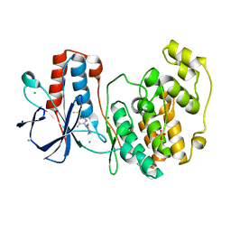
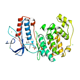
PanDDA analysis group deposition Form1 MAP kinase p38-alpha -- Fragment N09036b in complex with MAP kinase p38-alpha
Descriptor: 1-(3-methylpyridin-2-yl)-1,4-diazepane, CHLORIDE ION, DIMETHYL SULFOXIDE, ...
Authors:De Nicola, G.F, Nichols, C.E.
Deposit date:2020-03-04
Release date:2020-07-22
Last modified:2024-03-06
Method:X-RAY DIFFRACTION (1.57 Å)
Cite:Mining the PDB for Tractable Cases Where X-ray Crystallography Combined with Fragment Screens Can Be Used to Systematically Design Protein-Protein Inhibitors: Two Test Cases Illustrated by IL1 beta-IL1R and p38 alpha-TAB1 Complexes.
J.Med.Chem., 63, 2020
5WBM
DownloadVisualize
BU of 5wbm by Molmil
Structure of human Ketohexokinase complexed with hits from fragment screening
Descriptor: Ketohexokinase, SULFATE ION, [(3R)-1-(5-methyl-7H-pyrrolo[2,3-d]pyrimidin-4-yl)piperidin-3-yl]methanol
Authors:Pandit, J.
Deposit date:2017-06-29
Release date:2017-09-13
Last modified:2023-10-04
Method:X-RAY DIFFRACTION (2.16 Å)
Cite:Discovery of Fragment-Derived Small Molecules for in Vivo Inhibition of Ketohexokinase (KHK).
J. Med. Chem., 60, 2017
3GN2
DownloadVisualize
BU of 3gn2 by Molmil
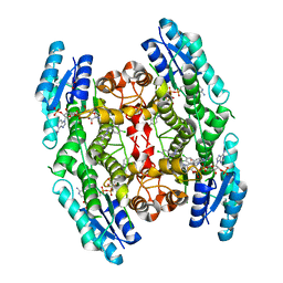
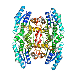
Structure of Pteridine Reductase 1 (PTR1) from TRYPANOSOMA BRUCEI in ternary complex with cofactor (NADP+) and inhibitor (DDD00066730)
Descriptor: 1-(3,4-dichlorobenzyl)-1H-benzimidazol-2-amine, ACETATE ION, NADP NICOTINAMIDE-ADENINE-DINUCLEOTIDE PHOSPHATE, ...
Authors:Tulloch, L.B, Brenk, R, Hunter, W.N.
Deposit date:2009-03-16
Release date:2009-12-29
Last modified:2024-02-21
Method:X-RAY DIFFRACTION (1.6 Å)
Cite:One scaffold, three binding modes: novel and selective pteridine reductase 1 inhibitors derived from fragment hits discovered by virtual screening.
J.Med.Chem., 52, 2009
3U4B
DownloadVisualize
BU of 3u4b by Molmil
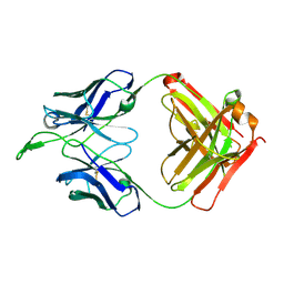
CH04H/CH02L Fab P4
Descriptor: CH02 Light chain, CH04 Heavy chain
Authors:Pancera, M, Louder, R, Mclellan, J.S, KWong, P.D.
Deposit date:2011-10-07
Release date:2011-11-30
Last modified:2011-12-21
Method:X-RAY DIFFRACTION (2.893 Å)
Cite:Structure of HIV-1 gp120 V1/V2 domain with broadly neutralizing antibody PG9.
Nature, 480, 2011
5R9I
DownloadVisualize
BU of 5r9i by Molmil
PanDDA analysis group deposition Form1 MAP kinase p38-alpha -- Fragment TCJ795 in complex with MAP kinase p38-alpha
Descriptor: 1,2-ETHANEDIOL, CHLORIDE ION, DIMETHYL SULFOXIDE, ...
Authors:De Nicola, G.F, Nichols, C.E.
Deposit date:2020-03-04
Release date:2020-07-22
Last modified:2024-03-06
Method:X-RAY DIFFRACTION (1.813 Å)
Cite:Mining the PDB for Tractable Cases Where X-ray Crystallography Combined with Fragment Screens Can Be Used to Systematically Design Protein-Protein Inhibitors: Two Test Cases Illustrated by IL1 beta-IL1R and p38 alpha-TAB1 Complexes.
J.Med.Chem., 63, 2020
3GN1
DownloadVisualize
BU of 3gn1 by Molmil
Structure of Pteridine Reductase 1 (PTR1) from TRYPANOSOMA BRUCEI in ternary complex with cofactor (NADP+) and inhibitor (DDD00067116)
Descriptor: 1H-benzimidazol-2-amine, ACETATE ION, NADP NICOTINAMIDE-ADENINE-DINUCLEOTIDE PHOSPHATE, ...
Authors:Tulloch, L.B, Brenk, R, Hunter, W.N.
Deposit date:2009-03-16
Release date:2009-12-29
Last modified:2011-09-07
Method:X-RAY DIFFRACTION (2 Å)
Cite:One scaffold, three binding modes: novel and selective pteridine reductase 1 inhibitors derived from fragment hits discovered by virtual screening.
J.Med.Chem., 52, 2009
2A0M
DownloadVisualize
BU of 2a0m by Molmil
Arginase superfamily protein from Trypanosoma cruzi
Descriptor: ARGINASE SUPERFAMILY PROTEIN, CHLORIDE ION
Authors:Arakaki, T.L, Merritt, E.A, Structural Genomics of Pathogenic Protozoa Consortium (SGPP)
Deposit date:2005-06-16
Release date:2005-07-05
Last modified:2024-04-03
Method:X-RAY DIFFRACTION (1.603 Å)
Cite:Structural genomics of pathogenic protozoa: an overview.
Methods Mol.Biol., 426, 2008
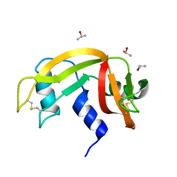
6ETL
DownloadVisualize
BU of 6etl by Molmil
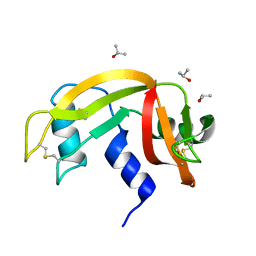
Atomic resolution structure of RNase A (data collection 2)
Descriptor: ISOPROPYL ALCOHOL, Ribonuclease pancreatic
Authors:Caterino, M, Vergara, A, Merlino, A.
Deposit date:2017-10-27
Release date:2018-02-21
Method:X-RAY DIFFRACTION (0.85 Å)
Cite:Raman-markers of X-ray radiation damage of proteins.
Int. J. Biol. Macromol., 111, 2018
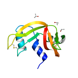
6ETM
DownloadVisualize
BU of 6etm by Molmil
Atomic resolution structure of RNase A (data collection 3)
Descriptor: ISOPROPYL ALCOHOL, Ribonuclease pancreatic
Authors:Caterino, M, Vergara, A, Merlino, A.
Deposit date:2017-10-27
Release date:2018-02-21
Method:X-RAY DIFFRACTION (0.92 Å)
Cite:Raman-markers of X-ray radiation damage of proteins.
Int. J. Biol. Macromol., 111, 2018
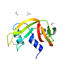
6ETR
DownloadVisualize
BU of 6etr by Molmil
Atomic resolution structure of RNase A (data collection 8)
Descriptor: ISOPROPYL ALCOHOL, Ribonuclease pancreatic
Authors:Caterino, M, Vergara, A, Merlino, A.
Deposit date:2017-10-27
Release date:2018-02-21
Last modified:2024-01-17
Method:X-RAY DIFFRACTION (1.17 Å)
Cite:Raman-markers of X-ray radiation damage of proteins.
Int. J. Biol. Macromol., 111, 2018
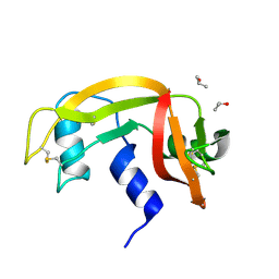
6ETP
DownloadVisualize
BU of 6etp by Molmil
Atomic resolution structure of RNase A (data collection 6)
Descriptor: ISOPROPYL ALCOHOL, Ribonuclease pancreatic
Authors:Caterino, M, Vergara, A, Merlino, A.
Deposit date:2017-10-27
Release date:2018-02-21
Last modified:2024-01-17
Method:X-RAY DIFFRACTION (1.02 Å)
Cite:Raman-markers of X-ray radiation damage of proteins.
Int. J. Biol. Macromol., 111, 2018
6ETK
DownloadVisualize
BU of 6etk by Molmil
Atomic resolution structure of RNase A (data collection 1)
Descriptor: ISOPROPYL ALCOHOL, Ribonuclease pancreatic
Authors:Caterino, M, Vergara, A, Merlino, A.
Deposit date:2017-10-27
Release date:2018-02-21
Last modified:2024-01-17
Method:X-RAY DIFFRACTION (0.85 Å)
Cite:Raman-markers of X-ray radiation damage of proteins.
Int. J. Biol. Macromol., 111, 2018

222624

數據於2024-07-17公開中

PDB statisticsPDBj update infoContact PDBjnumon