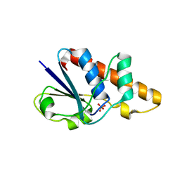
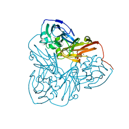
3UYV
 
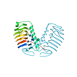 | | Crystal structure of a glycosylated ice-binding protein (LeIBP) from Arctic yeast | | Descriptor: | Antifreeze protein, alpha-D-mannopyranose-(1-6)-beta-D-mannopyranose-(1-4)-2-acetamido-2-deoxy-beta-D-glucopyranose-(1-4)-2-acetamido-2-deoxy-beta-D-glucopyranose | | Authors: | Lee, J.H, Park, A.K, Do, H, Park, K.S, Moh, S.H, Chi, Y.M, Kim, H.J. | | Deposit date: | 2011-12-06 | | Release date: | 2012-02-29 | | Last modified: | 2024-10-30 | | Method: | X-RAY DIFFRACTION (2.43 Å) | | Cite: | Structural basis for the antifreeze activity of an ice-binding protein from an Arctic yeast.
J.Biol.Chem., 2012
|
|
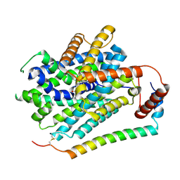
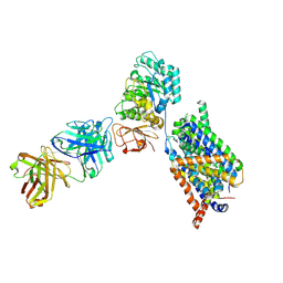
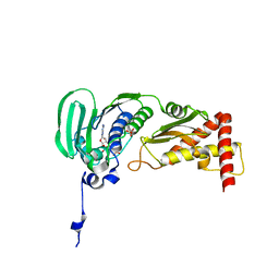
1L6W
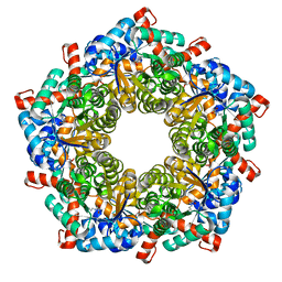 | | Fructose-6-phosphate aldolase | | Descriptor: | Fructose-6-phosphate aldolase 1, GLYCEROL | | Authors: | Thorell, S, Schuermann, M, Sprenger, G.A, Schneider, G. | | Deposit date: | 2002-03-14 | | Release date: | 2002-06-12 | | Last modified: | 2024-10-30 | | Method: | X-RAY DIFFRACTION (1.93 Å) | | Cite: | Crystal structure of decameric fructose-6-phosphate aldolase from Escherichia coli reveals inter-subunit helix swapping as a structural basis for assembly differences in the transaldolase family.
J.Mol.Biol., 319, 2002
|
|
1LD3
 | | Crystal Structure of B. subilis ferrochelatase with Zn(2+) bound at the active site. | | Descriptor: | Ferrochelatase, ZINC ION | | Authors: | Lecerof, D, Fodje, M.N, Leon, R.A, Olsson, U, Hansson, A, Sigfridsson, E, Ryde, U, Hansson, M, Al-Karadaghi, S. | | Deposit date: | 2002-04-08 | | Release date: | 2003-05-20 | | Last modified: | 2023-08-16 | | Method: | X-RAY DIFFRACTION (2.6 Å) | | Cite: | Metal binding to Bacillus subtilis ferrochelatase and interaction between metal sites
J.Biol.Inorg.Chem., 8, 2003
|
|
1L8X
 | | Crystal Structure of Ferrochelatase from the Yeast, Saccharomyces cerevisiae, with Cobalt(II) as the Substrate Ion | | Descriptor: | COBALT (II) ION, Ferrochelatase | | Authors: | Karlberg, T, Lecerof, D, Gora, M, Silvegren, G, Labbe-Bois, R, Hansson, M, Al-Karadaghi, S. | | Deposit date: | 2002-03-22 | | Release date: | 2002-11-20 | | Last modified: | 2023-08-16 | | Method: | X-RAY DIFFRACTION (2.7 Å) | | Cite: | Metal Binding to Saccharomyces cerevisiae Ferrochelatase
Biochemistry, 41, 2002
|
|
1JL3
 | | Crystal Structure of B. subtilis ArsC | | Descriptor: | ARSENATE REDUCTASE, SULFATE ION | | Authors: | Su, X.-D, Bennett, M.S. | | Deposit date: | 2001-07-15 | | Release date: | 2001-10-24 | | Last modified: | 2024-02-07 | | Method: | X-RAY DIFFRACTION (1.6 Å) | | Cite: | Bacillus subtilis arsenate reductase is structurally and functionally similar to low molecular weight protein tyrosine phosphatases.
Proc.Natl.Acad.Sci.USA, 98, 2001
|
|
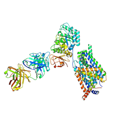
5JLH
 | | Cryo-EM structure of a human cytoplasmic actomyosin complex at near-atomic resolution | | Descriptor: | ADENOSINE-5'-DIPHOSPHATE, Actin, cytoplasmic 2, ... | | Authors: | von der Ecken, J, Heissler, S.M, Pathan-Chhatbar, S, Manstein, D.J, Raunser, S. | | Deposit date: | 2016-04-27 | | Release date: | 2016-06-15 | | Last modified: | 2024-07-10 | | Method: | ELECTRON MICROSCOPY (3.9 Å) | | Cite: | Cryo-EM structure of a human cytoplasmic actomyosin complex at near-atomic resolution.
Nature, 534, 2016
|
|
8KDN
 | | Structure of LAT1-CD98hc in complex with L-Phe, focused on TMD | | Descriptor: | 4F2 cell-surface antigen heavy chain, Large neutral amino acids transporter small subunit 1, PHENYLALANINE | | Authors: | Lee, Y. | | Deposit date: | 2023-08-09 | | Release date: | 2025-02-12 | | Last modified: | 2025-02-26 | | Method: | ELECTRON MICROSCOPY (4.12 Å) | | Cite: | Structural basis of anticancer drug recognition and amino acid transport by LAT1.
Nat Commun, 16, 2025
|
|
8KDJ
 | | Structure of apo inward-open LAT1-CD98h in nanodisc, focused on TMD | | Descriptor: | 1-palmitoyl-2-oleoyl-sn-glycero-3-phosphocholine, 4F2 cell-surface antigen heavy chain, CHOLESTEROL, ... | | Authors: | Lee, Y. | | Deposit date: | 2023-08-09 | | Release date: | 2025-02-12 | | Last modified: | 2025-02-26 | | Method: | ELECTRON MICROSCOPY (3.73 Å) | | Cite: | Structural basis of anticancer drug recognition and amino acid transport by LAT1.
Nat Commun, 16, 2025
|
|
8KDH
 | | Structure of LAT1-CD98hc in complex with BCH, focused on TMD | | Descriptor: | (1~{S},2~{R},4~{R})-2-azanylbicyclo[2.2.1]heptane-2-carboxylic acid, 1-palmitoyl-2-oleoyl-sn-glycero-3-phosphocholine, 4F2 cell-surface antigen heavy chain, ... | | Authors: | Lee, Y. | | Deposit date: | 2023-08-09 | | Release date: | 2025-02-12 | | Last modified: | 2025-02-26 | | Method: | ELECTRON MICROSCOPY (3.78 Å) | | Cite: | Structural basis of anticancer drug recognition and amino acid transport by LAT1.
Nat Commun, 16, 2025
|
|
8KDD
 | | Structure of LAT1-CD98hc-Fab170 in complex with JPH203, consensus map | | Descriptor: | 2-acetamido-2-deoxy-beta-D-glucopyranose, 4F2 cell-surface antigen heavy chain, Fab170 heavy chain, ... | | Authors: | Lee, Y. | | Deposit date: | 2023-08-09 | | Release date: | 2025-02-12 | | Last modified: | 2025-02-26 | | Method: | ELECTRON MICROSCOPY (3.83 Å) | | Cite: | Structural basis of anticancer drug recognition and amino acid transport by LAT1.
Nat Commun, 16, 2025
|
|
8KDP
 | | Structure of apo outward-open LAT1-CD98h in nanodisc, focused on TMD | | Descriptor: | 4F2 cell-surface antigen heavy chain, Large neutral amino acids transporter small subunit 1 | | Authors: | Lee, Y. | | Deposit date: | 2023-08-09 | | Release date: | 2025-02-12 | | Last modified: | 2025-02-26 | | Method: | ELECTRON MICROSCOPY (4.12 Å) | | Cite: | Structural basis of anticancer drug recognition and amino acid transport by LAT1.
Nat Commun, 16, 2025
|
|
8KDI
 | | Structure of apo inward-open LAT1-CD98hc-Fab170 in nanodisc, consensus map | | Descriptor: | 2-acetamido-2-deoxy-beta-D-glucopyranose, 4F2 cell-surface antigen heavy chain, Fab170 heavy chain, ... | | Authors: | Lee, Y. | | Deposit date: | 2023-08-09 | | Release date: | 2025-02-12 | | Last modified: | 2025-02-26 | | Method: | ELECTRON MICROSCOPY (3.58 Å) | | Cite: | Structural basis of anticancer drug recognition and amino acid transport by LAT1.
Nat Commun, 16, 2025
|
|
8KDF
 | | Structure of LAT1-CD98hc in complex with JPH203, focused on TMD | | Descriptor: | 1-palmitoyl-2-oleoyl-sn-glycero-3-phosphocholine, 4F2 cell-surface antigen heavy chain, CHOLESTEROL, ... | | Authors: | Lee, Y. | | Deposit date: | 2023-08-09 | | Release date: | 2025-02-12 | | Last modified: | 2025-02-26 | | Method: | ELECTRON MICROSCOPY (3.89 Å) | | Cite: | Structural basis of anticancer drug recognition and amino acid transport by LAT1.
Nat Commun, 16, 2025
|
|
8KDG
 | | Structure of LAT1-CD98hc-Fab170 in complex with BCH, consensus map | | Descriptor: | (1~{S},2~{R},4~{R})-2-azanylbicyclo[2.2.1]heptane-2-carboxylic acid, 2-acetamido-2-deoxy-beta-D-glucopyranose, 4F2 cell-surface antigen heavy chain, ... | | Authors: | Lee, Y. | | Deposit date: | 2023-08-09 | | Release date: | 2025-02-12 | | Last modified: | 2025-02-26 | | Method: | ELECTRON MICROSCOPY (3.68 Å) | | Cite: | Structural basis of anticancer drug recognition and amino acid transport by LAT1.
Nat Commun, 16, 2025
|
|
8KDO
 | | Structure of LAT1-CD98hc in complex with melphalan, focused on TMD | | Descriptor: | (2~{S})-2-azanyl-3-[4-[bis(2-chloroethyl)amino]phenyl]propanoic acid, 4F2 cell-surface antigen heavy chain, Large neutral amino acids transporter small subunit 1 | | Authors: | Lee, Y. | | Deposit date: | 2023-08-09 | | Release date: | 2025-02-12 | | Last modified: | 2025-02-26 | | Method: | ELECTRON MICROSCOPY (4.12 Å) | | Cite: | Structural basis of anticancer drug recognition and amino acid transport by LAT1.
Nat Commun, 16, 2025
|
|
2K3A
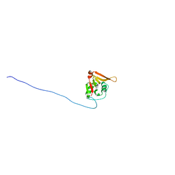 | | NMR solution structure of Staphylococcus saprophyticus CHAP (cysteine, histidine-dependent amidohydrolases/peptidases) domain protein. Northeast Structural Genomics Consortium target SyR11 | | Descriptor: | CHAP domain protein | | Authors: | Rossi, P, Aramini, J.M, Chen, C.X, Nwosu, C, Cunningham, K.C, Owens, L.A, Xiao, R, Liu, J, Baran, M.C, Swapna, G, Acton, T.B, Rost, B, Montelione, G.T, Northeast Structural Genomics Consortium (NESG) | | Deposit date: | 2008-04-29 | | Release date: | 2008-05-13 | | Last modified: | 2024-05-29 | | Method: | SOLUTION NMR | | Cite: | Structural elucidation of the Cys-His-Glu-Asn proteolytic relay in the secreted CHAP domain enzyme from the human pathogen Staphylococcus saprophyticus.
Proteins, 74, 2008
|
|
1GKA
 | | The molecular basis of the coloration mechanism in lobster shell. beta-crustacyanin at 3.2 A resolution | | Descriptor: | 2-AMINO-2-HYDROXYMETHYL-PROPANE-1,3-DIOL, 4-(2-HYDROXYETHYL)-1-PIPERAZINE ETHANESULFONIC ACID, ASTAXANTHIN, ... | | Authors: | Cianci, M, Rizkallah, P.J, Olczak, A, Raftery, J, Chayen, N.E, Zagalsky, P.F, Helliwell, J.R. | | Deposit date: | 2001-08-10 | | Release date: | 2002-08-08 | | Last modified: | 2024-10-16 | | Method: | X-RAY DIFFRACTION (3.23 Å) | | Cite: | The Molecular Basis of the Coloration Mechanism in Lobster Shell: Beta -Crustacyanin at 3.2-A Resolution
Proc.Natl.Acad.Sci.USA, 99, 2002
|
|
2JRS
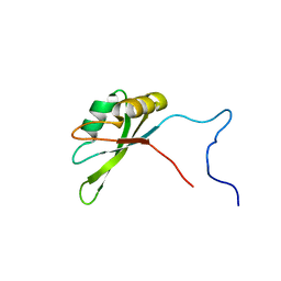 | | Solution NMR Structure of CAPER RRM2 Domain. Northeast Structural Genomics Target HR4730A | | Descriptor: | RNA-binding protein 39 | | Authors: | Rossi, P, Zhao, L, Nwosu, C, Cunningham, K, Owens, L, Xiao, R, Liu, J, Baran, M.C, Swapna, G, Acton, T.B, Rost, B, Montelione, G.T, Northeast Structural Genomics Consortium (NESG) | | Deposit date: | 2007-06-28 | | Release date: | 2007-09-04 | | Last modified: | 2024-05-01 | | Method: | SOLUTION NMR | | Cite: | Solution NMR Structure of CAPER RRM2 Domain.
To be Published
|
|
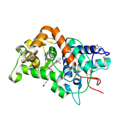
1OOW
 | | The crystal structure of the spinach plastocyanin double mutant G8D/L12E gives insight into its low reactivity towards photosystem 1 and cytochrome f | | Descriptor: | COPPER (II) ION, Plastocyanin, chloroplast | | Authors: | Jansson, H, Okvist, M, Jacobson, F, Ejdeback, M, Hansson, O, Sjolin, L. | | Deposit date: | 2003-03-04 | | Release date: | 2004-02-17 | | Last modified: | 2023-08-16 | | Method: | X-RAY DIFFRACTION (2 Å) | | Cite: | The crystal structure of the spinach plastocyanin double mutant G8D/L12E gives insight into its low reactivity towards photosystem 1 and cytochrome f.
Biochim.Biophys.Acta, 1607, 2003
|
|
1GW2
 | | RECOMBINANT HORSERADISH PEROXIDASE C1A THR171SER IN COMPLEX WITH FERULIC ACID | | Descriptor: | 3-(4-HYDROXY-3-METHOXYPHENYL)-2-PROPENOIC ACID, CALCIUM ION, PEROXIDASE C1A, ... | | Authors: | Henriksen, A, Meno, K, Brissett, N, Gajhede, M. | | Deposit date: | 2002-03-03 | | Release date: | 2003-03-28 | | Last modified: | 2024-11-13 | | Method: | X-RAY DIFFRACTION (2.15 Å) | | Cite: | Hrpc Heme Crevice Architecture
To be Published
|
|
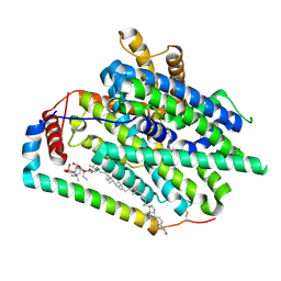
1MZY
 | | Crystal Structure of Nitrite Reductase | | Descriptor: | COPPER (II) ION, Copper-containing nitrite reductase, MAGNESIUM ION | | Authors: | Guo, H, Olesen, K, Xue, Y, Shapliegh, J, Sjolin, L. | | Deposit date: | 2002-10-10 | | Release date: | 2004-09-28 | | Last modified: | 2024-02-14 | | Method: | X-RAY DIFFRACTION (1.46 Å) | | Cite: | The High resolution Crystal Structures of Nitrite Reductase and its mutant Met182Thr from Rhodobacter Sphaeroides Reveal a Gating Mechanism for the Electron Transfer to the Type 1 Copper Center
To be Published
|
|
1S16
 | | Crystal Structure of E. coli Topoisomerase IV ParE 43kDa subunit complexed with ADPNP | | Descriptor: | MAGNESIUM ION, PHOSPHOAMINOPHOSPHONIC ACID-ADENYLATE ESTER, SULFATE ION, ... | | Authors: | Wei, Y, Gross, C.H. | | Deposit date: | 2004-01-05 | | Release date: | 2004-05-04 | | Last modified: | 2024-02-14 | | Method: | X-RAY DIFFRACTION (2.1 Å) | | Cite: | Crystal structures of Escherichia coli topoisomerase IV ParE subunit (24 and 43 kilodaltons): a single residue dictates differences in novobiocin potency against topoisomerase IV and DNA gyrase.
Antimicrob.Agents Chemother., 48, 2004
|
|
1GFY
 | |
1N0I
 | | Crystal Structure of Ferrochelatase with Cadmium bound at active site | | Descriptor: | CADMIUM ION, CHLORIDE ION, Ferrochelatase, ... | | Authors: | Lecerof, D, Fodje, M.N, Leon, R.A, Olsson, U, Hansson, A, Sigfridsson, E, Ryde, U, Hansson, M, Al-Karadaghi, S. | | Deposit date: | 2002-10-14 | | Release date: | 2003-05-27 | | Last modified: | 2024-02-14 | | Method: | X-RAY DIFFRACTION (2 Å) | | Cite: | Metal binding to Bacillus subtilis ferrochelatase and interaction between metal sites
J.Biol.Inorg.Chem., 8, 2003
|
|
1N70
 | |


