Loading
PDBj
MenuPDBj@FacebookPDBj@TwitterPDBj@YouTubewwPDB FoundationwwPDB
RCSB PDBPDBeBMRBAdv. SearchSearch help

3D2U
DownloadVisualize
BU of 3d2u by Molmil
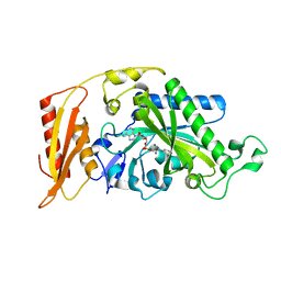
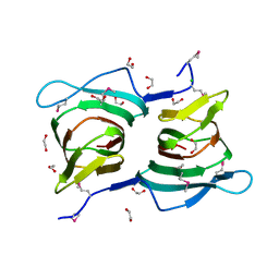
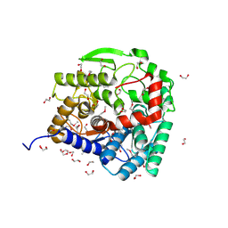
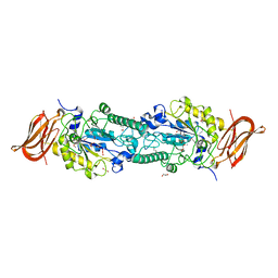
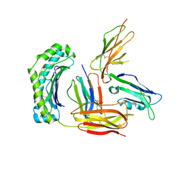
Structure of UL18, a Peptide-Binding Viral MHC Mimic, Bound to a Host Inhibitory Receptor
Descriptor: 2-acetamido-2-deoxy-beta-D-glucopyranose, Actin, Beta-2-microglobulin, ...
Authors:Yang, Z, Bjorkman, P.J.
Deposit date:2008-05-08
Release date:2008-07-08
Last modified:2023-08-30
Method:X-RAY DIFFRACTION (2.21 Å)
Cite:Structure of UL18, a peptide-binding viral MHC mimic, bound to a host inhibitory receptor
Proc.Natl.Acad.Sci.Usa, 105, 2008
3GZI
DownloadVisualize
BU of 3gzi by Molmil
CRYSTAL STRUCTURE OF a transcriptional regulator of the tetR family (SHEW_3567) FROM SHEWANELLA LOIHICA PV-4 AT 2.05 A RESOLUTION
Descriptor: 1,2-ETHANEDIOL, Transcriptional regulator, TetR family, ...
Authors:Joint Center for Structural Genomics (JCSG)
Deposit date:2009-04-07
Release date:2009-04-21
Last modified:2023-02-01
Method:X-RAY DIFFRACTION (2.05 Å)
Cite:Crystal structure of TETR family transcriptional regulator (YP_001095692.1) from SHEWANELLA SP. PV-4 at 2.05 A resolution
To be published
3GY9
DownloadVisualize
BU of 3gy9 by Molmil
Crystal structure of putative acetyltransferase (YP_001815201.1) from EXIGUOBACTERIUM SP. 255-15 at 1.52 A resolution
Descriptor: COENZYME A, GCN5-related N-acetyltransferase, GLYCEROL, ...
Authors:Joint Center for Structural Genomics (JCSG)
Deposit date:2009-04-03
Release date:2009-04-21
Last modified:2023-02-01
Method:X-RAY DIFFRACTION (1.52 Å)
Cite:Crystal structure of putative acetyltransferase (YP_001815201.1) from EXIGUOBACTERIUM SP. 255-15 at 1.52 A resolution
To be published
3H0N
DownloadVisualize
BU of 3h0n by Molmil
Crystal structure of a duf1470 family protein (jann_2411) from jannaschia sp. ccs1 at 1.45 A resolution
Descriptor: ACETATE ION, GLYCEROL, NICKEL (II) ION, ...
Authors:Joint Center for Structural Genomics (JCSG)
Deposit date:2009-04-09
Release date:2009-04-21
Last modified:2023-02-01
Method:X-RAY DIFFRACTION (1.45 Å)
Cite:The structure of Jann_2411 (DUF1470) from Jannaschia sp. at 1.45  resolution reveals a new fold (the ABATE domain) and suggests its possible role as a transcription regulator.
Acta Crystallogr.,Sect.F, 66, 2010
3GZ7
DownloadVisualize
BU of 3gz7 by Molmil
Crystal structure of Putative antibiotic biosynthesis monooxygenase (NP_888398.1) from BORDETELLA BRONCHISEPTICA at 2.15 A resolution
Descriptor: CITRIC ACID, Putative antibiotic biosynthesis monooxygenase
Authors:Joint Center for Structural Genomics (JCSG)
Deposit date:2009-04-06
Release date:2009-04-21
Last modified:2023-02-01
Method:X-RAY DIFFRACTION (2.15 Å)
Cite:Crystal structure of Putative antibiotic biosynthesis monooxygenase (NP_888398.1) from BORDETELLA BRONCHISEPTICA at 2.15 A resolution
To be published
3CLM
DownloadVisualize
BU of 3clm by Molmil
Crystal structure of transaldolase (YP_208650.1) from Neisseria gonorrhoeae FA 1090 at 1.14 A resolution
Descriptor: 1,2-ETHANEDIOL, CHLORIDE ION, SULFATE ION, ...
Authors:Joint Center for Structural Genomics (JCSG)
Deposit date:2008-03-19
Release date:2008-04-01
Last modified:2023-02-01
Method:X-RAY DIFFRACTION (1.14 Å)
Cite:Crystal structure of transaldolase (YP_208650.1) from Neisseria gonorrhoeae FA 1090 at 1.14 A resolution
To be published
3CMQ
DownloadVisualize
BU of 3cmq by Molmil
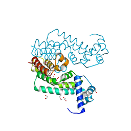
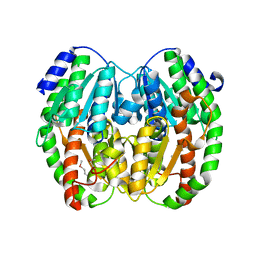
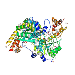
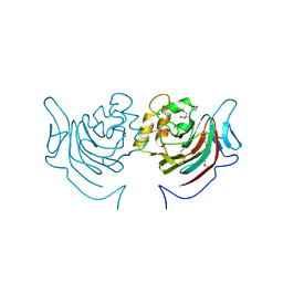
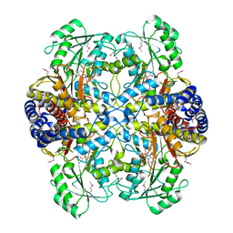
Crystal structure of human mitochondrial phenylalanine tRNA synthetase
Descriptor: ADENOSINE-5'-[PHENYLALANINYL-PHOSPHATE], MAGNESIUM ION, Phenylalanyl-tRNA synthetase, ...
Authors:Klipcan, L, Levin, I.L, Kessler, N, Moor, N, Finarov, I, Safro, M.
Deposit date:2008-03-24
Release date:2008-07-01
Last modified:2023-08-30
Method:X-RAY DIFFRACTION (2.2 Å)
Cite:The tRNA-Induced Conformational Activation of Human Mitochondrial Phenylalanyl-tRNA Synthetase.
Structure, 16, 2008
3EUA
DownloadVisualize
BU of 3eua by Molmil
CRYSTAL STRUCTURE OF A PUTATIVE PHOSPHOSUGAR ISOMERASE (BSU32610) FROM BACILLUS SUBTILIS AT 1.90 A RESOLUTION
Descriptor: CITRATE ANION, GLYCEROL, PUTATIVE FRUCTOSE-AMINOACID-6-PHOSPHATE DEGLYCASE
Authors:Joint Center for Structural Genomics (JCSG)
Deposit date:2008-10-09
Release date:2008-10-21
Last modified:2023-02-01
Method:X-RAY DIFFRACTION (1.9 Å)
Cite:Crystal structure of yurp protein (np_391141.1) from bacillus subtilis at 1.90 A resolution
To be published
3E5D
DownloadVisualize
BU of 3e5d by Molmil
Crystal structure of a putative glyoxalase i (lmof2365_0426) from listeria monocytogenes str. 4b f2365 at 2.70 A resolution
Descriptor: Putative Glyoxalase I, UNKNOWN LIGAND
Authors:Joint Center for Structural Genomics (JCSG)
Deposit date:2008-08-13
Release date:2008-08-26
Last modified:2023-02-01
Method:X-RAY DIFFRACTION (2.7 Å)
Cite:Crystal structure of Putative Glyoxalase I (YP_013033.1) from LISTERIA MONOCYTOGENES 4b F2365 at 2.70 A resolution
To be published
3ECQ
DownloadVisualize
BU of 3ecq by Molmil
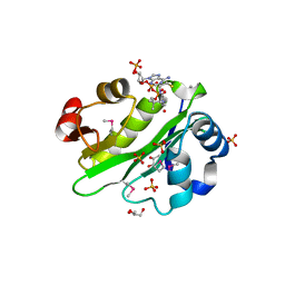
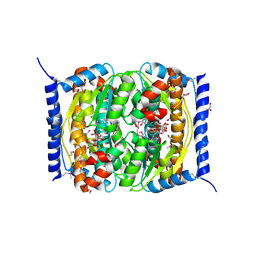
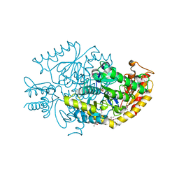
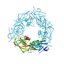
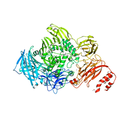
Endo-alpha-N-acetylgalactosaminidase from Streptococcus pneumoniae: SeMet structure
Descriptor: CALCIUM ION, Endo-alpha-N-acetylgalactosaminidase, GLYCEROL, ...
Authors:Caines, M.E.C, Zhu, H, Vuckovic, M, Strynadka, N.C.J.
Deposit date:2008-09-01
Release date:2008-09-09
Last modified:2011-07-13
Method:X-RAY DIFFRACTION (2.9 Å)
Cite:The structural basis for T-antigen hydrolysis by Streptococcus pneumoniae: a target for structure-based vaccine design.
J.Biol.Chem., 283, 2008
3ES4
DownloadVisualize
BU of 3es4 by Molmil
Crystal structure of Protein of unknown function (DUF861) with a RmlC-like cupin fold (17741406) from AGROBACTERIUM TUMEFACIENS str. C58 (Dupont) at 1.64 A resolution
Descriptor: 1,2-ETHANEDIOL, CHLORIDE ION, uncharacterized protein DUF861 with a RmlC-like cupin fold
Authors:Joint Center for Structural Genomics (JCSG)
Deposit date:2008-10-03
Release date:2008-10-14
Last modified:2023-02-01
Method:X-RAY DIFFRACTION (1.64 Å)
Cite:Crystal structure of Protein of unknown function (DUF861) with a RmlC-like cupin fold (17741406) from AGROBACTERIUM TUMEFACIENS str. C58 (Dupont) at 1.64 A resolution
To be published
3EQX
DownloadVisualize
BU of 3eqx by Molmil
CRYSTAL STRUCTURE OF A FIC FAMILY PROTEIN (SO_4266) FROM SHEWANELLA ONEIDENSIS AT 1.6 A RESOLUTION
Descriptor: FIC DOMAIN CONTAINING TRANSCRIPTIONAL REGULATOR, TRIETHYLENE GLYCOL
Authors:Joint Center for Structural Genomics (JCSG)
Deposit date:2008-10-01
Release date:2008-10-14
Last modified:2023-02-01
Method:X-RAY DIFFRACTION (1.6 Å)
Cite:Crystal structure of the Fic (Filamentation induced by cAMP) family protein SO4266 (gi|24375750) from Shewanella oneidensis MR-1 at 1.6 A resolution.
Proteins, 75, 2009
3ES1
DownloadVisualize
BU of 3es1 by Molmil
Crystal structure of protein with a cupin-like fold and unknown function (YP_001165807.1) from NOVOSPHINGOBIUM AROMATICIVORANS DSM 12444 at 1.91 A resolution
Descriptor: CHLORIDE ION, Cupin 2, conserved barrel domain protein
Authors:Joint Center for Structural Genomics (JCSG)
Deposit date:2008-10-03
Release date:2008-10-14
Last modified:2023-02-01
Method:X-RAY DIFFRACTION (1.91 Å)
Cite:Crystal structure of protein with a cupin-like fold and unknown function (YP_001165807.1) from NOVOSPHINGOBIUM AROMATICIVORANS DSM 12444 at 1.91 A resolution
To be published
3EUC
DownloadVisualize
BU of 3euc by Molmil
Crystal structure of histidinol-phosphate aminotransferase (YP_297314.1) from RALSTONIA EUTROPHA JMP134 at 2.05 A resolution
Descriptor: GLYCEROL, Histidinol-phosphate aminotransferase 2, SULFATE ION
Authors:Joint Center for Structural Genomics (JCSG)
Deposit date:2008-10-09
Release date:2008-11-11
Last modified:2023-02-01
Method:X-RAY DIFFRACTION (2.05 Å)
Cite:Crystal structure of histidinol-phosphate aminotransferase (YP_297314.1) from RALSTONIA EUTROPHA JMP134 at 2.05 A resolution
To be published
3ETN
DownloadVisualize
BU of 3etn by Molmil
Crystal structure of putative phosphosugar isomerase involved in capsule formation (YP_209877.1) from Bacteroides fragilis NCTC 9343 at 1.70 A resolution
Descriptor: 1,2-ETHANEDIOL, CYTIDINE 5'-MONOPHOSPHATE 3-DEOXY-BETA-D-GULO-OCT-2-ULO-PYRANOSONIC ACID, putative phosphosugar isomerase involved in capsule formation
Authors:Joint Center for Structural Genomics (JCSG)
Deposit date:2008-10-08
Release date:2008-10-21
Last modified:2023-02-01
Method:X-RAY DIFFRACTION (1.7 Å)
Cite:Structural analysis of arabinose-5-phosphate isomerase from Bacteroides fragilis and functional implications.
Acta Crystallogr.,Sect.D, 70, 2014
3EU8
DownloadVisualize
BU of 3eu8 by Molmil
Crystal structure of putative glucoamylase (YP_210071.1) from Bacteroides fragilis NCTC 9343 at 2.12 A resolution
Descriptor: 1,2-ETHANEDIOL, CHLORIDE ION, POTASSIUM ION, ...
Authors:Joint Center for Structural Genomics (JCSG)
Deposit date:2008-10-09
Release date:2008-10-21
Last modified:2023-02-01
Method:X-RAY DIFFRACTION (2.12 Å)
Cite:Crystal structure of putative glucoamylase (YP_210071.1) from Bacteroides fragilis NCTC 9343 at 2.12 A resolution
To be published
3GRD
DownloadVisualize
BU of 3grd by Molmil
Crystal structure of NTF2-superfamily protein with unknown function (NP_977240.1) from BACILLUS CEREUS ATCC 10987 at 1.25 A resolution
Descriptor: ACETATE ION, SODIUM ION, uncharacterized NTF2-superfamily protein
Authors:Joint Center for Structural Genomics (JCSG)
Deposit date:2009-03-25
Release date:2009-04-07
Last modified:2023-02-01
Method:X-RAY DIFFRACTION (1.25 Å)
Cite:Crystal structure of NTF2-superfamily protein with unknown function (NP_977240.1) from BACILLUS CEREUS ATCC 10987 at 1.25 A resolution
To be published
3H3L
DownloadVisualize
BU of 3h3l by Molmil
Crystal structure of PUTATIVE SUGAR HYDROLASE (YP_001304206.1) from Parabacteroides distasonis ATCC 8503 at 1.59 A resolution
Descriptor: 2-AMINO-2-HYDROXYMETHYL-PROPANE-1,3-DIOL, PUTATIVE SUGAR HYDROLASE
Authors:Joint Center for Structural Genomics (JCSG)
Deposit date:2009-04-16
Release date:2009-04-28
Last modified:2023-02-01
Method:X-RAY DIFFRACTION (1.59 Å)
Cite:Crystal structure of PUTATIVE SUGAR HYDROLASE (YP_001304206.1) from Parabacteroides distasonis ATCC 8503 at 1.59 A resolution
To be published
3H4Y
DownloadVisualize
BU of 3h4y by Molmil
Crystal structure of putative chemotaxis protein (YP_009526.1) from DESULFOVIBRIO VULGARIS HILDENBOROUGH at 1.55 A resolution
Descriptor: (4R)-2-METHYLPENTANE-2,4-DIOL, CITRIC ACID, putative chemotaxis protein
Authors:Joint Center for Structural Genomics (JCSG)
Deposit date:2009-04-21
Release date:2009-05-05
Last modified:2023-02-01
Method:X-RAY DIFFRACTION (1.55 Å)
Cite:Crystal structure of putative chemotaxis protein (YP_009526.1) from DESULFOVIBRIO VULGARIS HILDENBOROUGH at 1.55 A resolution
To be published
3GJU
DownloadVisualize
BU of 3gju by Molmil
Crystal structure of a putative aminotransferase (mll7127) from mesorhizobium loti maff303099 at 1.55 A resolution
Descriptor: (4S)-2-METHYL-2,4-PENTANEDIOL, PYRIDOXAL-5'-PHOSPHATE, Putative aminotransferase
Authors:Joint Center for Structural Genomics (JCSG)
Deposit date:2009-03-09
Release date:2009-03-17
Last modified:2023-02-01
Method:X-RAY DIFFRACTION (1.55 Å)
Cite:Crystal structure of putative aminotransferase (NP_107505.1) from Mesorhizobium loti at 1.55 A resolution
To be published
3GZA
DownloadVisualize
BU of 3gza by Molmil
Crystal structure of putative alpha-L-fucosidase (NP_812709.1) from BACTEROIDES THETAIOTAOMICRON VPI-5482 at 1.60 A resolution
Descriptor: 1,2-ETHANEDIOL, 4-(2-HYDROXYETHYL)-1-PIPERAZINE ETHANESULFONIC ACID, SODIUM ION, ...
Authors:Joint Center for Structural Genomics (JCSG)
Deposit date:2009-04-06
Release date:2009-04-21
Last modified:2023-02-01
Method:X-RAY DIFFRACTION (1.6 Å)
Cite:Crystal structure of putative alpha-L-fucosidase (NP_812709.1) from BACTEROIDES THETAIOTAOMICRON VPI-5482 at 1.60 A resolution
To be Published
3GWP
DownloadVisualize
BU of 3gwp by Molmil
Crystal structure of carbon-sulfur lyase involved in aluminum resistance (YP_878183.1) from CLOSTRIDIUM NOVYI NT at 2.90 A resolution
Descriptor: carbon-sulfur lyase involved in aluminum resistance
Authors:Joint Center for Structural Genomics (JCSG)
Deposit date:2009-04-01
Release date:2009-04-14
Last modified:2023-11-22
Method:X-RAY DIFFRACTION (2.9 Å)
Cite:Crystal structure of carbon-sulfur lyase involved in aluminum resistance (YP_878183.1) from CLOSTRIDIUM NOVYI NT at 2.90 A resolution
To be published
3GZB
DownloadVisualize
BU of 3gzb by Molmil
Crystal structure of putative SnoaL-like polyketide cyclase (YP_001182657.1) from Shewanella putrefaciens CN-32 at 1.44 A resolution
Descriptor: (4R)-2-METHYLPENTANE-2,4-DIOL, (4S)-2-METHYL-2,4-PENTANEDIOL, 1,2-ETHANEDIOL, ...
Authors:Joint Center for Structural Genomics (JCSG)
Deposit date:2009-04-06
Release date:2009-04-21
Last modified:2023-02-01
Method:X-RAY DIFFRACTION (1.44 Å)
Cite:Crystal structure of putative SnoaL-like polyketide cyclase (YP_001182657.1) from Shewanella putrefaciens CN-32 at 1.44 A resolution
To be published
3GS9
DownloadVisualize
BU of 3gs9 by Molmil
Crystal structure of prophage tail protein gp18 (NP_465809.1) from Listeria monocytogenes EGD-e at 1.70 A resolution
Descriptor: Protein gp18
Authors:Joint Center for Structural Genomics (JCSG)
Deposit date:2009-03-26
Release date:2009-04-14
Last modified:2023-02-01
Method:X-RAY DIFFRACTION (1.7 Å)
Cite:Crystal structure of prophage tail protein gp18 (NP_465809.1) from Listeria monocytogenes EGD-e at 1.70 A resolution
To be published
3HBA
DownloadVisualize
BU of 3hba by Molmil
Crystal structure of a putative phosphosugar isomerase (sden_2705) from shewanella denitrificans os217 at 2.00 A resolution
Descriptor: CITRIC ACID, GLYCEROL, Putative phosphosugar isomerase
Authors:Joint Center for Structural Genomics (JCSG)
Deposit date:2009-05-04
Release date:2009-05-19
Last modified:2023-02-01
Method:X-RAY DIFFRACTION (2 Å)
Cite:Crystal structure of Putative phosphosugar isomerase (YP_563707.1) from SHEWANELLA DENITRIFICANS OS-217 at 2.00 A resolution
To be published

222415

數據於2024-07-10公開中

PDB statisticsPDBj update infoContact PDBjnumon