Loading
PDBj
MenuPDBj@FacebookPDBj@TwitterPDBj@YouTubewwPDB FoundationwwPDB
RCSB PDBPDBeBMRBAdv. SearchSearch help

6JY9
DownloadVisualize
BU of 6jy9 by Molmil
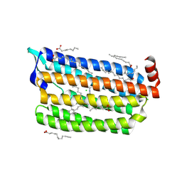
Structure of light-state marine bacterial chloride importer, NM-R3, with CW laser (ND-3%) at 95K.
Descriptor: CHLORIDE ION, Chloride pumping rhodopsin, OLEIC ACID, ...
Authors:Yun, J.H, Ohki, M, Park, S.Y, Lee, W.
Deposit date:2019-04-26
Release date:2020-03-04
Last modified:2023-11-22
Method:X-RAY DIFFRACTION (1.9 Å)
Cite:Pumping mechanism of NM-R3, a light-driven bacterial chloride importer in the rhodopsin family.
Sci Adv, 6, 2020
3WQJ
DownloadVisualize
BU of 3wqj by Molmil
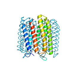
Crystal structure of archaerhodopsin-2 at 1.8 angstrom resolution
Descriptor: (6E,10E,14E,18E)-2,6,10,15,19,23-hexamethyltetracosa-2,6,10,14,18,22-hexaene, 2,3-DI-O-PHYTANLY-3-SN-GLYCERO-1-PHOSPHORYL-3'-SN-GLYCEROL-1'-PHOSPHATE, 2,3-DI-PHYTANYL-GLYCEROL, ...
Authors:Kouyama, T.
Deposit date:2014-01-27
Release date:2014-10-15
Last modified:2023-11-08
Method:X-RAY DIFFRACTION (1.8 Å)
Cite:Structure of archaerhodopsin-2 at 1.8 angstrom resolution.
Acta Crystallogr.,Sect.D, 70, 2014
1VGO
DownloadVisualize
BU of 1vgo by Molmil
Crystal Structure of Archaerhodopsin-2
Descriptor: Archaerhodopsin 2, RETINAL, SULFATE ION, ...
Authors:Yoshimura, K, Enami, N, Murakami, M, Okumura, H, Ihara, K, Kouyama, T.
Deposit date:2004-04-28
Release date:2005-10-04
Last modified:2023-10-25
Method:X-RAY DIFFRACTION (2.5 Å)
Cite:Crystal structures of archaerhodopsin-1 and -2: Common structural motif in archaeal light-driven proton pumps
J.Mol.Biol., 358, 2006
1UAZ
DownloadVisualize
BU of 1uaz by Molmil
Crystal structure of archaerhodopsin-1
Descriptor: RETINAL, archaerhodopsin-1
Authors:Enami, N, Okumua, H, Kouyama, T.
Deposit date:2003-03-26
Release date:2003-04-15
Last modified:2023-10-25
Method:X-RAY DIFFRACTION (3.4 Å)
Cite:X-ray crystallographic studies of archaerhodopsin
J.Photosci., 9, 2003
1GUE
DownloadVisualize
BU of 1gue by Molmil
SENSORY RHODOPSIN II
Descriptor: CHLORIDE ION, RETINAL, SENSORY RHODOPSIN II
Authors:Edman, K, Royant, A, Nollert, P, Maxwell, C.A, Pebay-Peyroula, E, Navarro, J, Neutze, R, Landau, E.M.
Deposit date:2002-01-24
Release date:2002-04-12
Last modified:2023-12-13
Method:X-RAY DIFFRACTION (2.27 Å)
Cite:Early Structural Rearrangements in the Photocycle of an Integral Membrane Sensory Receptor
Structure, 10, 2002
1GU8
DownloadVisualize
BU of 1gu8 by Molmil
SENSORY RHODOPSIN II
Descriptor: CHLORIDE ION, RETINAL, SENSORY RHODOPSIN II
Authors:Edman, K, Royant, A, Nollert, P, Maxwell, C.A, Pebay-Peyroula, E, Navarro, J, Neutze, R, Landau, E.M.
Deposit date:2002-01-24
Release date:2002-04-12
Last modified:2023-12-13
Method:X-RAY DIFFRACTION (2.27 Å)
Cite:Early Structural Rearrangements in the Photocycle of an Integral Membrane Sensory Receptor
Structure, 10, 2002
4YGH
DownloadVisualize
BU of 4ygh by Molmil
Crystal Structure of R111K:Y134F:T54V:R132Q:P39Y:R59Y mutant of human Cellular Retinoic Acid Binding Protein II with Retinal at 2.1 Angstrom resolution - UV irradiated crystal - 2nd cycle
Descriptor: Cellular retinoic acid-binding protein 2, RETINAL
Authors:Nosrati, M, Geiger, J.H.
Deposit date:2015-02-26
Release date:2016-03-02
Last modified:2023-09-27
Method:X-RAY DIFFRACTION (2.1 Å)
Cite:A Photoisomerizing Rhodopsin Mimic Observed at Atomic Resolution.
J.Am.Chem.Soc., 138, 2016
4YH0
DownloadVisualize
BU of 4yh0 by Molmil
Crystal structure of the R111K:Y134F:T54V:R132Q:P39Y:R59Y mutant of human Cellular Retinoic Acid Binding Protein II in complex with Retinal at 2.14 angstrom resolution - UV irradiated crystal - 3rd cycle
Descriptor: Cellular retinoic acid-binding protein 2, RETINAL
Authors:Nosrati, M, Geiger, J.H.
Deposit date:2015-02-26
Release date:2016-03-02
Last modified:2023-09-27
Method:X-RAY DIFFRACTION (2.144 Å)
Cite:A Photoisomerizing Rhodopsin Mimic Observed at Atomic Resolution.
J.Am.Chem.Soc., 138, 2016
4YDA
DownloadVisualize
BU of 4yda by Molmil
Crystal structure of R111K:Y134F:T54V:R132Q:P39Q:R59Y mutant of human cellular retinoic acid binding proteinii with retinal at 1.95 angstrom - after 1hour visible light irradiation - 3rd cycle
Descriptor: Cellular retinoic acid-binding protein 2, RETINAL
Authors:Nosrati, M, Geiger, J.H.
Deposit date:2015-02-21
Release date:2016-02-24
Last modified:2023-09-27
Method:X-RAY DIFFRACTION (1.951 Å)
Cite:A Photoisomerizing Rhodopsin Mimic Observed at Atomic Resolution.
J.Am.Chem.Soc., 138, 2016
4YDB
DownloadVisualize
BU of 4ydb by Molmil
Crystal structure of the R111K:Y134F:T54V:R132Q:P39Q:R59Y mutant of human Cellular Retinoic Acid Binding Protein II in complex with Retinal at 2.03 angstrom -UV irradiated crystal- 3rd cycle
Descriptor: Cellular retinoic acid-binding protein 2, RETINAL
Authors:Nosrati, M, Geiger, J.H.
Deposit date:2015-02-21
Release date:2016-02-24
Last modified:2023-09-27
Method:X-RAY DIFFRACTION (2.03 Å)
Cite:A Photoisomerizing Rhodopsin Mimic Observed at Atomic Resolution.
J.Am.Chem.Soc., 138, 2016
4YFQ
DownloadVisualize
BU of 4yfq by Molmil
CRYSTAL STRUCTURE OF THE R111K:Y134F:T54V:R132Q:P39Y:R59Y MUTANT OF HUMAN CELLULAR RETINOIC ACID BINDING PROTEINII WITH RETINAL AFTER 24 HOURS INCUBATION AT 1.62 ANGSTROM RESOLUTION - Thermodynamic product - 1st cycle
Descriptor: Cellular retinoic acid-binding protein 2, RETINAL
Authors:Nosrati, M, Geiger, J.H.
Deposit date:2015-02-25
Release date:2016-03-02
Last modified:2023-09-27
Method:X-RAY DIFFRACTION (1.62 Å)
Cite:A Photoisomerizing Rhodopsin Mimic Observed at Atomic Resolution.
J.Am.Chem.Soc., 138, 2016
4YCE
DownloadVisualize
BU of 4yce by Molmil
Crystal structure of the R111K:Y134F:T54V:R132Q:P39Q:R59Y mutant of human cellular retinoic acid binding proteinii with retinal at 1.95 angstrom- visible light irradiated crystal for 1 hour - 2nd cycle
Descriptor: Cellular retinoic acid-binding protein 2, RETINAL
Authors:Nosrati, M, Geiger, J.H.
Deposit date:2015-02-19
Release date:2016-02-24
Last modified:2023-09-27
Method:X-RAY DIFFRACTION (1.953 Å)
Cite:A Photoisomerizing Rhodopsin Mimic Observed at Atomic Resolution.
J.Am.Chem.Soc., 138, 2016
4YFR
DownloadVisualize
BU of 4yfr by Molmil
Crystal structure of the R111K:Y134F:T54V:R132Q:P39Y:R59Y mutant of human Cellular Retinoic Acid Binding Protein II with Retinal at 1.95 Angstrom Resolution - UV irradiated crystal for 30 minutes - 1st Cycle
Descriptor: Cellular retinoic acid-binding protein 2, RETINAL
Authors:Nosrati, M, Geiger, J.H.
Deposit date:2015-02-25
Release date:2016-03-02
Last modified:2023-09-27
Method:X-RAY DIFFRACTION (1.952 Å)
Cite:A Photoisomerizing Rhodopsin Mimic Observed at Atomic Resolution.
J.Am.Chem.Soc., 138, 2016
4YKM
DownloadVisualize
BU of 4ykm by Molmil
Crystal structure of the R111K:Y134F:T54V:R132Q:P39Q:R59Y:A32W:F3Q mutant of human Cellular Retinoic Acid Binding Protein II with Retinal at 1.58 angstrom resolution
Descriptor: Cellular retinoic acid-binding protein 2, RETINAL
Authors:Nosrati, M, Geiger, J.H.
Deposit date:2015-03-04
Release date:2016-03-09
Last modified:2023-09-27
Method:X-RAY DIFFRACTION (1.58 Å)
Cite:A Photoisomerizing Rhodopsin Mimic Observed at Atomic Resolution.
J.Am.Chem.Soc., 138, 2016
4YKO
DownloadVisualize
BU of 4yko by Molmil
Crystal structure of the R111K:Y134F:T54V:R132Q:P39Q:R59Y:A32Y:F3Q mutant of human Cellular Retinoic Acid Binding Protein II with Retinal at 1.58 angstrom resolution
Descriptor: Cellular retinoic acid-binding protein 2, RETINAL
Authors:Nosrati, M, Geiger, J.
Deposit date:2015-03-04
Release date:2016-03-09
Last modified:2023-09-27
Method:X-RAY DIFFRACTION (1.57 Å)
Cite:A Photoisomerizing Rhodopsin Mimic Observed at Atomic Resolution.
J.Am.Chem.Soc., 138, 2016
4YBP
DownloadVisualize
BU of 4ybp by Molmil
Crystal structure of the R111K:Y134F:T54V:R132Q:P39Q:R59Y mutant of human cellular retinoic acid binding proteinii with retinal after 24 hour incubation at 1.83 angstrom resolution - thermodynamic product - 1st cycle
Descriptor: Cellular retinoic acid-binding protein 2, RETINAL
Authors:Nosrati, M, Geiger, J.H.
Deposit date:2015-02-19
Release date:2016-02-24
Last modified:2023-09-27
Method:X-RAY DIFFRACTION (1.831 Å)
Cite:A Photoisomerizing Rhodopsin Mimic Observed at Atomic Resolution.
J.Am.Chem.Soc., 138, 2016
4YGZ
DownloadVisualize
BU of 4ygz by Molmil
CRYSTAL STRUCTURE OF THE R111K:Y134F:T54V:R132Q:P39Y:R59Y MUTANT OF HUMAN CELLULAR RETINOIC ACID BINDING PROTEIN II WITH RETINAL AT 2.06 ANGSTROM RESOLUTION - VISIBLE LIGHT IRRADIATED CRYSTAL -3RD CYCLE
Descriptor: Cellular retinoic acid-binding protein 2, RETINAL
Authors:Nosrati, M, Geiger, J.H.
Deposit date:2015-02-26
Release date:2016-03-02
Last modified:2023-09-27
Method:X-RAY DIFFRACTION (2.06 Å)
Cite:A Photoisomerizing Rhodopsin Mimic Observed at Atomic Resolution.
J.Am.Chem.Soc., 138, 2016
4YFP
DownloadVisualize
BU of 4yfp by Molmil
CRYSTAL STRUCTURE OF THE R111K:Y134F:T54V:R132Q:P39Y:R59Y MUTANT OF HUMAN CELLULAR RETINOIC ACID BINDING PROTEIN II WITH RETINAL AFTER 20 MINUTES INCUBATION AT 1.95 ANGSTROM RESOLUTION-Kinetic Product
Descriptor: Cellular retinoic acid-binding protein 2, RETINAL
Authors:Nosrati, M, Geiger, J.H.
Deposit date:2015-02-25
Release date:2016-03-02
Last modified:2023-09-27
Method:X-RAY DIFFRACTION (1.951 Å)
Cite:A Photoisomerizing Rhodopsin Mimic Observed at Atomic Resolution.
J.Am.Chem.Soc., 138, 2016
4YBU
DownloadVisualize
BU of 4ybu by Molmil
Crystal structure of the R111K:Y134F:T54V:R132Q:P39Q:R59Y mutant of human Cellular Retinoic Acid Binding ProteinII in complex with Retinal after 24 h incubation and 1 hour UV irradiation at 1.92 angstrom - 1st cycle
Descriptor: Cellular retinoic acid-binding protein 2, RETINAL
Authors:Nosrati, M, Geiger, J.H.
Deposit date:2015-02-19
Release date:2016-02-24
Last modified:2023-09-27
Method:X-RAY DIFFRACTION (1.924 Å)
Cite:A Photoisomerizing Rhodopsin Mimic Observed at Atomic Resolution.
J.Am.Chem.Soc., 138, 2016
4YCH
DownloadVisualize
BU of 4ych by Molmil
Crystal Structure of R111K:Y134F:T54V:R132Q:P39Q:R59Y mutant of human Cellular Retinoic Acid Binding ProteinII with Retinal at 1.96 Angstrom - UV irradiated Crystal for 1 hour - 2nd cycle
Descriptor: Cellular retinoic acid-binding protein 2, RETINAL
Authors:Nosrati, M, Geiger, J.H.
Deposit date:2015-02-20
Release date:2016-02-24
Last modified:2023-09-27
Method:X-RAY DIFFRACTION (1.96 Å)
Cite:A Photoisomerizing Rhodopsin Mimic Observed at Atomic Resolution.
J.Am.Chem.Soc., 138, 2016
5AX0
DownloadVisualize
BU of 5ax0 by Molmil
Crystal Structure of the Cell-Free Synthesized Membrane Protein, Acetabularia Rhodopsin I, at 1.52 angstrom
Descriptor: (2S)-2,3-dihydroxypropyl (9Z)-octadec-9-enoate, DECANE, DODECANE, ...
Authors:Furuse, M, Hosaka, T, Kimura-Someya, T, Yokoyama, S, Shirouzu, M.
Deposit date:2015-07-10
Release date:2015-08-26
Last modified:2023-11-08
Method:X-RAY DIFFRACTION (1.521 Å)
Cite:Structural basis for the slow photocycle and late proton release in Acetabularia rhodopsin I from the marine plant Acetabularia acetabulum
Acta Crystallogr.,Sect.D, 71, 2015
5AWZ
DownloadVisualize
BU of 5awz by Molmil
Crystal Structure of the Cell-Free Synthesized Membrane Protein, Acetabularia Rhodopsin I, at 1.57 angstrom
Descriptor: (2S)-2,3-dihydroxypropyl (9Z)-octadec-9-enoate, DECANE, DODECANE, ...
Authors:Furuse, M, Hosaka, T, Kimura-Someya, T, Yokoyama, S, Shirouzu, M.
Deposit date:2015-07-10
Release date:2015-08-26
Last modified:2023-11-08
Method:X-RAY DIFFRACTION (1.57 Å)
Cite:Structural basis for the slow photocycle and late proton release in Acetabularia rhodopsin I from the marine plant Acetabularia acetabulum
Acta Crystallogr.,Sect.D, 71, 2015
5AX1
DownloadVisualize
BU of 5ax1 by Molmil
Crystal Structure of the Cell-Free Synthesized Membrane Protein, Acetabularia Rhodopsin I, at 1.80 angstrom
Descriptor: (2S)-2,3-dihydroxypropyl (9Z)-octadec-9-enoate, DECANE, DODECANE, ...
Authors:Furuse, M, Hosaka, T, Kimura-Someya, T, Yokoyama, S, Shirouzu, M.
Deposit date:2015-07-10
Release date:2015-08-26
Last modified:2023-11-08
Method:X-RAY DIFFRACTION (1.803 Å)
Cite:Structural basis for the slow photocycle and late proton release in Acetabularia rhodopsin I from the marine plant Acetabularia acetabulum
Acta Crystallogr.,Sect.D, 71, 2015
4YGG
DownloadVisualize
BU of 4ygg by Molmil
CRYSTAL STRUCTURE OF R111K:Y134F:T54V:R132Q:P39Y:R59Y MUTANT OF HUMAN CELLULAR RETINOIC ACID BINDING PROTEIN II WITH RETINAL AT 1.9 ANGSTROM RESOLUTION - VISIBLE LIGHT IRRADIATED CRYSTAL - 2ND CYCLE
Descriptor: Cellular retinoic acid-binding protein 2, RETINAL
Authors:Nosrati, M, Geiger, J.H.
Deposit date:2015-02-26
Release date:2016-03-02
Last modified:2023-09-27
Method:X-RAY DIFFRACTION (1.9 Å)
Cite:A Photoisomerizing Rhodopsin Mimic Observed at Atomic Resolution.
J.Am.Chem.Soc., 138, 2016
5A3Z
DownloadVisualize
BU of 5a3z by Molmil
Structure of monoclinic Lysozyme obtained by multi crystal data collection
Descriptor: GLYCEROL, LYSOZYME, NITRATE ION
Authors:Zander, U, Bourenkov, G, Popov, A.N, de Sanctis, D, McCarthy, A.A, Svensson, O, Round, E.S, Gordeliy, V.I, Mueller-Dieckmann, C, Leonard, G.A.
Deposit date:2015-06-04
Release date:2015-11-11
Last modified:2015-11-25
Method:X-RAY DIFFRACTION (1.59 Å)
Cite:Meshandcollect: An Automated Multi-Crystal Data-Collection Workflow for Synchrotron Macromolecular Crystallography Beamlines.
Acta Crystallogr.,Sect.D, 71, 2015

222926

數據於2024-07-24公開中

PDB statisticsPDBj update infoContact PDBjnumon