Loading
PDBj
MenuPDBj@FacebookPDBj@TwitterPDBj@YouTubewwPDB FoundationwwPDB
RCSB PDBPDBeBMRBAdv. SearchSearch help

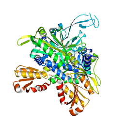
6DYG
DownloadVisualize
BU of 6dyg by Molmil
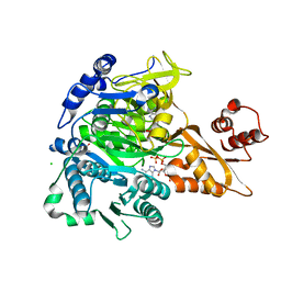
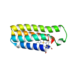
Fe(II)-bound structure of the engineered cyt cb562 variant, CH3Y
Descriptor: FE (III) ION, HEME C, MAGNESIUM ION, ...
Authors:Tezcan, F.A, Rittle, J.
Deposit date:2018-07-01
Release date:2019-04-24
Last modified:2024-10-16
Method:X-RAY DIFFRACTION (1.49 Å)
Cite:An efficient, step-economical strategy for the design of functional metalloproteins.
Nat.Chem., 11, 2019
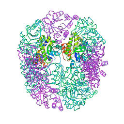
7MP4
DownloadVisualize
BU of 7mp4 by Molmil
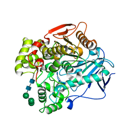
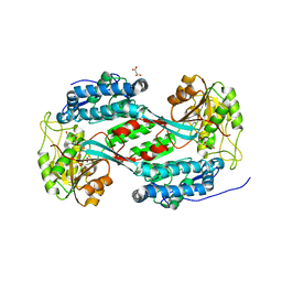
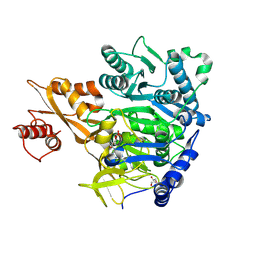
Crystal structure of Epiphyas postvittana antennal carboxylesterase 24
Descriptor: Carboxylesterase-24, alpha-D-mannopyranose-(1-2)-alpha-D-mannopyranose-(1-3)-beta-D-mannopyranose-(1-4)-2-acetamido-2-deoxy-beta-D-glucopyranose-(1-4)-2-acetamido-2-deoxy-beta-D-glucopyranose
Authors:Hamiaux, C, Carraher, C.
Deposit date:2021-05-04
Release date:2022-05-11
Last modified:2024-11-06
Method:X-RAY DIFFRACTION (2.43 Å)
Cite:Structure of an antennally-expressed carboxylesterase suggests lepidopteran odorant degrading enzymes are broadly tuned
Curr Res Insect Sci, 3, 2023
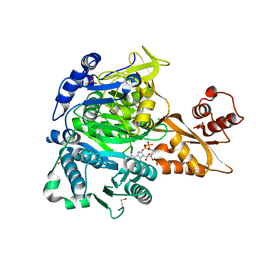
8UPZ
DownloadVisualize
BU of 8upz by Molmil
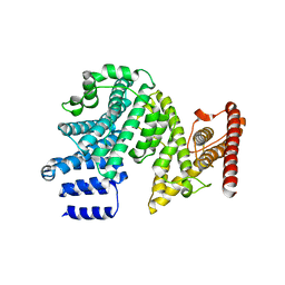
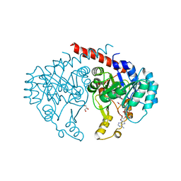
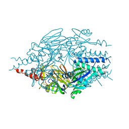
Minimal PutA proline dehydrogenase domain (design #2) complexed with (Prop-2-ynylthio)acetic acid
Descriptor: (Prop-2-ynylthio)acetic acid, Bifunctional protein PutA, DI(HYDROXYETHYL)ETHER, ...
Authors:Tanner, J.J.
Deposit date:2023-10-23
Release date:2024-10-30
Method:X-RAY DIFFRACTION (1.38 Å)
Cite:Noncovalent Inhibition and Covalent Inactivation of Proline Dehydrogenase by Analogs of N -Propargylglycine.
Biochemistry, 2024
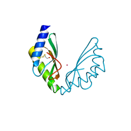
6H9P
DownloadVisualize
BU of 6h9p by Molmil
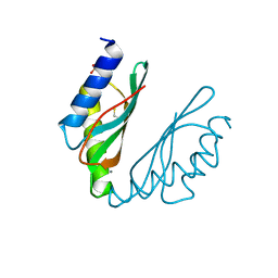
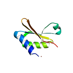
MamM CTD E289D - Manganese form
Descriptor: BETA-MERCAPTOETHANOL, MANGANESE (II) ION, Magnetosome protein MamM, ...
Authors:Barber-Zucker, S, Zarivach, R.
Deposit date:2018-08-05
Release date:2019-08-14
Last modified:2024-01-17
Method:X-RAY DIFFRACTION (2.1 Å)
Cite:MamM CTD E289D - Manganese form
To Be Published
8U2V
DownloadVisualize
BU of 8u2v by Molmil
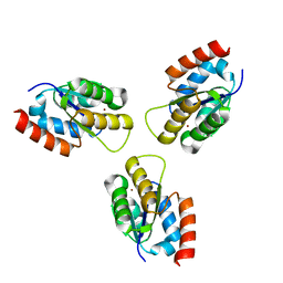
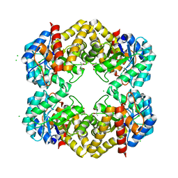
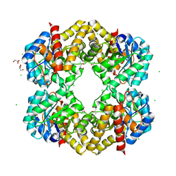
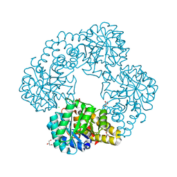
Crystal Structure of methylglyoxal synthase from Borrelia burgdorferi
Descriptor: BROMIDE ION, Methylglyoxal synthase
Authors:Seattle Structural Genomics Center for Infectious Disease, Seattle Structural Genomics Center for Infectious Disease (SSGCID)
Deposit date:2023-09-06
Release date:2023-09-13
Method:X-RAY DIFFRACTION (2.5 Å)
Cite:Crystal Structure of methylglyoxal synthase from Borrelia burgdorferi
To be published
7MNJ
DownloadVisualize
BU of 7mnj by Molmil
Crystal structure of the N-terminal domain of NUP358/RanBP2 (residues 145-673)
Descriptor: E3 SUMO-protein ligase RanBP2
Authors:Bley, C.J, Nie, S, Mobbs, G.W, Petrovic, S, Gres, A.T, Liu, X, Mukherjee, S, Harvey, S, Huber, F.M, Lin, D.H, Brown, B, Tang, A.W, Rundlet, E.J, Correia, A.R, Chen, S, Regmi, S.G, Stevens, T.A, Jette, C.A, Patke, A, Dasso, M, Palazzo, A.F, Kossiakoff, A.A, Hoelz, A.
Deposit date:2021-05-01
Release date:2022-06-15
Last modified:2024-05-22
Method:X-RAY DIFFRACTION (3.8 Å)
Cite:Architecture of the cytoplasmic face of the nuclear pore.
Science, 376, 2022
6DZX
DownloadVisualize
BU of 6dzx by Molmil
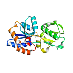
Crystal structure of the N. meningitides methionine-binding protein in its D-methionine bound conformation.
Descriptor: D-METHIONINE, Lipoprotein
Authors:Nguyen, P.T, Lai, J.Y, Kaiser, J.T, Rees, D.C.
Deposit date:2018-07-05
Release date:2019-07-10
Last modified:2024-03-13
Method:X-RAY DIFFRACTION (1.678 Å)
Cite:Structures of the Neisseria meningitides methionine-binding protein MetQ in substrate-free form and bound to l- and d-methionine isomers.
Protein Sci., 28, 2019
6H9Z
DownloadVisualize
BU of 6h9z by Molmil
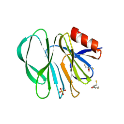
Molecular bases of histo-blood group antigen recognition by the most common human rotavirus
Descriptor: 2,3-DIHYDROXY-1,4-DITHIOBUTANE, Outer capsid protein VP4, SULFATE ION
Authors:Ciges-Tomas, J.R, Gozalbo-Rovira, R, Vila-Vicent, S, Buesa, J, Santiso-Bellon, C, Monedero, V, Yebra, M.J, Rodriguez-Diaz, J, Marina, A.
Deposit date:2018-08-06
Release date:2019-06-12
Last modified:2024-01-17
Method:X-RAY DIFFRACTION (1.51 Å)
Cite:Unraveling the role of the secretor antigen in human rotavirus attachment to histo-blood group antigens.
Plos Pathog., 15, 2019
8U91
DownloadVisualize
BU of 8u91 by Molmil
Crystal structure of N-acetylneuraminate lyase (NanA) from Klebsiella aerogenes (Apo, Orthorhombic P form)
Descriptor: (4S)-2-METHYL-2,4-PENTANEDIOL, CHLORIDE ION, N-acetylneuraminate lyase
Authors:Seattle Structural Genomics Center for Infectious Disease (SSGCID)
Deposit date:2023-09-18
Release date:2023-09-27
Method:X-RAY DIFFRACTION (2.65 Å)
Cite:Crystal structure of N-acetylneuraminate lyase (NanA) from Klebsiella aerogenes (Apo, Orthorhombic P form)
To be published
6E0N
DownloadVisualize
BU of 6e0n by Molmil
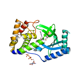
Structure of Elizabethkingia meningoseptica CdnE cyclic dinucleotide synthase with GTP and Apcpp
Descriptor: DIPHOSPHOMETHYLPHOSPHONIC ACID ADENOSYL ESTER, GUANOSINE-5'-TRIPHOSPHATE, MAGNESIUM ION, ...
Authors:Eaglesham, J.B, Whiteley, A.T, de Oliveira Mann, C.C, Morehouse, B.R, Nieminen, E.A, King, D.S, Lee, A.S.Y, Mekalanos, J.J, Kranzusch, P.J.
Deposit date:2018-07-06
Release date:2019-02-20
Last modified:2024-03-13
Method:X-RAY DIFFRACTION (1.5 Å)
Cite:Bacterial cGAS-like enzymes synthesize diverse nucleotide signals.
Nature, 567, 2019
8U2Q
DownloadVisualize
BU of 8u2q by Molmil
Crystal Structure of Glycine--tRNA ligase active site chimera from Mycobacterium thermoresistibile/tuberculosis (G5A bound)
Descriptor: 1,2-ETHANEDIOL, 5'-O-(glycylsulfamoyl)adenosine, CHLORIDE ION, ...
Authors:Seattle Structural Genomics Center for Infectious Disease, Seattle Structural Genomics Center for Infectious Disease (SSGCID)
Deposit date:2023-09-06
Release date:2023-09-13
Method:X-RAY DIFFRACTION (2.45 Å)
Cite:Crystal Structure of Glycine--tRNA ligase active site chimera from Mycobacterium thermoresistibile/tuberculosis (G5A bound)
To be published
6HYE
DownloadVisualize
BU of 6hye by Molmil
PDX1.2/PDX1.3 complex (PDX1.3:K97A)
Descriptor: Pyridoxal 5'-phosphate synthase subunit PDX1.3, Pyridoxal 5'-phosphate synthase-like subunit PDX1.2, SULFATE ION
Authors:Robinson, G.C, Kaufmann, M, Roux, C, Martinez-Font, J, Hothorn, M, Thore, S, Fitzpatrick, T.B.
Deposit date:2018-10-20
Release date:2019-04-24
Last modified:2024-01-24
Method:X-RAY DIFFRACTION (2.53 Å)
Cite:Crystal structure of the pseudoenzyme PDX1.2 in complex with its cognate enzyme PDX1.3: a total eclipse.
Acta Crystallogr D Struct Biol, 75, 2019
8U2T
DownloadVisualize
BU of 8u2t by Molmil
Crystal Structure of Acetyl-coenzyme A synthetase from Leishmania infantum (CoA and AMP bound)
Descriptor: ADENOSINE MONOPHOSPHATE, Acetyl-coenzyme A synthetase, CHLORIDE ION, ...
Authors:Seattle Structural Genomics Center for Infectious Disease, Seattle Structural Genomics Center for Infectious Disease (SSGCID)
Deposit date:2023-09-06
Release date:2023-09-13
Last modified:2024-10-23
Method:X-RAY DIFFRACTION (1.65 Å)
Cite:A single Leishmania adenylate forming enzyme of the ANL superfamily generates both acetyl- and acetoacetyl-CoA.
J.Biol.Chem., 2024
6HAO
DownloadVisualize
BU of 6hao by Molmil
MamM CTD H264E - Cadmium form 2
Descriptor: BETA-MERCAPTOETHANOL, CADMIUM ION, Magnetosome protein MamM, ...
Authors:Barber-Zucker, S, Zarivach, R.
Deposit date:2018-08-08
Release date:2019-08-21
Last modified:2024-01-17
Method:X-RAY DIFFRACTION (2.4 Å)
Cite:MamM CTD H264E - Cadmium form 2
To Be Published
8U92
DownloadVisualize
BU of 8u92 by Molmil
Crystal structure of N-acetylneuraminate lyase (NanA) from Klebsiella aerogenes (pyruvate bound, Orthorhombic P form)
Descriptor: (4S)-2-METHYL-2,4-PENTANEDIOL, CHLORIDE ION, GLYCEROL, ...
Authors:Seattle Structural Genomics Center for Infectious Disease (SSGCID)
Deposit date:2023-09-18
Release date:2023-09-27
Last modified:2023-11-29
Method:X-RAY DIFFRACTION (2.1 Å)
Cite:Crystal structure of N-acetylneuraminate lyase (NanA) from Klebsiella aerogenes (pyruvate bound, Orthorhombic P form)
To be published
6HAS
DownloadVisualize
BU of 6has by Molmil
Crystal Structure of the small subunit-like domain of Rubisco activase from Nostoc sp. (strain PCC 7120)
Descriptor: NICKEL (II) ION, Ribulose bisphosphate carboxylase/oxygenase activase
Authors:Popilka, L, Bracher, A.
Deposit date:2018-08-08
Release date:2020-01-15
Last modified:2024-11-06
Method:X-RAY DIFFRACTION (1.38 Å)
Cite:Dual Functions of a Rubisco Activase in Metabolic Repair and Recruitment to Carboxysomes.
Cell, 183, 2020
6E2A
DownloadVisualize
BU of 6e2a by Molmil
Crystal structure of NADH:quinone reductase PA1024 from Pseudomonas aeruginosa PAO1 in complex with NAD+
Descriptor: FLAVIN MONONUCLEOTIDE, GLYCEROL, NICOTINAMIDE-ADENINE-DINUCLEOTIDE, ...
Authors:Reis, R.A.G, Ball, J, Agniswamy, J, Weber, I, Gadda, G.
Deposit date:2018-07-10
Release date:2019-02-06
Last modified:2023-10-11
Method:X-RAY DIFFRACTION (2.2 Å)
Cite:Steric hindrance controls pyridine nucleotide specificity of a flavin-dependent NADH:quinone oxidoreductase.
Protein Sci., 28, 2019
8U9B
DownloadVisualize
BU of 8u9b by Molmil
Crystal Structure of Betaine aldehyde dehydrogenase (BetB) from Klebsiella aerogenes (Apo, P21 Form)
Descriptor: 2-[3-(2-HYDROXY-1,1-DIHYDROXYMETHYL-ETHYLAMINO)-PROPYLAMINO]-2-HYDROXYMETHYL-PROPANE-1,3-DIOL, 4-(2-HYDROXYETHYL)-1-PIPERAZINE ETHANESULFONIC ACID, Betaine aldehyde dehydrogenase, ...
Authors:Seattle Structural Genomics Center for Infectious Disease, Seattle Structural Genomics Center for Infectious Disease (SSGCID)
Deposit date:2023-09-18
Release date:2023-09-27
Method:X-RAY DIFFRACTION (1.9 Å)
Cite:Crystal Structure of Betaine aldehyde dehydrogenase (BetB) from Klebsiella aerogenes (Apo, P21 Form)
To be published
8U2U
DownloadVisualize
BU of 8u2u by Molmil
Crystal Structure of Acetyl-coenzyme A synthetase from Leishmania infantum (CoA, AMP and potassium bound)
Descriptor: ADENOSINE MONOPHOSPHATE, Acetyl-coenzyme A synthetase, CHLORIDE ION, ...
Authors:Seattle Structural Genomics Center for Infectious Disease, Seattle Structural Genomics Center for Infectious Disease (SSGCID)
Deposit date:2023-09-06
Release date:2023-09-13
Last modified:2024-10-23
Method:X-RAY DIFFRACTION (1.97 Å)
Cite:A single Leishmania adenylate forming enzyme of the ANL superfamily generates both acetyl- and acetoacetyl-CoA.
J.Biol.Chem., 2024
8U2R
DownloadVisualize
BU of 8u2r by Molmil
Crystal Structure of Acetyl-coenzyme A synthetase from Leishmania infantum (Ethyl AMP bound)
Descriptor: 5'-O-[(S)-ethoxy(hydroxy)phosphoryl]adenosine, Acetyl-coenzyme A synthetase, DI(HYDROXYETHYL)ETHER
Authors:Seattle Structural Genomics Center for Infectious Disease, Seattle Structural Genomics Center for Infectious Disease (SSGCID)
Deposit date:2023-09-06
Release date:2023-09-13
Last modified:2024-10-23
Method:X-RAY DIFFRACTION (1.55 Å)
Cite:A single Leishmania adenylate forming enzyme of the ANL superfamily generates both acetyl- and acetoacetyl-CoA.
J.Biol.Chem., 2024
8U98
DownloadVisualize
BU of 8u98 by Molmil
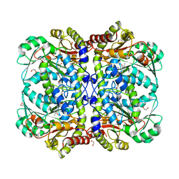
Crystal Structure of Cystathionine beta lyase from Klebsiella aerogenes (PLP-Glycine adduct)
Descriptor: 1,2-ETHANEDIOL, Cystathionine beta-lyase, GLYCINE, ...
Authors:Seattle Structural Genomics Center for Infectious Disease, Seattle Structural Genomics Center for Infectious Disease (SSGCID)
Deposit date:2023-09-18
Release date:2023-09-27
Method:X-RAY DIFFRACTION (1.4 Å)
Cite:Crystal Structure of Cystathionine beta lyase from Klebsiella aerogenes (PLP-Glycine adduct)
To be published
6HBT
DownloadVisualize
BU of 6hbt by Molmil
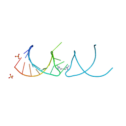
The structure of the G. violaceus guanidine II riboswitch P2 stem-loop with arcaine
Descriptor: 1-(4-carbamimidamidobutyl)guanidine, RNA (5'-R(*GP*GP*UP*GP*GP*GP*GP*AP*CP*GP*AP*CP*CP*CP*CP*AP*(CBV)P*C)-3'), SULFATE ION
Authors:Huang, L, Lilley, D.M.J.
Deposit date:2018-08-13
Release date:2019-02-20
Last modified:2024-05-15
Method:X-RAY DIFFRACTION (1.66 Å)
Cite:Structure-guided design of a high-affinity ligand for a riboswitch.
Rna, 25, 2019
8U90
DownloadVisualize
BU of 8u90 by Molmil
Crystal structure of N-acetylneuraminate lyase (NanA) from Klebsiella aerogenes (Apo, hexagonal form)
Descriptor: CHLORIDE ION, GLYCEROL, IODIDE ION, ...
Authors:Seattle Structural Genomics Center for Infectious Disease (SSGCID)
Deposit date:2023-09-18
Release date:2023-09-27
Method:X-RAY DIFFRACTION (1.85 Å)
Cite:Crystal structure of N-acetylneuraminate lyase (NanA) from Klebsiella aerogenes (Apo, hexagonal form)
To be published
7O39
DownloadVisualize
BU of 7o39 by Molmil
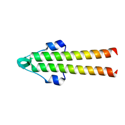
Crystal structure of S. aureus DivIVA N terminal domain
Descriptor: DivIVA
Authors:Rao, V.A, Booth, S, Lewis, R.J.
Deposit date:2021-04-01
Release date:2021-04-14
Last modified:2024-01-31
Method:X-RAY DIFFRACTION (1.3 Å)
Cite:Crystal structure of S. aureus DivIVA N terminal domain
To Be Published
6EAG
DownloadVisualize
BU of 6eag by Molmil
CRYSTAL STRUCTURE OF FUSION INHIBITOR JNJ-2408068 IN COMPLEX WITH HUMAN RESPIRATORY SYNCYTIAL VIRUS FUSION GLYCOPROTEIN ESCAPE VARIANT G143S STABILIZED IN THE PREFUSION STATE
Descriptor: 2-[N-CYCLOHEXYLAMINO]ETHANE SULFONIC ACID, 2-[[2-[[1-(2-azanylethyl)piperidin-4-yl]amino]-4-methyl-benzimidazol-1-yl]methyl]-6-methyl-pyridin-3-ol, D(-)-TARTARIC ACID, ...
Authors:Battles, M.B, McLellan, J.S.
Deposit date:2018-08-03
Release date:2019-08-07
Last modified:2023-10-11
Method:X-RAY DIFFRACTION (3.302 Å)
Cite:Structural Basis for Respiratory Syncytial Virus Fusion Inhibitor Resistance
To be published

227111

數據於2024-11-06公開中

PDB statisticsPDBj update infoContact PDBjnumon