Loading
PDBj
MenuPDBj@FacebookPDBj@TwitterPDBj@YouTubewwPDB FoundationwwPDB
RCSB PDBPDBeBMRBAdv. SearchSearch help
PDB: 40 results

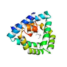
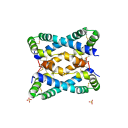
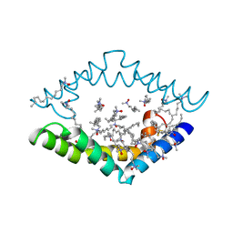
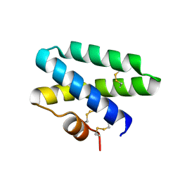
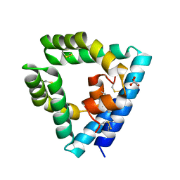
6SLR
DownloadVisualize
BU of 6slr by Molmil
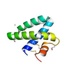
Structure of saposin B in complex with atovaquone
Descriptor: 2-[trans-4-(4-chlorophenyl)cyclohexyl]-3-hydroxynaphthalene-1,4-dione, GLYCEROL, Prosaposin
Authors:Zubieta, C, Milliken, B, Doyle, R.
Deposit date:2019-08-20
Release date:2020-09-09
Last modified:2024-01-24
Method:X-RAY DIFFRACTION (2.38 Å)
Cite:Structure of saposin B in complex with atovaquone
To Be Published
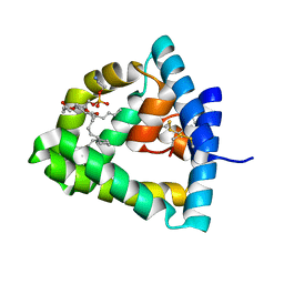
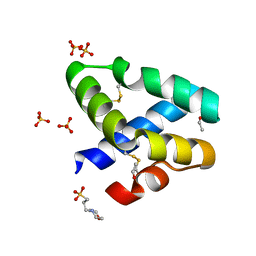
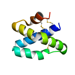
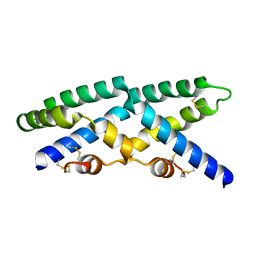
9AXG
DownloadVisualize
BU of 9axg by Molmil
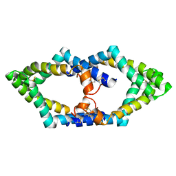
Human saposin B in the presence of globotriaosylceramide-NBD
Descriptor: MALONATE ION, Saposin-B
Authors:Sawyer, T.K, Garman, S.C.
Deposit date:2024-03-06
Release date:2024-03-13
Last modified:2024-09-04
Method:X-RAY DIFFRACTION (2.68 Å)
Cite:Human Saposin B Ligand Binding and Presentation to alpha-Galactosidase A.
Biorxiv, 2024
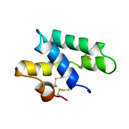
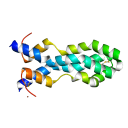
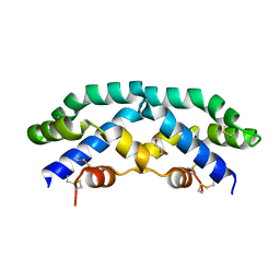
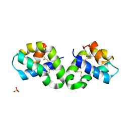
2JSA
DownloadVisualize
BU of 2jsa by Molmil
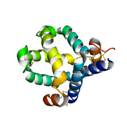
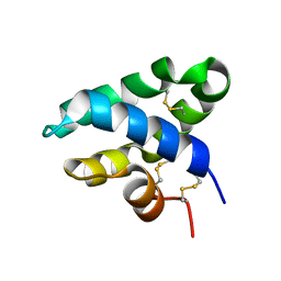
Solution structure of Caenopore-5 (81 Pro Trans confomer)
Descriptor: Saposin-like protein family protein 5
Authors:Mysliwy, J, Grotzinger, J.
Deposit date:2007-07-02
Release date:2008-10-14
Last modified:2024-10-16
Method:SOLUTION NMR
Cite:Caenopore-5: the three-dimensional structure of an antimicrobial protein from Caenorhabditis elegans.
Dev.Comp.Immunol., 34, 2010
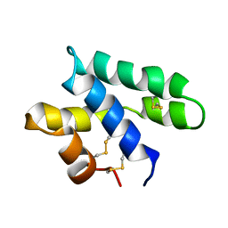
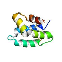
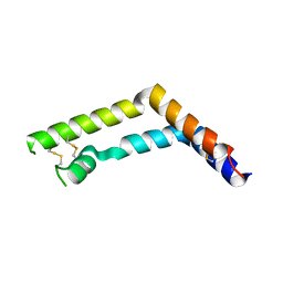
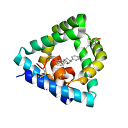
2JS9
DownloadVisualize
BU of 2js9 by Molmil
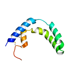
Structure of caenopore-5 (81 Pro cis conformer)
Descriptor: Saposin-like protein family protein 5
Authors:Mysliwy, J, Grotzinger, J.
Deposit date:2007-07-02
Release date:2008-10-14
Last modified:2022-03-16
Method:SOLUTION NMR
Cite:Caenopore-5: the three-dimensional structure of an antimicrobial protein from Caenorhabditis elegans.
Dev.Comp.Immunol., 34, 2010
4V2O
DownloadVisualize
BU of 4v2o by Molmil
Structure of saposin B in complex with chloroquine
Descriptor: N4-(7-CHLORO-QUINOLIN-4-YL)-N1,N1-DIETHYL-PENTANE-1,4-DIAMINE, SAPOSIN-B
Authors:Zubieta, C, Lai, X, Doyle, R.P.
Deposit date:2014-10-13
Release date:2015-12-09
Last modified:2024-10-23
Method:X-RAY DIFFRACTION (2.13 Å)
Cite:The Lysosomal Protein Saposin B Binds Chloroquine.
Chemmedchem, 11, 2016
1N69
DownloadVisualize
BU of 1n69 by Molmil
Crystal structure of human saposin B
Descriptor: 1,2-Distearoyl-sn-glycerophosphoethanolamine, SAPOSIN B
Authors:Ahn, V.E, Faull, K.F, Whitelegge, J.P, Fluharty, A.L, Prive, G.G.
Deposit date:2002-11-08
Release date:2003-01-07
Last modified:2024-10-30
Method:X-RAY DIFFRACTION (2.2 Å)
Cite:Crystal Structure of saposin B reveals a dimeric shell for lipid binding
Proc.Natl.Acad.Sci.USA, 100, 2003
6VZE
DownloadVisualize
BU of 6vze by Molmil
C-terminal domain of mouse surfactant protein B crystallized at low pH
Descriptor: Pulmonary surfactant-associated protein B, SULFATE ION
Authors:Rapoport, T.A, Bodnar, N.O.
Deposit date:2020-02-28
Release date:2020-11-25
Last modified:2024-11-06
Method:X-RAY DIFFRACTION (1.9 Å)
Cite:Mechanism of Lamellar Body Formation by Lung Surfactant Protein B.
Mol.Cell, 81, 2021
1L9L
DownloadVisualize
BU of 1l9l by Molmil
GRANULYSIN FROM HUMAN CYTOLYTIC T LYMPHOCYTES
Descriptor: 3[N-MORPHOLINO]PROPANE SULFONIC ACID, ETHANOL, Granulysin, ...
Authors:Anderson, D.H, Sawaya, M.R, Cascio, D, Ernst, W, Krensky, A, Modlin, R, Eisenberg, D.
Deposit date:2002-03-25
Release date:2002-11-06
Last modified:2024-11-06
Method:X-RAY DIFFRACTION (0.92 Å)
Cite:Granulysin Crystal Structure and a Structure-Derived Lytic Mechanism
J.Mol.Biol., 325, 2002
6VZ0
DownloadVisualize
BU of 6vz0 by Molmil
C-terminal domain of mouse surfactant protein B crystallized at high pH
Descriptor: Pulmonary surfactant-associated protein B, ZINC ION
Authors:Rapoport, T.A, Bodnar, N.O.
Deposit date:2020-02-27
Release date:2020-11-25
Last modified:2024-10-23
Method:X-RAY DIFFRACTION (1.75 Å)
Cite:Mechanism of Lamellar Body Formation by Lung Surfactant Protein B.
Mol.Cell, 81, 2021
1OF9
DownloadVisualize
BU of 1of9 by Molmil
Solution structure of the pore forming toxin of entamoeba histolytica (Amoebapore A)
Descriptor: PORE-FORMING PEPTIDE AMEOBAPORE A
Authors:Hecht, O, Schleinkofer, K, Bruhn, H, Leippe, M, Van Nuland, N, Dingley, A.J, Grotzinger, J.
Deposit date:2003-04-09
Release date:2004-02-26
Last modified:2024-10-16
Method:SOLUTION NMR
Cite:Solution Structure of the Pore-Forming Protein of Entamoeba Histolytica
J.Biol.Chem., 279, 2004
8EQU
DownloadVisualize
BU of 8equ by Molmil
Structure of SARS-CoV-2 Orf3a in late endosome/lysosome-like environment, Saposin A nanodisc
Descriptor: 1,2-dioleoyl-sn-glycero-3-phosphoethanolamine, ORF3a protein, Saposin A, ...
Authors:Miller, A.N, Houlihan, P.R, Matamala, E, Cabezas-Bratesco, D, Lee, G.Y, Cristofori-Armstrong, B, Dilan, T.L, Sanchez-Martinez, S, Matthies, D, Yan, R, Yu, Z, Ren, D, Brauchi, S.E, Clapham, D.E.
Deposit date:2022-10-09
Release date:2023-02-08
Method:ELECTRON MICROSCOPY (2.8 Å)
Cite:The SARS-CoV-2 accessory protein Orf3a is not an ion channel, but does interact with trafficking proteins.
Elife, 12, 2023
2DOB
DownloadVisualize
BU of 2dob by Molmil
Crystal Structure of Human Saposin A
Descriptor: CALCIUM ION, Proactivator polypeptide
Authors:Prive, G.G, Ahn, V.E.
Deposit date:2006-04-28
Release date:2006-07-25
Last modified:2011-07-13
Method:X-RAY DIFFRACTION (2 Å)
Cite:Crystal structures of saposins A and C.
Protein Sci., 15, 2006
7MBK
DownloadVisualize
BU of 7mbk by Molmil
N-terminal domain of mouse surfactant protein B, 6W mutant
Descriptor: Pulmonary surfactant-associated protein B
Authors:Milicic, G, Rapoport, T.A.
Deposit date:2021-03-31
Release date:2021-04-14
Last modified:2024-11-06
Method:X-RAY DIFFRACTION (2.17 Å)
Cite:Mechanism of Lamellar Body Formation by Lung Surfactant Protein B.
Mol.Cell, 81, 2021
7P4T
DownloadVisualize
BU of 7p4t by Molmil
Tetrameric structure of murine SapA
Descriptor: Saposin-A
Authors:Shamin, M, Deane, J.E.
Deposit date:2021-07-13
Release date:2022-05-25
Last modified:2024-10-16
Method:X-RAY DIFFRACTION (3.17 Å)
Cite:A Tetrameric Assembly of Saposin A: Increasing Structural Diversity in Lipid Transfer Proteins.
Contact, 4, 2021
4DDJ
DownloadVisualize
BU of 4ddj by Molmil
Crystal structure of saposin A in complex with lauryldimethylamine-N-oxide (LDAO)
Descriptor: LAURYL DIMETHYLAMINE-N-OXIDE, Saposin-A
Authors:Popovic, K, Prive, G.G.
Deposit date:2012-01-18
Release date:2012-02-15
Last modified:2020-01-29
Method:X-RAY DIFFRACTION (1.9 Å)
Cite:Structure of saposin A lipoprotein discs.
Proc.Natl.Acad.Sci.USA, 109, 2012
2GTG
DownloadVisualize
BU of 2gtg by Molmil
Crystal Structure of Human Saposin C
Descriptor: Proactivator polypeptide
Authors:Prive, G.G, Ahn, V.E.
Deposit date:2006-04-28
Release date:2006-07-25
Last modified:2024-10-30
Method:X-RAY DIFFRACTION (2.4 Å)
Cite:Crystal structures of saposins A and C.
Protein Sci., 15, 2006
2Z9A
DownloadVisualize
BU of 2z9a by Molmil
Crystal Structure of Human Saposin C Dimer in Open Conformation
Descriptor: GLYCEROL, Proactivator polypeptide
Authors:Rossmann, M, Saenger, W, Maier, T.
Deposit date:2007-09-18
Release date:2008-04-29
Last modified:2023-08-30
Method:X-RAY DIFFRACTION (2.5 Å)
Cite:Crystal structures of human saposins C and d: implications for lipid recognition and membrane interactions.
Structure, 16, 2008
3RFI
DownloadVisualize
BU of 3rfi by Molmil
Crystal structure of the saposin-like domain of plant aspartic protease from Solanum tuberosum
Descriptor: Asp
Authors:Bhaumik, P, Wlodawer, A.
Deposit date:2011-04-06
Release date:2011-06-15
Last modified:2024-11-06
Method:X-RAY DIFFRACTION (1.9 Å)
Cite:Structure and Mechanism of the Saposin-like Domain of a Plant Aspartic Protease.
J.Biol.Chem., 286, 2011
9AVS
DownloadVisualize
BU of 9avs by Molmil
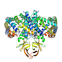
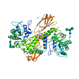
Human alpha-galactosidase A in complex with saposin B
Descriptor: 2-acetamido-2-deoxy-beta-D-glucopyranose, 2-acetamido-2-deoxy-beta-D-glucopyranose-(1-4)-2-acetamido-2-deoxy-beta-D-glucopyranose, Alpha-galactosidase A, ...
Authors:Sawyer, T.K, Garman, S.C.
Deposit date:2024-03-04
Release date:2024-04-17
Last modified:2024-10-23
Method:X-RAY DIFFRACTION (3.53 Å)
Cite:Human Saposin B Ligand Binding and Presentation to alpha-Galactosidase A.
Biorxiv, 2024
3BQQ
DownloadVisualize
BU of 3bqq by Molmil
Crystal Structure of Human Saposin D (triclinic)
Descriptor: PROACTIVATOR POLYPEPTIDE
Authors:Prive, G.G, Popovic, K.
Deposit date:2007-12-20
Release date:2008-02-05
Last modified:2024-11-06
Method:X-RAY DIFFRACTION (2 Å)
Cite:Structures of the human ceramide activator protein saposin D.
Acta Crystallogr.,Sect.D, 64, 2008
3BQP
DownloadVisualize
BU of 3bqp by Molmil
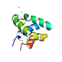
Crystal Structure of Human Saposin D (orthorhombic)
Descriptor: MAGNESIUM ION, Proactivator polypeptide
Authors:Prive, G.G, Popovic, K.
Deposit date:2007-12-20
Release date:2008-02-05
Last modified:2024-10-30
Method:X-RAY DIFFRACTION (1.3 Å)
Cite:Structures of the human ceramide activator protein saposin D.
Acta Crystallogr.,Sect.D, 64, 2008
1SN6
DownloadVisualize
BU of 1sn6 by Molmil
NMR solution structure of human Saposin C in SDS micelles
Descriptor: Proactivator polypeptide
Authors:Hawkins, C.A, de Alba, E, Tjandra, N.
Deposit date:2004-03-10
Release date:2005-03-01
Last modified:2022-03-02
Method:SOLUTION NMR
Cite:Solution structure of human saposin C in a detergent environment.
J.Mol.Biol., 346, 2005
4UEX
DownloadVisualize
BU of 4uex by Molmil
Structure of human Saposin A at lysosomal pH
Descriptor: PROSAPOSIN
Authors:Hill, C.H, Read, R.J, Deane, J.E.
Deposit date:2014-12-20
Release date:2015-07-15
Last modified:2023-12-20
Method:X-RAY DIFFRACTION (1.8 Å)
Cite:Structure of Human Saposin a at Lysosomal Ph.
Acta Crystallogr.,Sect.D, 71, 2015
2QYP
DownloadVisualize
BU of 2qyp by Molmil
Orthorhombic Crystal Structure of Human Saposin C Dimer in Open Conformation
Descriptor: Proactivator polypeptide
Authors:Rossmann, M, Saenger, W, Maier, T.
Deposit date:2007-08-15
Release date:2008-04-29
Last modified:2024-10-30
Method:X-RAY DIFFRACTION (2.45 Å)
Cite:Crystal structures of human saposins C and d: implications for lipid recognition and membrane interactions.
Structure, 16, 2008
2R0R
DownloadVisualize
BU of 2r0r by Molmil
Crystal Structure of Human Saposin D variant SapD K9E
Descriptor: Proactivator polypeptide, SULFATE ION
Authors:Rossmann, M, Saenger, W, Maier, T.
Deposit date:2007-08-21
Release date:2008-04-29
Last modified:2024-11-06
Method:X-RAY DIFFRACTION (2.5 Å)
Cite:Crystal structures of human saposins C and d: implications for lipid recognition and membrane interactions.
Structure, 16, 2008


12>

227111

數據於2024-11-06公開中

PDB statisticsPDBj update infoContact PDBjnumon