6ICS
 
 | |
1D1K
 | |
6DG2
 | |
1CZG
 | |
4OMQ
 | |
4OMM
 | |
6KK9
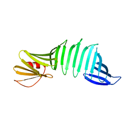
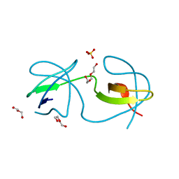
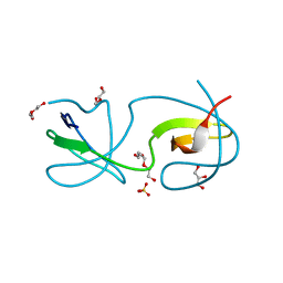
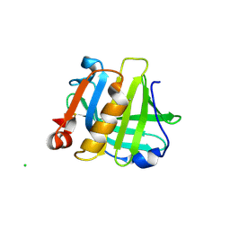
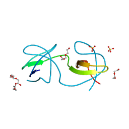
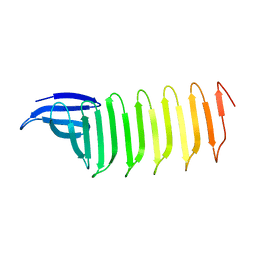 | | A Crystal structure of OspA mutant | | Descriptor: | Outer Surface Protein A | | Authors: | Shiga, S, Makabe, K. | | Deposit date: | 2019-07-24 | | Release date: | 2020-07-29 | | Last modified: | 2023-11-22 | | Method: | X-RAY DIFFRACTION (2.2 Å) | | Cite: | Structural analysis of the beta-sheet edge of peptide self-assembly using a model protein.
Proteins, 89, 2021
|
|
4ZNX
 | |
4ZNY
 | |
4F16
 | |
3M0Q
 | |
5IO5
 | |
3M0T
 | |
3I4W
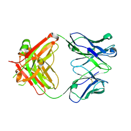
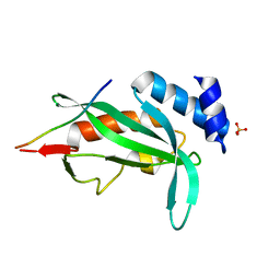
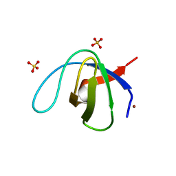
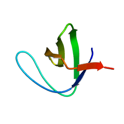
 | | Crystal Structure of the third PDZ domain of PSD-95 | | Descriptor: | ACETATE ION, Disks large homolog 4 | | Authors: | Camara-Artigas, A, Gavira, J.A. | | Deposit date: | 2009-07-03 | | Release date: | 2010-04-07 | | Last modified: | 2024-11-06 | | Method: | X-RAY DIFFRACTION (1.35 Å) | | Cite: | Novel conformational aspects of the third PDZ domain of the neuronal post-synaptic density-95 protein revealed from two 1.4A X-ray structures
J.Struct.Biol., 170, 2010
|
|
3I9Q
 | |
4RTY
 | |
4RTV
 | |
4RTX
 | |
4RTU
 | |
3NGP
 | |
4F17
 | |
4RTZ
 | |
3M0P
 | |
3M0S
 | |
4RTW
 | |


