Loading
PDBj
MenuPDBj@FacebookPDBj@TwitterPDBj@YouTubewwPDB FoundationwwPDB
RCSB PDBPDBeBMRBAdv. SearchSearch help

7A2U
DownloadVisualize
BU of 7a2u by Molmil
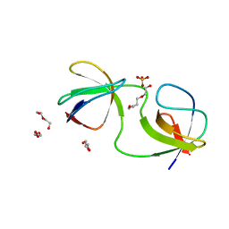
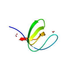
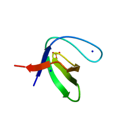
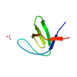
Crystal structure of the Fyn SH3 domain L112V-S114N-S115T at pH 4.5
Descriptor: ACETATE ION, Tyrosine-protein kinase Fyn
Authors:Camara-Artigas, A, Plaza-Garrido, M, Salinas-Garcia, M.C.
Deposit date:2020-08-18
Release date:2021-08-25
Last modified:2024-01-31
Method:X-RAY DIFFRACTION (1.7 Å)
Cite:Crystal structure of the Fyn SH3 domain L112V-S114N-S115T at pH 4.5
To be published
7A2O
DownloadVisualize
BU of 7a2o by Molmil
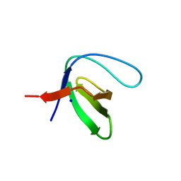
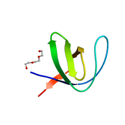
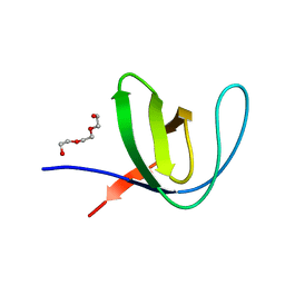
Crystal structure of the Fyn SH3 domain L112V-S114N-S115T-E121L-R123H mutant at pH 4.5
Descriptor: TRIETHYLENE GLYCOL, Tyrosine-protein kinase Fyn
Authors:Camara-Artigas, A, Plaza-Garrido, M, Salinas-Garcia, M.C.
Deposit date:2020-08-18
Release date:2021-08-25
Last modified:2024-01-31
Method:X-RAY DIFFRACTION (0.94 Å)
Cite:Crystal structure of the Fyn SH3 domain L112V-S114N-S115T-E121L-R123H mutant at pH 4.5
To be published
7A3D
DownloadVisualize
BU of 7a3d by Molmil
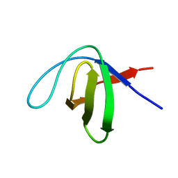
Intertwined dimer of the c-Src SH3 domain mutant T125S
Descriptor: DI(HYDROXYETHYL)ETHER, GLYCEROL, Proto-oncogene tyrosine-protein kinase Src, ...
Authors:Camara-Artigas, A, Plaza-Garrido, M, Salinas-Garcia, M.C.
Deposit date:2020-08-18
Release date:2021-08-25
Last modified:2024-01-31
Method:X-RAY DIFFRACTION (2.2 Å)
Cite:Intertwined dimer of the c-Src SH3 domain mutant T125S
To be published
7A2J
DownloadVisualize
BU of 7a2j by Molmil
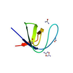
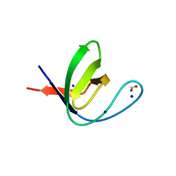
Crystal structure of the Fyn SH3 domain in space group C21 at pH 7.5
Descriptor: SODIUM ION, Tyrosine-protein kinase Fyn
Authors:Camara-Artigas, A, Plaza-Garrido, M, Salinas-Garcia, M.C.
Deposit date:2020-08-18
Release date:2021-08-25
Last modified:2024-01-31
Method:X-RAY DIFFRACTION (1.5 Å)
Cite:Crystal structure of the Fyn SH3 domain in space group C21 at pH 7.5
To be published
7A2V
DownloadVisualize
BU of 7a2v by Molmil
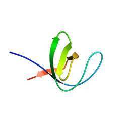
Crystal structure of the Fyn SH3 domain L112V-S114N-S115T-E129Q at pH 4.0
Descriptor: Tyrosine-protein kinase Fyn
Authors:Camara-Artigas, A, Plaza-Garrido, M, Salinas-Garcia, M.C.
Deposit date:2020-08-18
Release date:2021-08-25
Last modified:2024-01-31
Method:X-RAY DIFFRACTION (1.81 Å)
Cite:Crystal structure of the Fyn SH3 domain L112V-S114N-S115T-E129Q at pH 4.0
To be published
7A37
DownloadVisualize
BU of 7a37 by Molmil
Crystal structure of the c-Src SH3 domain mutant V111L-N113S-T114S in 3 M urea
Descriptor: Proto-oncogene tyrosine-protein kinase Src, UREA
Authors:Camara-Artigas, A, Plaza-Garrido, M, Salinas-Garcia, M.C.
Deposit date:2020-08-18
Release date:2021-08-25
Last modified:2024-01-31
Method:X-RAY DIFFRACTION (1.52 Å)
Cite:Crystal structure of the c-Src SH3 domain mutant V111L-N113S-T114S in 3 M urea
To be published
7A2Q
DownloadVisualize
BU of 7a2q by Molmil
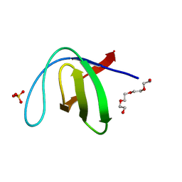
Crystal structure of the Fyn SH3 domain L112V-S114N-S115T-E121L-R123H mutant at pH 3.0 with PEG
Descriptor: SULFATE ION, TETRAETHYLENE GLYCOL, Tyrosine-protein kinase Fyn
Authors:Camara-Artigas, A, Plaza-Garrido, M, Salinas-Garcia, M.C.
Deposit date:2020-08-18
Release date:2021-08-25
Last modified:2024-01-31
Method:X-RAY DIFFRACTION (0.94 Å)
Cite:Crystal structure of the Fyn SH3 domain L112V-S114N-S115T-E121L-R123H mutant at pH 3.0 with PEG
To be published
7A33
DownloadVisualize
BU of 7a33 by Molmil
Crystal structure of the c-Src SH3 domain mutant S94A-T98D-V111L-N113S-T114S at pH 3.0
Descriptor: FORMIC ACID, Proto-oncogene tyrosine-protein kinase Src, SODIUM ION
Authors:Camara-Artigas, A, Plaza-Garrido, M, Salinas-Garcia, M.C.
Deposit date:2020-08-18
Release date:2021-08-25
Last modified:2024-01-31
Method:X-RAY DIFFRACTION (0.96 Å)
Cite:Crystal structure of the c-Src SH3 domain mutant S94A-T98D-V111L-N113S-T114S at pH 3.0
To be published
7A2W
DownloadVisualize
BU of 7a2w by Molmil
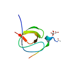
Crystal structure of the Fyn SH3 domain L112V-S114N-S115T-E121L-R123H mutant in complex with VSL12 at pH 3.0
Descriptor: CITRIC ACID, Tyrosine-protein kinase Fyn, VSL12
Authors:Camara-Artigas, A, Plaza-Garrido, M, Salinas-Garcia, M.C.
Deposit date:2020-08-18
Release date:2021-08-25
Last modified:2024-01-31
Method:X-RAY DIFFRACTION (0.99 Å)
Cite:Crystal structure of the Fyn SH3 domain L112V-S114N-S115T-E121L-R123H mutant in complex with VSL12 at pH 3.0
To be published
7A3A
DownloadVisualize
BU of 7a3a by Molmil
Intertwined dimer of the c-Src SH3 domain mutant V111L-N113S-T114S-Q128E at pH 6.0
Descriptor: 1,2-ETHANEDIOL, DI(HYDROXYETHYL)ETHER, Proto-oncogene tyrosine-protein kinase Src, ...
Authors:Camara-Artigas, A, Plaza-Garrido, M, Salinas-Garcia, M.C.
Deposit date:2020-08-18
Release date:2021-08-25
Last modified:2024-01-31
Method:X-RAY DIFFRACTION (1.8 Å)
Cite:Intertwined dimer of the c-Src SH3 domain mutant V111L-N113S-T114S-Q128E at pH 6.0
To be published
7A2P
DownloadVisualize
BU of 7a2p by Molmil
Crystal structure of the Fyn SH3 domain L112V-S114N-S115T-E121L-R123H mutant at pH 5.0 with PEG
Descriptor: TRIETHYLENE GLYCOL, Tyrosine-protein kinase Fyn
Authors:Camara-Artigas, A, Plaza-Garrido, M, Salinas-Garcia, M.C.
Deposit date:2020-08-18
Release date:2021-08-25
Last modified:2024-01-31
Method:X-RAY DIFFRACTION (0.9 Å)
Cite:Crystal structure of the Fyn SH3 domain L112V-S114N-S115T-E121L-R123H mutant at pH 5.0 with PEG
To be published
7A2N
DownloadVisualize
BU of 7a2n by Molmil
Crystal structure of the Fyn SH3 domain A95S-D99T mutant in space group P21
Descriptor: FORMIC ACID, SODIUM ION, Tyrosine-protein kinase Fyn
Authors:Camara-Artigas, A, Plaza-Garrido, M, Salinas-Garcia, M.C.
Deposit date:2020-08-18
Release date:2021-08-25
Last modified:2024-01-31
Method:X-RAY DIFFRACTION (1.4 Å)
Cite:Crystal structure of the Fyn SH3 domain A95S-D99T mutant in space group P21
To be published
7A2M
DownloadVisualize
BU of 7a2m by Molmil
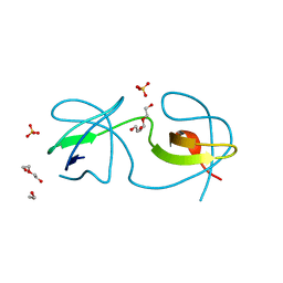
Crystal structure of the Fyn SH3 domain A95S-D99T mutant in C21 space group
Descriptor: FORMIC ACID, SODIUM ION, SULFATE ION, ...
Authors:Camara-Artigas, A, Plaza-Garrido, M, Salinas-Garcia, M.C.
Deposit date:2020-08-18
Release date:2021-08-25
Last modified:2024-01-31
Method:X-RAY DIFFRACTION (1.5 Å)
Cite:Crystal structure of the Fyn SH3 domain A95S-D99T mutant in C21 space group
To be published
7A2R
DownloadVisualize
BU of 7a2r by Molmil
Crystal structure of the Fyn SH3 domain L112V-S114N-S115T-E121L-R123H mutant at pH 6.0
Descriptor: Tyrosine-protein kinase Fyn
Authors:Camara-Artigas, A, Plaza-Garrido, M, Salinas-Garcia, M.C.
Deposit date:2020-08-18
Release date:2021-08-25
Last modified:2024-01-31
Method:X-RAY DIFFRACTION (1.05 Å)
Cite:Crystal structure of the Fyn SH3 domain L112V-S114N-S115T-E121L-R123H mutant at pH 6.0
To be published
5X5S
DownloadVisualize
BU of 5x5s by Molmil
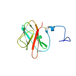
Ligand induced structure of AmyP-SBD
Descriptor: Amylase
Authors:Li, X, Yu, J, Sun, H, Zhang, X.
Deposit date:2017-02-17
Release date:2017-04-05
Last modified:2023-06-14
Method:SOLUTION NMR
Cite:Ligand binding induced folding of a novel CBM69 starch binding domain
To Be Published
7SWD
DownloadVisualize
BU of 7swd by Molmil
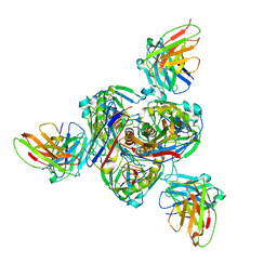
Structure of EBOV GP lacking the mucin-like domain with 1C11 scFv and 1C3 Fab bound
Descriptor: 1C11 scFv, 1C3 heavy chain, 1C3 light chain, ...
Authors:Milligan, J.C, Yu, X, Saphire, E.O.
Deposit date:2021-11-19
Release date:2022-04-06
Last modified:2022-08-10
Method:ELECTRON MICROSCOPY (3.59 Å)
Cite:Asymmetric and non-stoichiometric glycoprotein recognition by two distinct antibodies results in broad protection against ebolaviruses.
Cell, 185, 2022
4YHN
DownloadVisualize
BU of 4yhn by Molmil
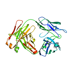
Dabigatran Reversal Agent
Descriptor: aDabi-Fab3 heavy chain, aDabi-Fab3 light chain
Authors:Schiele, F, Nar, H.
Deposit date:2015-02-27
Release date:2016-02-10
Last modified:2024-01-10
Method:X-RAY DIFFRACTION (2.31 Å)
Cite:Structure-guided residence time optimization of a dabigatran reversal agent.
Mabs, 7, 2015
6QJJ
DownloadVisualize
BU of 6qjj by Molmil
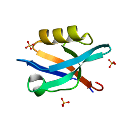
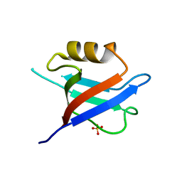
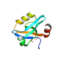
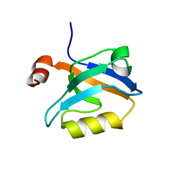
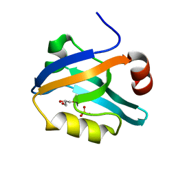
Crystal Structure of the third PDZ domain of PSD-95 protein: space group P3221
Descriptor: Disks large homolog 4, SULFATE ION
Authors:Camara-Artigas, A.
Deposit date:2019-01-24
Release date:2019-04-17
Last modified:2024-01-24
Method:X-RAY DIFFRACTION (1.7 Å)
Cite:Conformational changes in the third PDZ domain of the neuronal postsynaptic density protein 95.
Acta Crystallogr D Struct Biol, 75, 2019
6QJD
DownloadVisualize
BU of 6qjd by Molmil
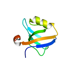
Crystal Structure of the truncated form of the third PDZ domain of PSD-95: residues 302-392
Descriptor: Disks large homolog 4, SULFATE ION
Authors:Camara-Artigas, A.
Deposit date:2019-01-24
Release date:2019-04-17
Last modified:2024-01-24
Method:X-RAY DIFFRACTION (1.551 Å)
Cite:Conformational changes in the third PDZ domain of the neuronal postsynaptic density protein 95.
Acta Crystallogr D Struct Biol, 75, 2019
6QJN
DownloadVisualize
BU of 6qjn by Molmil
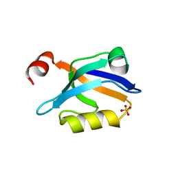
Crystal Structure of the third PDZ domain of PSD-95 protein D332G mutant: space group I4122
Descriptor: Disks large homolog 4
Authors:Camara-Artigas, A.
Deposit date:2019-01-24
Release date:2019-04-17
Last modified:2024-01-24
Method:X-RAY DIFFRACTION (1.8 Å)
Cite:Conformational changes in the third PDZ domain of the neuronal postsynaptic density protein 95.
Acta Crystallogr D Struct Biol, 75, 2019
6QJI
DownloadVisualize
BU of 6qji by Molmil
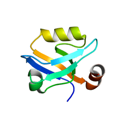
Crystal Structure of the third PDZ domain of PSD-95 protein: space group P3112
Descriptor: Disks large homolog 4, SULFATE ION
Authors:Camara-Artigas, A.
Deposit date:2019-01-24
Release date:2019-04-17
Last modified:2024-01-24
Method:X-RAY DIFFRACTION (1.5 Å)
Cite:Conformational changes in the third PDZ domain of the neuronal postsynaptic density protein 95.
Acta Crystallogr D Struct Biol, 75, 2019
6QJF
DownloadVisualize
BU of 6qjf by Molmil
Crystal Structure of the third PDZ domain of PSD-95 protein D332P mutant: space group C121, structure 1
Descriptor: Disks large homolog 4
Authors:Camara-Artigas, A.
Deposit date:2019-01-24
Release date:2019-04-17
Last modified:2024-01-24
Method:X-RAY DIFFRACTION (1.5 Å)
Cite:Conformational changes in the third PDZ domain of the neuronal postsynaptic density protein 95.
Acta Crystallogr D Struct Biol, 75, 2019
6QJL
DownloadVisualize
BU of 6qjl by Molmil
Crystal Structure of the third PDZ domain of PSD-95 protein D332G mutant: space group P21
Descriptor: Disks large homolog 4, GLYCEROL, SULFATE ION
Authors:Camara-Artigas, A.
Deposit date:2019-01-24
Release date:2019-04-17
Last modified:2024-01-24
Method:X-RAY DIFFRACTION (1.043 Å)
Cite:Conformational changes in the third PDZ domain of the neuronal postsynaptic density protein 95.
Acta Crystallogr D Struct Biol, 75, 2019
6QJG
DownloadVisualize
BU of 6qjg by Molmil
Crystal Structure of the third PDZ domain of PSD-95 protein D332P mutant: space group C121, structure 2
Descriptor: Disks large homolog 4
Authors:Camara-Artigas, A.
Deposit date:2019-01-24
Release date:2019-04-17
Last modified:2024-01-24
Method:X-RAY DIFFRACTION (2.003 Å)
Cite:Conformational changes in the third PDZ domain of the neuronal postsynaptic density protein 95.
Acta Crystallogr D Struct Biol, 75, 2019
6QJK
DownloadVisualize
BU of 6qjk by Molmil
Crystal Structure of the third PDZ domain of PSD-95 protein D332G mutant: space group P43
Descriptor: Disks large homolog 4, GLYCEROL, SULFATE ION
Authors:Camara-Artigas, A.
Deposit date:2019-01-24
Release date:2019-04-17
Last modified:2024-01-24
Method:X-RAY DIFFRACTION (1.046 Å)
Cite:Conformational changes in the third PDZ domain of the neuronal postsynaptic density protein 95.
Acta Crystallogr D Struct Biol, 75, 2019

224201

数据于2024-08-28公开中

PDB statisticsPDBj update infoContact PDBjnumon