Loading
PDBj
MenuPDBj@FacebookPDBj@TwitterPDBj@YouTubewwPDB FoundationwwPDB
RCSB PDBPDBeBMRBAdv. SearchSearch help

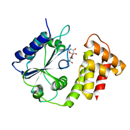
6XZ0
DownloadVisualize
BU of 6xz0 by Molmil
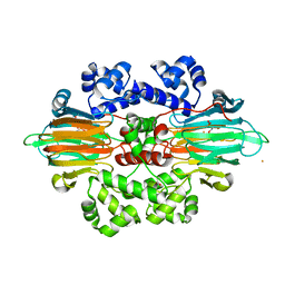
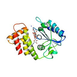
Crystal structure of spectinomycin adenyltransferase AAD(9) from Enterococcus faecialis with spectinomycin
Descriptor: SPECTINOMYCIN, Streptomycin 3''-adenylyltransferase
Authors:Kanchugal P, S, Selmer, M.
Deposit date:2020-01-31
Release date:2020-04-15
Last modified:2024-01-24
Method:X-RAY DIFFRACTION (2.8 Å)
Cite:Structural Recognition of Spectinomycin by Resistance Enzyme ANT(9) from Enterococcus faecalis.
Antimicrob.Agents Chemother., 64, 2020
1FJG
DownloadVisualize
BU of 1fjg by Molmil
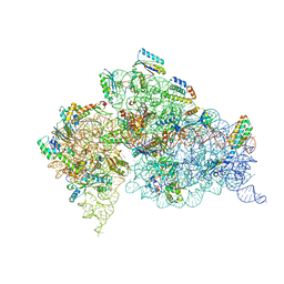
STRUCTURE OF THE THERMUS THERMOPHILUS 30S RIBOSOMAL SUBUNIT IN COMPLEX WITH THE ANTIBIOTICS STREPTOMYCIN, SPECTINOMYCIN, AND PAROMOMYCIN
Descriptor: 16S RIBOSOMAL RNA, 30S RIBOSOMAL PROTEIN S10, 30S RIBOSOMAL PROTEIN S11, ...
Authors:Carter, A.P, Clemons Jr, W.M, Brodersen, D.E, Wimberly, B.T, Morgan-Warren, R.J, Ramakrishnan, V.
Deposit date:2000-08-08
Release date:2000-09-25
Last modified:2011-07-13
Method:X-RAY DIFFRACTION (3 Å)
Cite:Functional insights from the structure of the 30S ribosomal subunit and its interactions with antibiotics
Nature, 407, 2000
5BV0
DownloadVisualize
BU of 5bv0 by Molmil
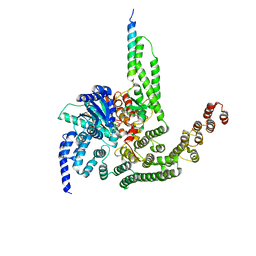
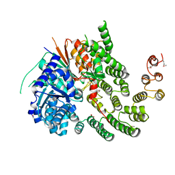
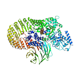
Crystal Structure of a Complex Between the SNARE Nyv1 and the HOPS Vps33-Vps16 subcomplex from Chaetomium thermophilum
Descriptor: SM (Sec1/Munc18-like) protein, SNARE domain, Vps16
Authors:Baker, R.W, Jeffrey, P.D, Hughson, F.M.
Deposit date:2015-06-04
Release date:2015-08-05
Last modified:2023-09-27
Method:X-RAY DIFFRACTION (3.1 Å)
Cite:A direct role for the Sec1/Munc18-family protein Vps33 as a template for SNARE assembly.
Science, 349, 2015
5BUZ
DownloadVisualize
BU of 5buz by Molmil
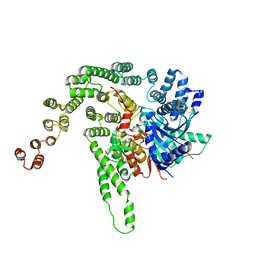
Crystal Structure of a Complex Between the SNARE Vam3 and the HOPS Vps33-Vps16 subcomplex from Chaetomium thermophilum
Descriptor: Putative vacuolar protein sorting-associated protein, SM (Sec1/Munc18-like) protein, SNAP receptor-like protein
Authors:Baker, R.W, Jeffrey, P.D, Hughson, F.M.
Deposit date:2015-06-04
Release date:2015-08-05
Last modified:2023-09-27
Method:X-RAY DIFFRACTION (3.1 Å)
Cite:A direct role for the Sec1/Munc18-family protein Vps33 as a template for SNARE assembly.
Science, 349, 2015
5BV1
DownloadVisualize
BU of 5bv1 by Molmil
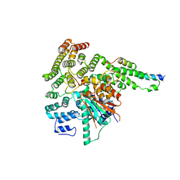
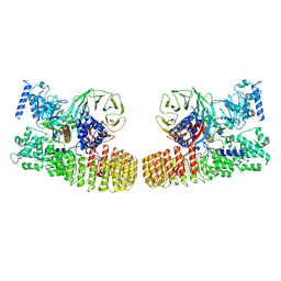
Crystal Structure of a Vps33-Vps16 Complex from Chaetomium thermophilum
Descriptor: D-MALATE, Putative vacuolar protein sorting-associated protein, VPS33
Authors:Baker, R.W, Jeffrey, P.D, Hughson, F.M.
Deposit date:2015-06-04
Release date:2015-08-12
Last modified:2023-09-27
Method:X-RAY DIFFRACTION (2.902 Å)
Cite:A direct role for the Sec1/Munc18-family protein Vps33 as a template for SNARE assembly.
Science, 349, 2015
8P5E
DownloadVisualize
BU of 8p5e by Molmil
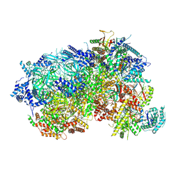
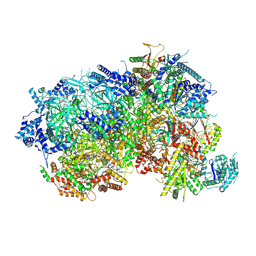
S. cerevisiae nexus-sCMGE after DNA replication initiation
Descriptor: ADENOSINE-5'-DIPHOSPHATE, ADENOSINE-5'-TRIPHOSPHATE, Cell division control protein 45, ...
Authors:Henrikus, S.S, Willhoft, O.
Deposit date:2023-05-24
Release date:2024-05-29
Method:ELECTRON MICROSCOPY (3.9 Å)
Cite:Unwinding of a eukaryotic origin of replication visualized by cryo-EM.
Nat.Struct.Mol.Biol., 2024
4N5X
DownloadVisualize
BU of 4n5x by Molmil
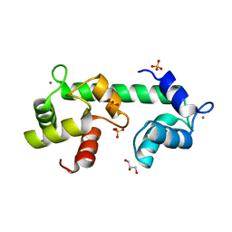
Crystal structure of N-terminal calmodulin-like Calcium sensor of human mitochondrial ATP-Mg/Pi carrier SCaMC1
Descriptor: CALCIUM ION, Calcium-binding mitochondrial carrier protein SCaMC-1, DI(HYDROXYETHYL)ETHER
Authors:Yang, Q, Bruschweiler, S, Chou, J.
Deposit date:2013-10-10
Release date:2013-12-18
Last modified:2023-09-20
Method:X-RAY DIFFRACTION (2.1 Å)
Cite:A Self-Sequestered Calmodulin-like Ca(2+) Sensor of Mitochondrial SCaMC Carrier and Its Implication to Ca(2+)-Dependent ATP-Mg/Pi Transport.
Structure, 22, 2014
6B9T
DownloadVisualize
BU of 6b9t by Molmil
Crystal structure of MPnS with substrate 2-hydroxyethylphosphonate (2-HEP) and Fe(II) bound
Descriptor: (2-hydroxyethyl)phosphonic acid, FE (III) ION, FORMIC ACID, ...
Authors:Born, D.A, Drennan, C.L.
Deposit date:2017-10-11
Release date:2017-12-20
Last modified:2024-03-13
Method:X-RAY DIFFRACTION (2.35 Å)
Cite:Structural basis for methylphosphonate biosynthesis.
Science, 358, 2017
6VJR
DownloadVisualize
BU of 6vjr by Molmil
Oxygen tolerant Archeal 4hydroxybutyrylCoA dehydratase (4HBD) from N. maritimus
Descriptor: FE (III) ION, FLAVIN-ADENINE DINUCLEOTIDE, IRON/SULFUR CLUSTER, ...
Authors:DeMirci, H.
Deposit date:2020-01-17
Release date:2021-01-27
Last modified:2023-10-11
Method:X-RAY DIFFRACTION (1.55 Å)
Cite:Structural adaptation of oxygen tolerance in a key archaeal carbon fixation enzyme
To Be Published
1CHZ
DownloadVisualize
BU of 1chz by Molmil
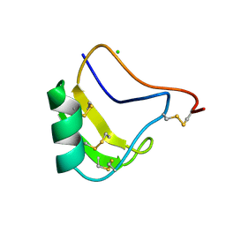
A NEW NEUROTOXIN FROM BUTHUS MARTENSII KARSCH
Descriptor: CHLORIDE ION, PROTEIN (BMK M2)
Authors:He, X.L, Deng, J.P, Li, H.M, Wang, D.C.
Deposit date:1999-03-31
Release date:2000-03-31
Last modified:2023-08-09
Method:X-RAY DIFFRACTION (1.76 Å)
Cite:Structure of a new neurotoxin from the scorpion Buthus martensii Karsch at 1.76 A.
Acta Crystallogr.,Sect.D, 56, 2000
8C8R
DownloadVisualize
BU of 8c8r by Molmil
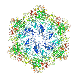
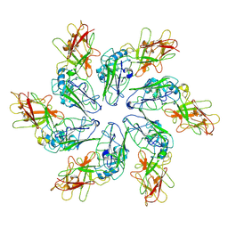
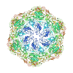
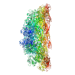
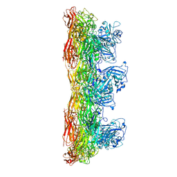
In situ structure of the Nitrosopumilus maritimus S-layer - Composite map between C2 and C6
Descriptor: Cell surface protein
Authors:von Kuegelgen, A, Bharat, T.
Deposit date:2023-01-20
Release date:2024-04-17
Last modified:2024-06-19
Method:ELECTRON MICROSCOPY (3.5 Å)
Cite:Membraneless channels sieve cations in ammonia-oxidizing marine archaea.
Nature, 630, 2024
1ZIS
DownloadVisualize
BU of 1zis by Molmil
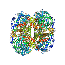
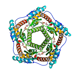
Recombinant Lumazine synthase (hexagonal form)
Descriptor: 5-NITRO-6-RIBITYL-AMINO-2,4(1H,3H)-PYRIMIDINEDIONE, 6,7-dimethyl-8-ribityllumazine synthase, PHOSPHATE ION
Authors:Lopez-Jaramillo, F.J.
Deposit date:2005-04-27
Release date:2006-10-03
Last modified:2023-08-23
Method:X-RAY DIFFRACTION (2.9 Å)
Cite:Crystal structure of recombinant Lumazine synthase (hexagonal form)
To be Published
4ZCV
DownloadVisualize
BU of 4zcv by Molmil
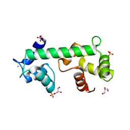
Structure of calcium-bound regulatory domain of the human ATP-Mg/Pi carrier in the P212121 form
Descriptor: CALCIUM ION, Calcium-binding mitochondrial carrier protein SCaMC-1, GLYCEROL, ...
Authors:Harborne, S.P.D, Ruprecht, J.J, Kunji, E.R.S.
Deposit date:2015-04-16
Release date:2015-07-22
Last modified:2024-01-10
Method:X-RAY DIFFRACTION (2.8 Å)
Cite:Calcium-induced conformational changes in the regulatory domain of the human mitochondrial ATP-Mg/Pi carrier.
Biochim.Biophys.Acta, 1847, 2015
4ZCU
DownloadVisualize
BU of 4zcu by Molmil
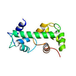
Structure of calcium-bound regulatory domain of the human ATP-Mg/Pi carrier in the P2 form
Descriptor: CALCIUM ION, Calcium-binding mitochondrial carrier protein SCaMC-1, GLYCEROL, ...
Authors:Harborne, S.P.D, Ruprecht, J.J, Kunji, E.R.S.
Deposit date:2015-04-16
Release date:2015-07-22
Last modified:2024-01-10
Method:X-RAY DIFFRACTION (2.1 Å)
Cite:Calcium-induced conformational changes in the regulatory domain of the human mitochondrial ATP-Mg/Pi carrier.
Biochim.Biophys.Acta, 1847, 2015
8P62
DownloadVisualize
BU of 8p62 by Molmil
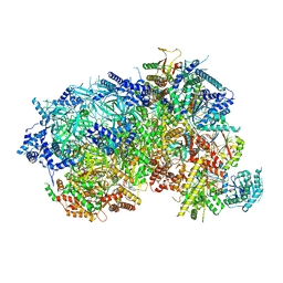
S. cerevisiae ssDNA-sCMGE after DNA replication initiation
Descriptor: ADENOSINE-5'-DIPHOSPHATE, ADENOSINE-5'-TRIPHOSPHATE, Cell division control protein 45, ...
Authors:Henrikus, S.S, Willhoft, O.
Deposit date:2023-05-25
Release date:2024-05-29
Method:ELECTRON MICROSCOPY (3.9 Å)
Cite:Unwinding of a eukaryotic origin of replication visualized by cryo-EM.
Nat.Struct.Mol.Biol., 2024
8P63
DownloadVisualize
BU of 8p63 by Molmil
S. cerevisiae consensus-sCMGE on ssDNA after DNA replication initiation
Descriptor: ADENOSINE-5'-DIPHOSPHATE, ADENOSINE-5'-TRIPHOSPHATE, Cell division control protein 45, ...
Authors:Henrikus, S.S, Willhoft, O.
Deposit date:2023-05-25
Release date:2024-05-29
Method:ELECTRON MICROSCOPY (3.7 Å)
Cite:Unwinding of a eukaryotic origin of replication visualized by cryo-EM.
Nat.Struct.Mol.Biol., 2024
7EUM
DownloadVisualize
BU of 7eum by Molmil
Crystal structure of apo Nmar_1308 protein at cryogenic temperature
Descriptor: Enoyl-CoA hydratase/isomerase
Authors:DeMirci, H, Destan, E, Yuksel, B, Ayan, E.
Deposit date:2021-05-18
Release date:2021-09-29
Last modified:2024-05-29
Method:X-RAY DIFFRACTION (2.15 Å)
Cite:Crystal structure of apo Nmar_1308 protein at cryogenic temperature
To Be Published
8WZU
DownloadVisualize
BU of 8wzu by Molmil
4-hydroxybutyryl-CoA Synthetase (ADP-forming) from Nitrosopumilus maritimus.
Descriptor: 4-hydroxybutyrate--CoA ligase [ADP-forming], SULFATE ION
Authors:Johnson, J, Demirci, H.
Deposit date:2023-11-02
Release date:2024-06-12
Method:X-RAY DIFFRACTION (2.8 Å)
Cite:Crystal Structure of 4-hydroxybutyryl CoA synthetase (ADP-forming): A Key Enzyme in the Thaumarchaeal Hydroxypropionate/Hydroxybutyrate cycle.
To Be Published
6B9S
DownloadVisualize
BU of 6b9s by Molmil
MPnS crystallized in the absence of substrate
Descriptor: FE (III) ION, Methylphosphonate synthase
Authors:Born, D.A, Drennan, C.L.
Deposit date:2017-10-11
Release date:2017-12-20
Last modified:2024-03-13
Method:X-RAY DIFFRACTION (2.373 Å)
Cite:Structural basis for methylphosphonate biosynthesis.
Science, 358, 2017
8C8K
DownloadVisualize
BU of 8c8k by Molmil
In vitro structure of the Nitrosopumilus maritimus S-layer - Six-fold symmetry (C6)
Descriptor: Cell surface protein
Authors:von Kuegelgen, A, Bharat, T.
Deposit date:2023-01-20
Release date:2024-04-10
Last modified:2024-06-19
Method:ELECTRON MICROSCOPY (2.87 Å)
Cite:Membraneless channels sieve cations in ammonia-oxidizing marine archaea.
Nature, 630, 2024
8C8N
DownloadVisualize
BU of 8c8n by Molmil
In situ structure of the Nitrosopumilus maritimus S-layer - Two-fold symmetry (C2)
Descriptor: Cell surface protein
Authors:von Kuegelgen, A, Bharat, T.
Deposit date:2023-01-20
Release date:2024-04-10
Last modified:2024-06-19
Method:ELECTRON MICROSCOPY (3.4 Å)
Cite:Membraneless channels sieve cations in ammonia-oxidizing marine archaea.
Nature, 630, 2024
8C8M
DownloadVisualize
BU of 8c8m by Molmil
In vitro structure of the Nitrosopumilus maritimus S-layer - Composite map between two and six-fold symmetrised
Descriptor: Cell surface protein
Authors:von Kuegelgen, A, Bharat, T.
Deposit date:2023-01-20
Release date:2024-04-10
Last modified:2024-06-19
Method:ELECTRON MICROSCOPY (2.87 Å)
Cite:Membraneless channels sieve cations in ammonia-oxidizing marine archaea.
Nature, 630, 2024
8C8L
DownloadVisualize
BU of 8c8l by Molmil
In vitro structure of the Nitrosopumilus maritimus S-layer - Two-fold symmetry (C2)
Descriptor: Cell surface protein
Authors:von Kuegelgen, A, Bharat, T.
Deposit date:2023-01-20
Release date:2024-04-10
Last modified:2024-06-19
Method:ELECTRON MICROSCOPY (2.71 Å)
Cite:Membraneless channels sieve cations in ammonia-oxidizing marine archaea.
Nature, 630, 2024
8C8O
DownloadVisualize
BU of 8c8o by Molmil
In situ structure of the Nitrosopumilus maritimus S-layer - Six-fold symmetry (C6)
Descriptor: Cell surface protein
Authors:von Kuegelgen, A, Bharat, T.
Deposit date:2023-01-20
Release date:2024-04-17
Last modified:2024-06-19
Method:ELECTRON MICROSCOPY (3.4 Å)
Cite:Membraneless channels sieve cations in ammonia-oxidizing marine archaea.
Nature, 630, 2024
8DIT
DownloadVisualize
BU of 8dit by Molmil
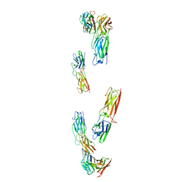
Cryo-EM structure of a HOPS core complex containing Vps33, Vps16, and Vps18
Descriptor: Vacuolar protein sorting-associated protein 16, Vacuolar protein sorting-associated protein 18, Vacuolar protein sorting-associated protein 33
Authors:Port, S.A, Farrell, P.D, Jeffrey, P.D, DiMaio, F, Hughson, F.M.
Deposit date:2022-06-29
Release date:2022-08-31
Last modified:2024-02-14
Method:ELECTRON MICROSCOPY (5.1 Å)
Cite:Cryo-EM structure of the HOPS core complex and its implication for SNARE assembly
To Be Published

221371

数据于2024-06-19公开中

PDB statisticsPDBj update infoContact PDBjnumon