3P28
 
 | |
3GEX
 | |
3I19
 | |
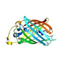
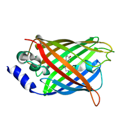
6AA2
 | | X-ray structure of ReQy1 (oxidized form) | | Descriptor: | Green fluorescent protein | | Authors: | Sugiura, K, Yasuda, A, Tabushi, N, Tanaka, H, Kurisu, G, Hisabori, T. | | Deposit date: | 2018-07-17 | | Release date: | 2019-05-29 | | Last modified: | 2023-11-22 | | Method: | X-RAY DIFFRACTION (2.3 Å) | | Cite: | Multicolor redox sensor proteins can visualize redox changes in various compartments of the living cell.
Biochim Biophys Acta Gen Subj, 1863, 2019
|
|
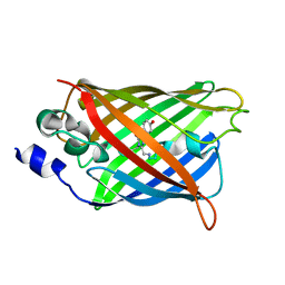
6AA6
 | |
4KW9
 | |
4KW8
 | |
4KW4
 | |
6T90
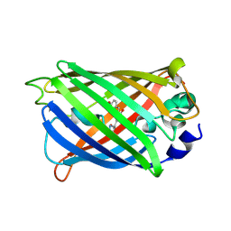
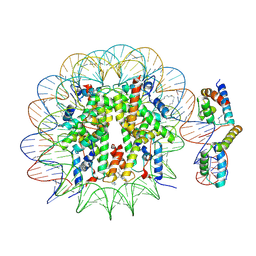 | | OCT4-SOX2-bound nucleosome - SHL-6 | | Descriptor: | DNA (146-MER), Green fluorescent protein,POU domain, class 5, ... | | Authors: | Michael, A.K, Kempf, G, Cavadini, S, Bunker, R.D, Thoma, N.H. | | Deposit date: | 2019-10-25 | | Release date: | 2020-05-06 | | Last modified: | 2020-07-08 | | Method: | ELECTRON MICROSCOPY (3.05 Å) | | Cite: | Mechanisms of OCT4-SOX2 motif readout on nucleosomes.
Science, 368, 2020
|
|
3DPW
 | |
3DQ4
 | |
3DQF
 | |
3DQO
 | |
3DPX
 | |
3DQ5
 | |
3DQE
 | |
3DQI
 | |
3DQU
 | |
3DQJ
 | |
3DQ7
 | |
3DQK
 | |
3DPZ
 | |
3DQ8
 | |
3DQ1
 | |
3DQA
 | |


