6JEH
 
 | |
6JEG
 | |
6JCO
 | |
6KWZ
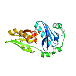
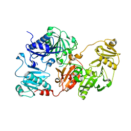
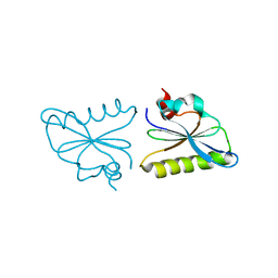 | | Crystal structure of fragmin F3 domain with calcium ion | | Descriptor: | Actin-binding protein fragmin P, CALCIUM ION | | Authors: | Takeda, S. | | Deposit date: | 2019-09-09 | | Release date: | 2020-01-01 | | Last modified: | 2023-11-22 | | Method: | X-RAY DIFFRACTION (1.551 Å) | | Cite: | Novel inter-domain Ca2+-binding site in the gelsolin superfamily protein fragmin.
J.Muscle Res.Cell.Motil., 41, 2020
|
|
6LJD
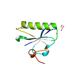
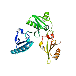
 | | Crystal structure of fragmin F2-F3 domains (calcium condition) | | Descriptor: | 2-(N-MORPHOLINO)-ETHANESULFONIC ACID, Actin-binding protein fragmin P, CALCIUM ION, ... | | Authors: | Takeda, S. | | Deposit date: | 2019-12-14 | | Release date: | 2020-01-01 | | Last modified: | 2023-11-22 | | Method: | X-RAY DIFFRACTION (2.15 Å) | | Cite: | Novel inter-domain Ca2+-binding site in the gelsolin superfamily protein fragmin.
J.Muscle Res.Cell.Motil., 41, 2020
|
|
6LJF
 | |
6LJC
 | |
6LJE
 | |
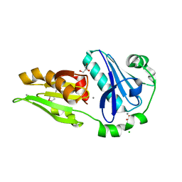
5ZZ0
 | | HUMAN GELSOLIN FROM RESIDUES GLU28 TO ARG161 WITH CALCIUM | | Descriptor: | 2-(2-METHOXYETHOXY)ETHANOL, CALCIUM ION, Gelsolin | | Authors: | Sharma, P, Badmalia, M, Yadav, S.P.S, Singh, S. | | Deposit date: | 2018-05-29 | | Release date: | 2019-06-05 | | Last modified: | 2023-11-22 | | Method: | X-RAY DIFFRACTION (2.635 Å) | | Cite: | Bonsai Gelsolin Survives Heat Induced Denaturation by Forming beta-Amyloids which Leach Out Functional Monomer.
Sci Rep, 8, 2018
|
|
6H1F
 | | Structure of the nanobody-stabilized gelsolin D187N variant (second domain) | | Descriptor: | Gelsolin, THIOCYANATE ION, gelsolin nanobody, ... | | Authors: | Hassan, A, Milani, M, Mastrangelo, E, de Rosa, M. | | Deposit date: | 2018-07-11 | | Release date: | 2019-01-23 | | Last modified: | 2024-01-17 | | Method: | X-RAY DIFFRACTION (1.9 Å) | | Cite: | Nanobody interaction unveils structure, dynamics and proteotoxicity of the Finnish-type amyloidogenic gelsolin variant.
Biochim Biophys Acta Mol Basis Dis, 1865, 2019
|
|
1NPH
 | | Gelsolin Domains 4-6 in Active, Actin Free Conformation Identifies Sites of Regulatory Calcium Ions | | Descriptor: | CALCIUM ION, Gelsolin | | Authors: | Kolappan, S, Gooch, J.T, Weeds, A.G, McLaughlin, P.J. | | Deposit date: | 2003-01-17 | | Release date: | 2003-05-13 | | Last modified: | 2023-08-16 | | Method: | X-RAY DIFFRACTION (3 Å) | | Cite: | Gelsolin Domains 4-6 in Active, Actin-Free Conformation Identifies Sites of Regulatory Calcium Ions
J.Mol.Biol., 329, 2003
|
|
1P8X
 | |
1SVQ
 | |
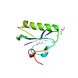
1SVY
 | | SEVERIN DOMAIN 2, 1.75 ANGSTROM CRYSTAL STRUCTURE | | Descriptor: | CALCIUM ION, SEVERIN, SODIUM ION | | Authors: | Puius, Y.A, Fedorov, E.V, Eichinger, L, Sullivan, M, Schleicher, M, Almo, S.C. | | Deposit date: | 1998-08-10 | | Release date: | 1999-08-10 | | Last modified: | 2011-07-13 | | Method: | X-RAY DIFFRACTION (1.75 Å) | | Cite: | Mapping the functional surface of domain 2 in the gelsolin superfamily.
Biochemistry, 39, 2000
|
|
1SVR
 | |
3FG7
 | |
3FFN
 | |
3FG6
 | | Structure of the C-terminus of Adseverin | | Descriptor: | Adseverin, CALCIUM ION | | Authors: | Robinson, R.C. | | Deposit date: | 2008-12-05 | | Release date: | 2009-08-11 | | Last modified: | 2023-11-01 | | Method: | X-RAY DIFFRACTION (3 Å) | | Cite: | The crystal structure of the C-terminus of adseverin reveals the actin-binding interface.
Proc.Natl.Acad.Sci.USA, 106, 2009
|
|
2NUT
 | |
2NUP
 | |
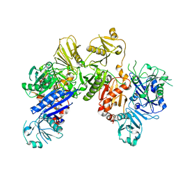
5KYY
 | | Crystal structure of Sec23 and TANGO1 peptide4 complex | | Descriptor: | Protein transport protein Sec23A, Protein transport protein Sec24D, ZINC ION | | Authors: | Ma, W, Goldberg, J. | | Deposit date: | 2016-07-22 | | Release date: | 2016-09-07 | | Last modified: | 2017-08-23 | | Method: | X-RAY DIFFRACTION (3.403 Å) | | Cite: | TANGO1/cTAGE5 receptor as a polyvalent template for assembly of large COPII coats.
Proc.Natl.Acad.Sci.USA, 113, 2016
|
|
5KYX
 | | crystal structure of Sec23 and TANGO1 peptide1 complex | | Descriptor: | Protein transport protein Sec23A, Protein transport protein Sec24D, ZINC ION | | Authors: | Ma, W, Goldberg, J. | | Deposit date: | 2016-07-22 | | Release date: | 2016-09-07 | | Last modified: | 2017-08-23 | | Method: | X-RAY DIFFRACTION (3.516 Å) | | Cite: | TANGO1/cTAGE5 receptor as a polyvalent template for assembly of large COPII coats.
Proc.Natl.Acad.Sci.USA, 113, 2016
|
|
5KYN
 | | Structure of Sec23 and TANGO1 complex | | Descriptor: | Melanoma inhibitory activity protein 3, Protein transport protein Sec23A, ZINC ION | | Authors: | Ma, W, Goldberg, J. | | Deposit date: | 2016-07-21 | | Release date: | 2016-09-07 | | Last modified: | 2023-10-04 | | Method: | X-RAY DIFFRACTION (2.552 Å) | | Cite: | TANGO1/cTAGE5 receptor as a polyvalent template for assembly of large COPII coats.
Proc.Natl.Acad.Sci.USA, 113, 2016
|
|
5KYW
 | | crystal structure of Sec23 and TANGO1 peptide3 complex | | Descriptor: | Protein transport protein Sec23A, Protein transport protein Sec24D, TANGO1 peptide3, ... | | Authors: | Ma, W, Goldberg, J. | | Deposit date: | 2016-07-22 | | Release date: | 2016-09-07 | | Last modified: | 2017-08-23 | | Method: | X-RAY DIFFRACTION (3.2 Å) | | Cite: | TANGO1/cTAGE5 receptor as a polyvalent template for assembly of large COPII coats.
Proc.Natl.Acad.Sci.USA, 113, 2016
|
|
5KYU
 | | crystal structure of Sec23 and TANGO1 peptide2 complex | | Descriptor: | Protein transport protein Sec23A, Protein transport protein Sec24D, TANGO1 peptide2, ... | | Authors: | Ma, W, Goldberg, J. | | Deposit date: | 2016-07-22 | | Release date: | 2016-09-14 | | Last modified: | 2023-10-04 | | Method: | X-RAY DIFFRACTION (3.512 Å) | | Cite: | TANGO1/cTAGE5 receptor as a polyvalent template for assembly of large COPII coats.
Proc.Natl.Acad.Sci.USA, 113, 2016
|
|


