Loading
PDBj
MenuPDBj@FacebookPDBj@X(formerly Twitter)PDBj@BlueSkyPDBj@YouTubewwPDB FoundationwwPDB
RCSB PDBPDBeBMRBAdv. SearchSearch help
PDB: 165 results

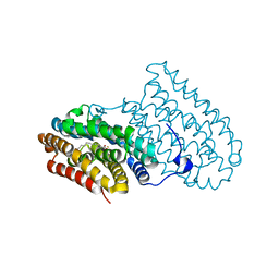
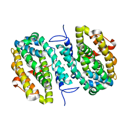
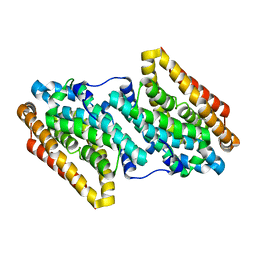
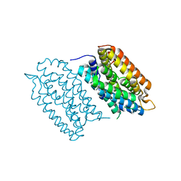
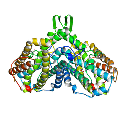
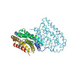
7BET
DownloadVisualize
BU of 7bet by Molmil
Structure of Ribonucleotide reductase R2 from Escherichia coli collected by femtosecond serial crystallography on a COC membrane
Descriptor: FE (III) ION, Ribonucleoside-diphosphate reductase 1 subunit beta
Authors:Aurelius, O, John, J, Martiel, I, Marsh, M, Vera, L, Huang, C.Y, Olieric, V, Leonarski, P, Nass, K, Padeste, C, Karpik, A, Hogbom, M, Wang, M, Pedrini, B.
Deposit date:2020-12-24
Release date:2022-01-12
Last modified:2024-01-31
Method:X-RAY DIFFRACTION (2.3 Å)
Cite:Commissioning results from the SwissMX instrument for fixed target macromolecular crystallography at SwissFEL
To Be Published
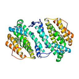
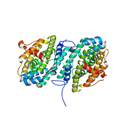
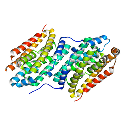
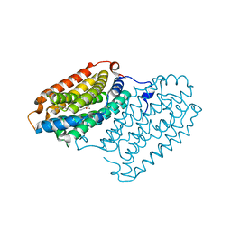
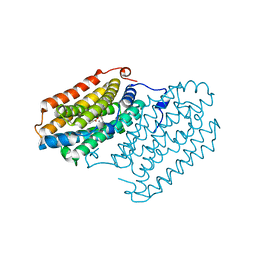
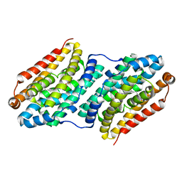
5DCO
DownloadVisualize
BU of 5dco by Molmil
R2-like ligand-binding oxidase with aerobically reconstituted diiron cofactor (short soak)
Descriptor: FE (III) ION, PALMITIC ACID, Ribonucleotide reductase small subunit
Authors:Griese, J.J, Hogbom, M.
Deposit date:2015-08-24
Release date:2015-09-09
Last modified:2024-01-10
Method:X-RAY DIFFRACTION (2.326 Å)
Cite:Structural Basis for Oxygen Activation at a Heterodinuclear Manganese/Iron Cofactor.
J.Biol.Chem., 290, 2015
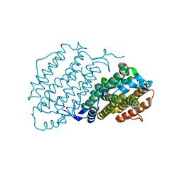
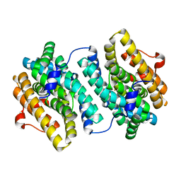
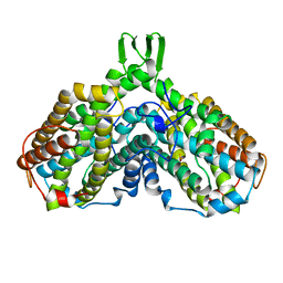
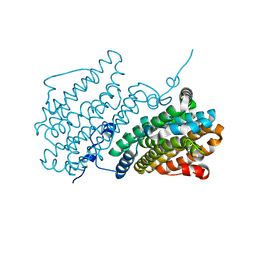
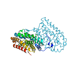
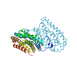
3HF1
DownloadVisualize
BU of 3hf1 by Molmil
Crystal structure of human p53R2
Descriptor: FE (III) ION, Ribonucleoside-diphosphate reductase subunit M2 B, SULFATE ION
Authors:Smith, P, Zhou, B, Yuan, Y.-C, Su, L, Tsai, S.-C, Yen, Y.
Deposit date:2009-05-10
Release date:2009-10-13
Last modified:2023-09-06
Method:X-RAY DIFFRACTION (2.6 Å)
Cite:2.6 A X-ray crystal structure of human p53R2, a p53-inducible ribonucleotide reductase .
Biochemistry, 48, 2009
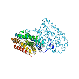
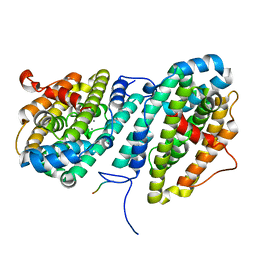
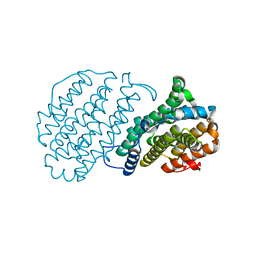
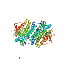
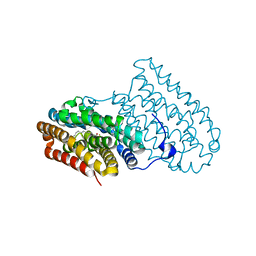
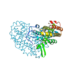
6Y2N
DownloadVisualize
BU of 6y2n by Molmil
Crystal structure of ribonucleotide reductase R2 subunit solved by serial synchrotron crystallography
Descriptor: FE (III) ION, MANGANESE (III) ION, Ribonucleoside-diphosphate reductase subunit beta
Authors:Shilova, A, Lebrette, H, Aurelius, O, Hogbom, M, Mueller, U.
Deposit date:2020-02-17
Release date:2020-10-07
Last modified:2024-05-01
Method:X-RAY DIFFRACTION (2.4 Å)
Cite:Current status and future opportunities for serial crystallography at MAX IV Laboratory.
J.Synchrotron Radiat., 27, 2020
5EKB
DownloadVisualize
BU of 5ekb by Molmil
R2-like ligand-binding oxidase with aerobically reconstituted Mn/Fe cofactor (reconstituted in solution)
Descriptor: FE (III) ION, MANGANESE (III) ION, PALMITIC ACID, ...
Authors:Griese, J.J, Hogbom, M.
Deposit date:2015-11-03
Release date:2016-04-27
Last modified:2024-01-10
Method:X-RAY DIFFRACTION (2.074 Å)
Cite:Divergent assembly mechanisms of the manganese/iron cofactors in R2lox and R2c proteins.
J.Inorg.Biochem., 162, 2016
6CWQ
DownloadVisualize
BU of 6cwq by Molmil
X-ray crystal structure of Flavobacterium johnsoniae dimanganese(II) ribonucleotide reductase beta subunit (as-isolated)
Descriptor: MAGNESIUM ION, MANGANESE (II) ION, Ribonucleotide reductase
Authors:Rose, H.R, Maggiolo, A.O, Boal, A.K.
Deposit date:2018-03-30
Release date:2018-04-18
Last modified:2024-03-13
Method:X-RAY DIFFRACTION (1.9 Å)
Cite:Structural Basis for Superoxide Activation of Flavobacterium johnsoniae Class I Ribonucleotide Reductase and for Radical Initiation by Its Dimanganese Cofactor.
Biochemistry, 57, 2018
6CWO
DownloadVisualize
BU of 6cwo by Molmil
X-ray crystal structure of Flavobacterium johnsoniae dimanganese(II) ribonucleotide reductase beta subunit (aerobic)
Descriptor: MAGNESIUM ION, MANGANESE (II) ION, Ribonucleotide reductase
Authors:Maggiolo, A.O, Rose, H.R, Boal, A.K.
Deposit date:2018-03-30
Release date:2018-06-27
Last modified:2024-03-13
Method:X-RAY DIFFRACTION (1.87 Å)
Cite:Structural Basis for Superoxide Activation of Flavobacterium johnsoniae Class I Ribonucleotide Reductase and for Radical Initiation by Its Dimanganese Cofactor.
Biochemistry, 57, 2018
3DHZ
DownloadVisualize
BU of 3dhz by Molmil
Apo (iron free) structure of C. ammoniagenes R2 protein
Descriptor: FE (II) ION, Ribonucleotide reductase subunit R2F
Authors:Hogbom, M, Nordlund, P.
Deposit date:2008-06-19
Release date:2008-07-01
Last modified:2024-02-21
Method:X-RAY DIFFRACTION (1.63 Å)
Cite:Structural and mutational studies of the carboxylate cluster in Iron-free Ribonucleotide Reductase R2.
Biochemistry, 43, 2004
6CWP
DownloadVisualize
BU of 6cwp by Molmil
X-ray crystal structure of Flavobacterium johnsoniae dimanganese(II) ribonucleotide reductase beta subunit (anaerobic)
Descriptor: MANGANESE (II) ION, Ribonucleotide reductase, VAL-GLU-TYR-THR-LYS-HIS
Authors:Maggiolo, A.O, Rose, H.R, Boal, A.K.
Deposit date:2018-03-30
Release date:2018-04-18
Last modified:2024-03-13
Method:X-RAY DIFFRACTION (1.92 Å)
Cite:Structural Basis for Superoxide Activation of Flavobacterium johnsoniae Class I Ribonucleotide Reductase and for Radical Initiation by Its Dimanganese Cofactor.
Biochemistry, 57, 2018
6QK2
DownloadVisualize
BU of 6qk2 by Molmil
R2-like ligand-binding oxidase Y715F mutant with anaerobically reconstituted Mn/Fe cofactor
Descriptor: FE (II) ION, MANGANESE (II) ION, PALMITIC ACID, ...
Authors:Griese, J.J, Hogbom, M.
Deposit date:2019-01-28
Release date:2020-02-19
Last modified:2024-01-24
Method:X-RAY DIFFRACTION (1.596 Å)
Cite:Key Structural Motifs Balance Metal Binding and Oxidative Reactivity in a Heterobimetallic Mn/Fe Protein.
J.Am.Chem.Soc., 142, 2020
6QK0
DownloadVisualize
BU of 6qk0 by Molmil
R2-like ligand-binding oxidase E69D mutant with anaerobically reconstituted Mn/Fe cofactor
Descriptor: FE (II) ION, MANGANESE (II) ION, PALMITIC ACID, ...
Authors:Griese, J.J, Hogbom, M.
Deposit date:2019-01-28
Release date:2020-02-19
Last modified:2024-01-24
Method:X-RAY DIFFRACTION (2.091 Å)
Cite:Key Structural Motifs Balance Metal Binding and Oxidative Reactivity in a Heterobimetallic Mn/Fe Protein.
J.Am.Chem.Soc., 142, 2020
1AV8
DownloadVisualize
BU of 1av8 by Molmil
RIBONUCLEOTIDE REDUCTASE R2 SUBUNIT FROM E. COLI
Descriptor: MU-OXO-DIIRON, RIBONUCLEOTIDE REDUCTASE R2
Authors:Han, S, Arvai, A, Tainer, J.A.
Deposit date:1997-09-30
Release date:1998-10-28
Last modified:2024-05-22
Method:X-RAY DIFFRACTION (2.8 Å)
Cite:Characterization of Y122F R2 of Escherichia coli ribonucleotide reductase by time-resolved physical biochemical methods and X-ray crystallography.
Biochemistry, 37, 1998
1H0O
DownloadVisualize
BU of 1h0o by Molmil
Cobalt substitution of mouse R2 ribonucleotide reductase to model the reactive diferrous state
Descriptor: COBALT (II) ION, RIBONUCLEOSIDE-DIPHOSPHATE REDUCTASE
Authors:Strand, K.R, Karlsen, S, Andersson, K.K.
Deposit date:2002-06-26
Release date:2002-07-25
Last modified:2023-12-13
Method:X-RAY DIFFRACTION (2.2 Å)
Cite:Cobalt Substitution of Mouse R2 Ribonucleotide Reductase as a Model for Thereactive Diferrous State. Spectroscopic and Structural Evidence for a Ferromagnetically Coupled Dinuclear Cobalt Cluster
J.Biol.Chem., 277, 2002
1H0N
DownloadVisualize
BU of 1h0n by Molmil
Cobalt substitution of mouse R2 ribonucleotide reductase to model the reactive diferrous state
Descriptor: COBALT (II) ION, RIBONUCLEOSIDE-DIPHOSPHATE REDUCTASE
Authors:Strand, K.R, Karlsen, S, Andersson, K.K.
Deposit date:2002-06-26
Release date:2002-07-25
Last modified:2023-12-13
Method:X-RAY DIFFRACTION (2.4 Å)
Cite:Cobalt Substitution of Mouse R2 Ribonucleotide Reductase as a Model for Thereactive Diferrous State. Spectroscopic and Structural Evidence for a Ferromagnetically Coupled Dinuclear Cobalt Cluster
J.Biol.Chem., 277, 2002
7QBK
DownloadVisualize
BU of 7qbk by Molmil
Crystal structure of a second homolog of R2-like ligand-binding oxidase in Sulfolobus acidocaldarius (SaR2loxII)
Descriptor: FE (III) ION, MANGANESE (III) ION, R2-like ligand-binding oxidase (homolog II) from Sulfolobus acidocaldarius
Authors:Lebrette, H, Diamanti, R, Srinivas, V, Hogbom, M.
Deposit date:2021-11-19
Release date:2022-04-06
Last modified:2024-01-31
Method:X-RAY DIFFRACTION (2.26 Å)
Cite:Comparative structural analysis provides new insights into the function of R2-like ligand-binding oxidase.
Febs Lett., 596, 2022
7QBP
DownloadVisualize
BU of 7qbp by Molmil
Crystal structure of R2-like ligand-binding oxidase from Saccharopolyspora Erythraea
Descriptor: FE (III) ION, MANGANESE (III) ION, PALMITIC ACID, ...
Authors:Srinivas, V, Diamanti, R, Lebrette, H, Hogbom, M.
Deposit date:2021-11-19
Release date:2022-04-06
Last modified:2024-01-31
Method:X-RAY DIFFRACTION (1.38 Å)
Cite:Comparative structural analysis provides new insights into the function of R2-like ligand-binding oxidase.
Febs Lett., 596, 2022
8BT3
DownloadVisualize
BU of 8bt3 by Molmil
Ribonucleotide Reductase class Ie R2 from Mesoplasma florum, catalytically active radical state solved by XFEL
Descriptor: Ribonucleoside-diphosphate reductase beta chain
Authors:Lebrette, H, Srinivas, V, Hogbom, M.
Deposit date:2022-11-27
Release date:2023-11-01
Last modified:2024-11-13
Method:X-RAY DIFFRACTION (1.5 Å)
Cite:Structure of a ribonucleotide reductase R2 protein radical.
Science, 382, 2023
8BT4
DownloadVisualize
BU of 8bt4 by Molmil
Ribonucleotide Reductase class Ie R2 from Mesoplasma florum, radical-lost ground state
Descriptor: CALCIUM ION, GLYCEROL, Ribonucleoside-diphosphate reductase
Authors:Lebrette, H, Srinivas, V, Hogbom, M.
Deposit date:2022-11-27
Release date:2023-11-01
Last modified:2024-11-13
Method:X-RAY DIFFRACTION (1.35 Å)
Cite:Structure of a ribonucleotide reductase R2 protein radical.
Science, 382, 2023
1BIQ
DownloadVisualize
BU of 1biq by Molmil
RIBONUCLEOSIDE-DIPHOSPHATE REDUCTASE 1 BETA CHAIN MUTANT E238A
Descriptor: FE (II) ION, FE (III) ION, HYDROXIDE ION, ...
Authors:Logan, D.T, Demare, F, Persson, B.O, Slaby, A, Sjoberg, B.M, Nordlund, P.
Deposit date:1998-06-18
Release date:1999-01-13
Last modified:2023-08-02
Method:X-RAY DIFFRACTION (2.05 Å)
Cite:Crystal structures of two self-hydroxylating ribonucleotide reductase protein R2 mutants: structural basis for the oxygen-insertion step of hydroxylation reactions catalyzed by diiron proteins.
Biochemistry, 37, 1998
3EE4
DownloadVisualize
BU of 3ee4 by Molmil
R2-like ligand binding Mn/Fe oxidase from M. tuberculosis
Descriptor: FE (III) ION, MANGANESE (III) ION, MYRISTIC ACID, ...
Authors:Andersson, C.S, Jones, T.A, Hogbom, M.
Deposit date:2008-09-04
Release date:2009-04-07
Last modified:2011-07-13
Method:X-RAY DIFFRACTION (1.9 Å)
Cite:A Mycobacterium tuberculosis ligand-binding Mn/Fe protein reveals a new cofactor in a remodeled R2-protein scaffold
Proc.Natl.Acad.Sci.Usa, 106, 2009
5OMJ
DownloadVisualize
BU of 5omj by Molmil
R2-like ligand-binding oxidase with aerobically reconstituted metal cofactor after photoconversion
Descriptor: FE (III) ION, PALMITIC ACID, Ribonucleotide reductase small subunit
Authors:Griese, J.J, Hogbom, M.
Deposit date:2017-07-31
Release date:2018-01-03
Last modified:2024-01-17
Method:X-RAY DIFFRACTION (2.005 Å)
Cite:Driving Protein Conformational Changes with Light: Photoinduced Structural Rearrangement in a Heterobimetallic Oxidase.
J. Am. Chem. Soc., 140, 2018
4XB9
DownloadVisualize
BU of 4xb9 by Molmil
R2-like ligand-binding oxidase with aerobically reconstituted diiron cofactor
Descriptor: FE (III) ION, PALMITIC ACID, Ribonuleotide reductase small subunit
Authors:Griese, J.J, Hogbom, M.
Deposit date:2014-12-16
Release date:2015-09-09
Last modified:2024-01-10
Method:X-RAY DIFFRACTION (1.798 Å)
Cite:Structural Basis for Oxygen Activation at a Heterodinuclear Manganese/Iron Cofactor.
J.Biol.Chem., 290, 2015
4XBW
DownloadVisualize
BU of 4xbw by Molmil
R2-like ligand-binding oxidase with aerobically reconstituted dimanganese cofactor
Descriptor: MANGANESE (II) ION, PALMITIC ACID, Ribonucleotide reductase small subunit
Authors:Griese, J.J, Hogbom, M.
Deposit date:2014-12-17
Release date:2015-09-09
Last modified:2024-01-10
Method:X-RAY DIFFRACTION (1.991 Å)
Cite:Structural Basis for Oxygen Activation at a Heterodinuclear Manganese/Iron Cofactor.
J.Biol.Chem., 290, 2015
4XBV
DownloadVisualize
BU of 4xbv by Molmil
R2-like ligand-binding oxidase with anaerobically reconstituted diiron cofactor
Descriptor: FE (II) ION, PALMITIC ACID, Ribonuleotide reductase small subunit
Authors:Griese, J.J, Hogbom, M.
Deposit date:2014-12-17
Release date:2015-09-09
Last modified:2024-01-10
Method:X-RAY DIFFRACTION (1.799 Å)
Cite:Structural Basis for Oxygen Activation at a Heterodinuclear Manganese/Iron Cofactor.
J.Biol.Chem., 290, 2015
4HR4
DownloadVisualize
BU of 4hr4 by Molmil
R2-like ligand-binding oxidase with anaerobically reconstituted metal cofactor
Descriptor: FE (II) ION, MANGANESE (II) ION, PALMITIC ACID, ...
Authors:Griese, J.J, Hogbom, M.
Deposit date:2012-10-26
Release date:2013-10-16
Last modified:2024-03-20
Method:X-RAY DIFFRACTION (1.901 Å)
Cite:Direct observation of structurally encoded metal discrimination and ether bond formation in a heterodinuclear metalloprotein
Proc.Natl.Acad.Sci.USA, 110, 2013

227344

数据于2024-11-13公开中

PDB statisticsPDBj update infoContact PDBjnumon