Loading
PDBj
MenuPDBj@FacebookPDBj@TwitterPDBj@YouTubewwPDB FoundationwwPDB
RCSB PDBPDBeBMRBAdv. SearchSearch help

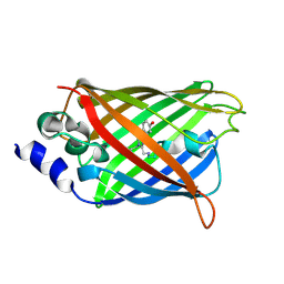
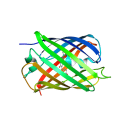
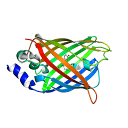
3DQ9
DownloadVisualize
BU of 3dq9 by Molmil
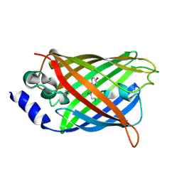
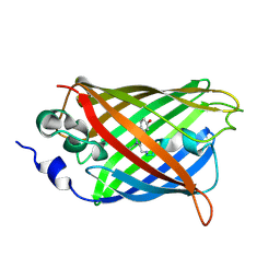
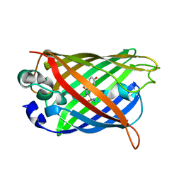
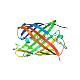
Structure of the Yellow Fluorescent Protein Citrine Frozen at 1500 Atmospheres Number 1: Structure 15 in a Series of 26 High Pressure Structures
Descriptor: Green fluorescent protein
Authors:Barstow, B, Kim, C.U.
Deposit date:2008-07-09
Release date:2008-09-23
Last modified:2023-11-15
Method:X-RAY DIFFRACTION (1.4 Å)
Cite:Alteration of citrine structure by hydrostatic pressure explains the accompanying spectral shift.
Proc.Natl.Acad.Sci.Usa, 105, 2008
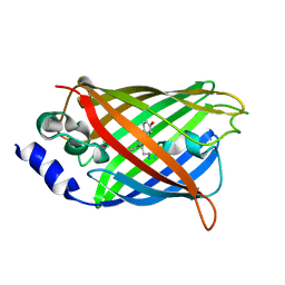
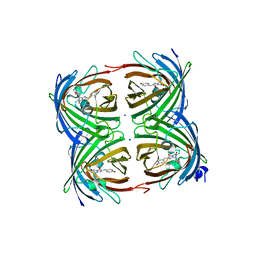
3DQM
DownloadVisualize
BU of 3dqm by Molmil
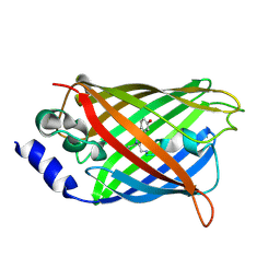
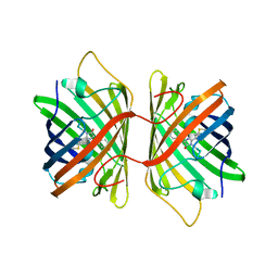
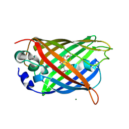
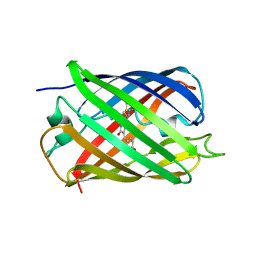
Structure of the Yellow Fluorescent Protein Citrine Frozen at 750 Atmospheres: Structure 4 in a Series of 26 High Pressure Structures
Descriptor: Green fluorescent protein
Authors:Barstow, B, Kim, C.U.
Deposit date:2008-07-09
Release date:2008-09-23
Last modified:2023-11-15
Method:X-RAY DIFFRACTION (1.44 Å)
Cite:Alteration of citrine structure by hydrostatic pressure explains the accompanying spectral shift.
Proc.Natl.Acad.Sci.Usa, 105, 2008
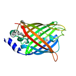
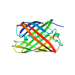
3DQH
DownloadVisualize
BU of 3dqh by Molmil
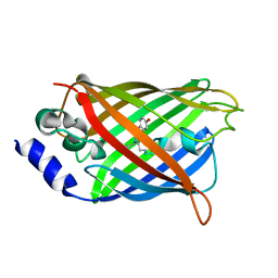
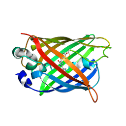
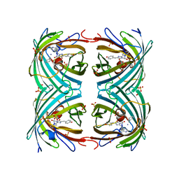
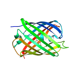
Structure of the Yellow Fluorescent Protein Citrine Frozen at 1000 Atmospheres Number 5: Structure 9 in a Series of 26 High Pressure Structures
Descriptor: Green fluorescent protein
Authors:Barstow, B, Kim, C.U.
Deposit date:2008-07-09
Release date:2008-09-23
Last modified:2023-11-15
Method:X-RAY DIFFRACTION (1.45 Å)
Cite:Alteration of citrine structure by hydrostatic pressure explains the accompanying spectral shift.
Proc.Natl.Acad.Sci.Usa, 105, 2008
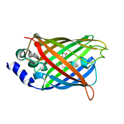
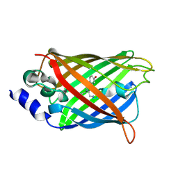
3DQF
DownloadVisualize
BU of 3dqf by Molmil
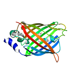
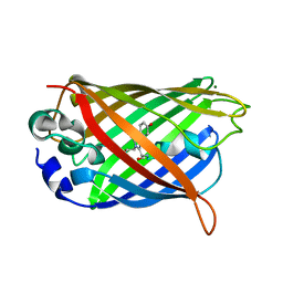
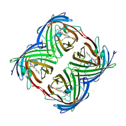
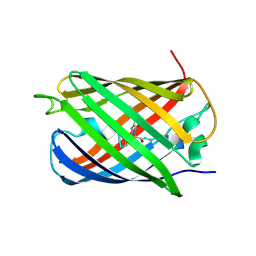
Structure of the Yellow Fluorescent Protein Citrine Frozen at 1000 Atmospheres Number 6: Structure 10 in a Series of 26 High Pressure Structures
Descriptor: Green fluorescent protein
Authors:Barstow, B, Kim, C.U.
Deposit date:2008-07-09
Release date:2008-09-23
Last modified:2023-11-15
Method:X-RAY DIFFRACTION (1.46 Å)
Cite:Alteration of citrine structure by hydrostatic pressure explains the accompanying spectral shift.
Proc.Natl.Acad.Sci.Usa, 105, 2008
3DQO
DownloadVisualize
BU of 3dqo by Molmil
Structure of the Yellow Fluorescent Protein Citrine Frozen at 1 Atmosphere Number 2: Structure 2 in a Series of 26 High Pressure Structures
Descriptor: Green fluorescent protein
Authors:Barstow, B, Kim, C.U.
Deposit date:2008-07-09
Release date:2008-09-23
Last modified:2023-11-15
Method:X-RAY DIFFRACTION (1.5 Å)
Cite:Alteration of citrine structure by hydrostatic pressure explains the accompanying spectral shift.
Proc.Natl.Acad.Sci.Usa, 105, 2008
3DPX
DownloadVisualize
BU of 3dpx by Molmil
Structure of the Yellow Fluorescent Protein Citrine Frozen at 5000 Atmospheres: Structure 26 in a Series of 26 High Pressure Structures
Descriptor: Green fluorescent protein
Authors:Barstow, B, Kim, C.U.
Deposit date:2008-07-09
Release date:2008-09-23
Last modified:2023-11-15
Method:X-RAY DIFFRACTION (1.5 Å)
Cite:Alteration of citrine structure by hydrostatic pressure explains the accompanying spectral shift.
Proc.Natl.Acad.Sci.Usa, 105, 2008
3DQ5
DownloadVisualize
BU of 3dq5 by Molmil
Structure of the Yellow Fluorescent Protein Citrine Frozen at 1960 Atmospheres: Structure 19 in a Series of 26 High Pressure Structures
Descriptor: Green fluorescent protein
Authors:Barstow, B, Kim, C.U.
Deposit date:2008-07-09
Release date:2008-09-23
Last modified:2023-11-15
Method:X-RAY DIFFRACTION (1.5 Å)
Cite:Alteration of citrine structure by hydrostatic pressure explains the accompanying spectral shift.
Proc.Natl.Acad.Sci.Usa, 105, 2008
3DQE
DownloadVisualize
BU of 3dqe by Molmil
Structure of the Yellow Fluorescent Protein Citrine Frozen at 1250 Atmospheres Number 1: Structure 11 in a Series of 26 High Pressure Structures
Descriptor: Green fluorescent protein
Authors:Barstow, B, Kim, C.U.
Deposit date:2008-07-09
Release date:2008-09-23
Last modified:2023-11-15
Method:X-RAY DIFFRACTION (1.43 Å)
Cite:Alteration of citrine structure by hydrostatic pressure explains the accompanying spectral shift.
Proc.Natl.Acad.Sci.Usa, 105, 2008
3DQI
DownloadVisualize
BU of 3dqi by Molmil
Structure of the Yellow Fluorescent Protein Citrine Frozen at 1000 Atmospheres Number 4: Structure 8 in a Series of 26 High Pressure Structures
Descriptor: Green fluorescent protein
Authors:Barstow, B, Kim, C.U.
Deposit date:2008-07-09
Release date:2008-09-23
Last modified:2023-11-15
Method:X-RAY DIFFRACTION (1.42 Å)
Cite:Alteration of citrine structure by hydrostatic pressure explains the accompanying spectral shift.
Proc.Natl.Acad.Sci.Usa, 105, 2008
3DQJ
DownloadVisualize
BU of 3dqj by Molmil
Structure of the Yellow Fluorescent Protein Citrine Frozen at 1000 Atmospheres Number 3: Structure 7 in a Series of 26 High Pressure Structures
Descriptor: Green fluorescent protein
Authors:Barstow, B, Kim, C.U.
Deposit date:2008-07-09
Release date:2008-09-23
Last modified:2023-11-15
Method:X-RAY DIFFRACTION (1.51 Å)
Cite:Alteration of citrine structure by hydrostatic pressure explains the accompanying spectral shift.
Proc.Natl.Acad.Sci.Usa, 105, 2008
3DQU
DownloadVisualize
BU of 3dqu by Molmil
Structure of the Yellow Fluorescent Protein Citrine Frozen at 2000 Atmospheres Number 1: Structure 20 in a Series of 26 High Pressure Structures
Descriptor: Green fluorescent protein
Authors:Barstow, B, Kim, C.U.
Deposit date:2008-07-09
Release date:2008-09-23
Last modified:2023-11-15
Method:X-RAY DIFFRACTION (1.42 Å)
Cite:Alteration of citrine structure by hydrostatic pressure explains the accompanying spectral shift.
Proc.Natl.Acad.Sci.Usa, 105, 2008
3E5W
DownloadVisualize
BU of 3e5w by Molmil
Crystal Structure Analysis of FP611
Descriptor: Red fluorescent protein eqFP611
Authors:Nienhaus, K, Nar, H, Heilker, R, Wiedenmann, J, Nienhaus, G.U.
Deposit date:2008-08-14
Release date:2008-09-23
Last modified:2023-11-15
Method:X-RAY DIFFRACTION (1.71 Å)
Cite:Trans-cis isomerization is responsible for the red-shifted fluorescence in variants of the red fluorescent protein eqFP611.
J.Am.Chem.Soc., 130, 2008
3CD1
DownloadVisualize
BU of 3cd1 by Molmil
Development of a family of redox-sensitive green fluorescent protein indicators for use in relatively oxidizing subcellular environments
Descriptor: Green fluorescent protein
Authors:Lohman, J.R.
Deposit date:2008-02-26
Release date:2008-08-19
Last modified:2024-10-09
Method:X-RAY DIFFRACTION (1.312 Å)
Cite:Development of a family of redox-sensitive green fluorescent protein indicators for use in relatively oxidizing subcellular environments
Biochemistry, 47, 2008
3CBE
DownloadVisualize
BU of 3cbe by Molmil
Development of a family of redox-sensitive green fluorescent protein indicators for use in relatively oxidizing subcellular environments
Descriptor: Green fluorescent protein, MAGNESIUM ION
Authors:Lohman, J.R.
Deposit date:2008-02-21
Release date:2008-08-19
Last modified:2024-10-16
Method:X-RAY DIFFRACTION (1.488 Å)
Cite:Development of a family of redox-sensitive green fluorescent protein indicators for use in relatively oxidizing subcellular environments
Biochemistry, 47, 2008
3CD9
DownloadVisualize
BU of 3cd9 by Molmil
Development of a family of redox-sensitive green fluorescent protein indicators for use in relatively oxidizing subcellular environments
Descriptor: Green fluorescent protein
Authors:Lohman, J.R.
Deposit date:2008-02-26
Release date:2008-08-19
Last modified:2021-10-20
Method:X-RAY DIFFRACTION (1.502 Å)
Cite:Development of a family of redox-sensitive green fluorescent protein indicators for use in relatively oxidizing subcellular environments
Biochemistry, 47, 2008
3CB9
DownloadVisualize
BU of 3cb9 by Molmil
Development of a family of redox-sensitive green fluorescent protein indicators for use in relatively oxidizing subcellular environments
Descriptor: Green fluorescent protein, MAGNESIUM ION
Authors:Lohman, J.R.
Deposit date:2008-02-21
Release date:2008-08-19
Last modified:2023-11-15
Method:X-RAY DIFFRACTION (1.31 Å)
Cite:Development of a family of redox-sensitive green fluorescent protein indicators for use in relatively oxidizing subcellular environments
Biochemistry, 47, 2008
2VVJ
DownloadVisualize
BU of 2vvj by Molmil
IrisFP fluorescent protein in its red form, cis conformation
Descriptor: Green to red photoconvertible GFP-like protein EosFP, SULFATE ION, SULFITE ION
Authors:Adam, V, Lelimousin, M, Boehme, S, Desfonds, G, Nienhaus, K, Field, M.J, Wiedenmann, J, McSweeney, S, Nienhaus, G.U, Bourgeois, D.
Deposit date:2008-06-09
Release date:2008-08-12
Last modified:2023-12-13
Method:X-RAY DIFFRACTION (2 Å)
Cite:Structural Characterization of Irisfp, an Optical Highlighter Undergoing Multiple Photo-Induced Transformations.
Proc.Natl.Acad.Sci.USA, 105, 2008
2Z6X
DownloadVisualize
BU of 2z6x by Molmil
Crystal structure of 22G, the wild-type protein of the photoswitchable GFP-like protein Dronpa
Descriptor: photochromic protein Dronpa
Authors:Kikuchi, A, Jeyakanthan, J, Taka, J, Shiro, Y, Mizuno, H, Miyawaki, A.
Deposit date:2007-08-09
Release date:2008-07-22
Last modified:2023-11-15
Method:X-RAY DIFFRACTION (2.3 Å)
Cite:Light-dependent regulation of structural flexibility in a photochromic fluorescent protein.
Proc.Natl.Acad.Sci.Usa, 105, 2008
2Z6Y
DownloadVisualize
BU of 2z6y by Molmil
Crystal structure of a photoswitchable GFP-like protein Dronpa in the bright-state
Descriptor: Fluorescent protein Dronpa
Authors:Kikuchi, A, Jeyakanthan, J, Taka, J, Shiro, Y, Mizuno, H, Miyawaki, A.
Deposit date:2007-08-09
Release date:2008-07-22
Last modified:2023-11-15
Method:X-RAY DIFFRACTION (2 Å)
Cite:Light-dependent regulation of structural flexibility in a photochromic fluorescent protein.
Proc.Natl.Acad.Sci.Usa, 105, 2008
2QLG
DownloadVisualize
BU of 2qlg by Molmil
mPlum
Descriptor: Fluorescent protein plum
Authors:Shu, X, Remington, S.J.
Deposit date:2007-07-12
Release date:2008-07-22
Last modified:2023-11-15
Method:X-RAY DIFFRACTION (1.8 Å)
Cite:Structural Studies of Far-Red Emission in mPlum, a Monomeric Red Fluorescent Protein
To be Published
2QLH
DownloadVisualize
BU of 2qlh by Molmil
mPlum I65L mutant
Descriptor: Fluorescent protein plum
Authors:Shu, X, Remington, S.J.
Deposit date:2007-07-12
Release date:2008-07-22
Last modified:2023-11-15
Method:X-RAY DIFFRACTION (1.9 Å)
Cite:Structural Studies of Far-Red Emission in mPlum, a Monomeric Red Fluorescent Protein
To be Published
2Z6Z
DownloadVisualize
BU of 2z6z by Molmil
Crystal structure of a photoswitchable GFP-like protein Dronpa in the bright-state
Descriptor: Fluorescent protein Dronpa
Authors:Kikuchi, A, Jeyakanthan, J, Taka, J, Shiro, Y, Mizuno, H, Miyawaki, A.
Deposit date:2007-08-09
Release date:2008-07-22
Last modified:2023-11-15
Method:X-RAY DIFFRACTION (1.8 Å)
Cite:Light-dependent regulation of structural flexibility in a photochromic fluorescent protein.
Proc.Natl.Acad.Sci.Usa, 105, 2008
2QLI
DownloadVisualize
BU of 2qli by Molmil
mPlum E16Q mutant
Descriptor: Fluorescent protein plum
Authors:Shu, X, Remington, S.J.
Deposit date:2007-07-12
Release date:2008-07-22
Last modified:2023-11-15
Method:X-RAY DIFFRACTION (1.34 Å)
Cite:Structural Studies of Far-Red Emission in mPlum, a Monomeric Red Fluorescent Protein
To be Published
3CGL
DownloadVisualize
BU of 3cgl by Molmil
Crystal Structure and Raman Studies of dsFP483, a Cyan Fluorescent Protein from Discosoma striata
Descriptor: GFP-like fluorescent chromoprotein dsFP483, SODIUM ION
Authors:Malo, G.D.
Deposit date:2008-03-05
Release date:2008-07-01
Last modified:2024-10-09
Method:X-RAY DIFFRACTION (2.09 Å)
Cite:Crystal structure and Raman studies of dsFP483, a cyan fluorescent protein from Discosoma striata.
J.Mol.Biol., 378, 2008
2Z1O
DownloadVisualize
BU of 2z1o by Molmil
Crystal structure of a photoswitchable GFP-like protein Dronpa in the bright-state
Descriptor: Fluorescent protein Dronpa
Authors:Kikuchi, A, Jeyakanthan, J, Taka, J, Shiro, Y, Mizuno, H, Miyawaki, A.
Deposit date:2007-05-10
Release date:2008-05-13
Last modified:2023-11-15
Method:X-RAY DIFFRACTION (1.75 Å)
Cite:Light-dependent regulation of structural flexibility in a photochromic fluorescent protein
Proc.Natl.Acad.Sci.Usa, 105, 2008

226262

건을2024-10-16부터공개중

PDB statisticsPDBj update infoContact PDBjnumon