Loading
PDBj
MenuPDBj@FacebookPDBj@TwitterPDBj@YouTubewwPDB FoundationwwPDB
RCSB PDBPDBeBMRBAdv. SearchSearch help

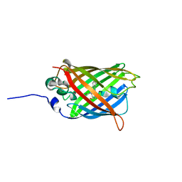
3W1D
DownloadVisualize
BU of 3w1d by Molmil
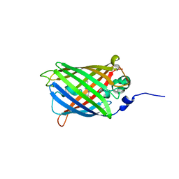
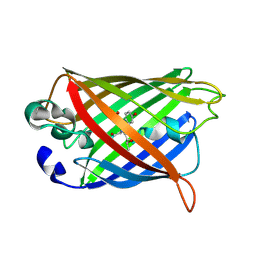
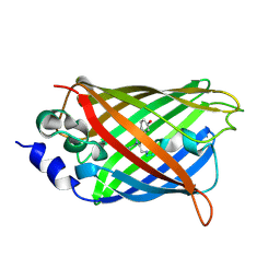
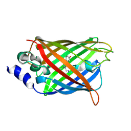
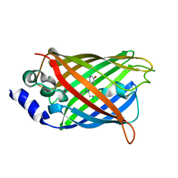
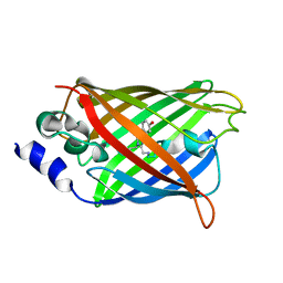
Structure of a pressure sensitive YFP variant YFP-G3
Descriptor: Green Fluorescent protein
Authors:Imada, K, Yoshizawa, K, Kinoshita, M, Watanabe, T.M.
Deposit date:2012-11-14
Release date:2013-10-02
Last modified:2023-12-06
Method:X-RAY DIFFRACTION (1.5 Å)
Cite:Glycine insertion makes yellow fluorescent protein sensitive to hydrostatic pressure.
Plos One, 8, 2013
3UFZ
DownloadVisualize
BU of 3ufz by Molmil
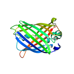
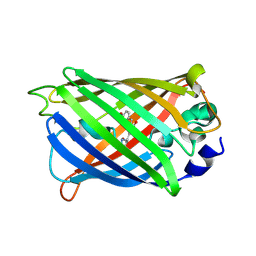
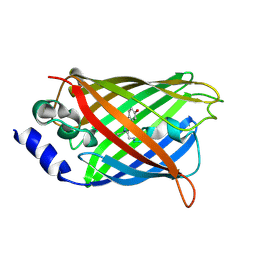
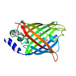
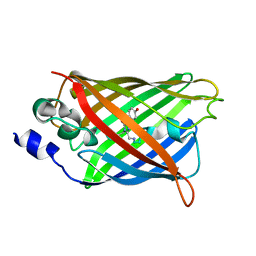
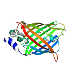
Crystal structure of a Trp-less green fluorescent protein translated by the universal genetic code
Descriptor: Green fluorescent protein
Authors:Kawahara-Kobayashi, A, Araiso, Y, Matsuda, T, Yokoyama, S, Kigawa, T, Nureki, O, Kiga, D.
Deposit date:2011-11-02
Release date:2012-10-17
Last modified:2023-12-06
Method:X-RAY DIFFRACTION (1.85 Å)
Cite:Simplification of the genetic code: restricted diversity of genetically encoded amino acids.
Nucleic Acids Res., 40, 2012
5FJI
DownloadVisualize
BU of 5fji by Molmil
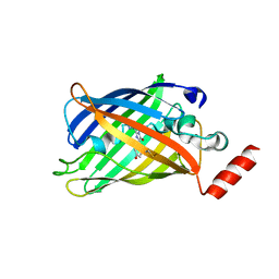
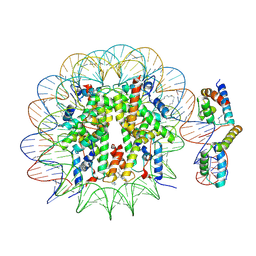
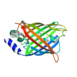
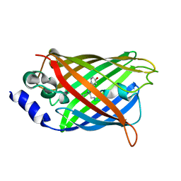
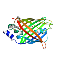
Three-dimensional structures of two heavily N-glycosylated Aspergillus sp. Family GH3 beta-D-glucosidases
Descriptor: 1,2-ETHANEDIOL, 2-acetamido-2-deoxy-beta-D-glucopyranose, BETA-GLUCOSIDASE, ...
Authors:Agirre, J, Ariza, A, Offen, W.A, Turkenburg, J.P, Roberts, S.M, McNicholas, S, Harris, P.V, McBrayer, B, Dohnalek, J, Cowtan, K.D, Davies, G.J, Wilson, K.S.
Deposit date:2015-10-09
Release date:2016-02-10
Last modified:2024-01-10
Method:X-RAY DIFFRACTION (1.95 Å)
Cite:Three-Dimensional Structures of Two Heavily N-Glycosylated Aspergillus Sp. Family Gh3 Beta-D-Glucosidases
Acta Crystallogr.,Sect.D, 72, 2016
7AMB
DownloadVisualize
BU of 7amb by Molmil
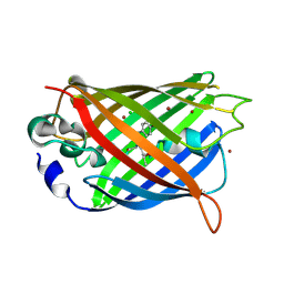
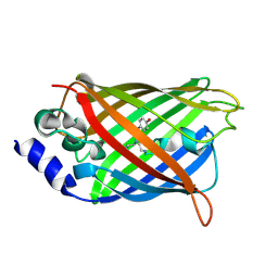
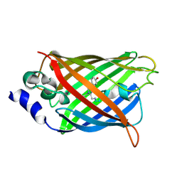
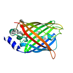
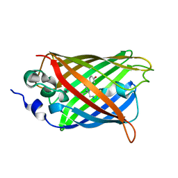
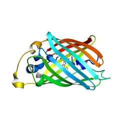
Crystal structure of rsFolder2 in its fluorescent on-state
Descriptor: GLYCEROL, Green fluorescent protein
Authors:Moreno-Chicano, T, El Khatib, M, Colletier, J.-P.
Deposit date:2020-10-08
Release date:2022-08-24
Last modified:2024-01-31
Method:X-RAY DIFFRACTION (1.63 Å)
Cite:Rational Control of Off-State Heterogeneity in a Photoswitchable Fluorescent Protein Provides Switching Contrast Enhancement.
Chemphyschem, 23, 2022
7AMF
DownloadVisualize
BU of 7amf by Molmil
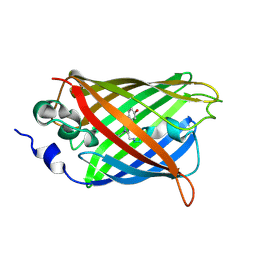
Crystal structure of rsFolder2 in its non-fluorescent off-state
Descriptor: GLYCEROL, Green fluorescent protein
Authors:Moreno-Chicano, T, El Khatib, M, Colletier, J.-P.
Deposit date:2020-10-08
Release date:2022-08-24
Last modified:2024-01-31
Method:X-RAY DIFFRACTION (1.63 Å)
Cite:Rational Control of Off-State Heterogeneity in a Photoswitchable Fluorescent Protein Provides Switching Contrast Enhancement.
Chemphyschem, 23, 2022
2P4M
DownloadVisualize
BU of 2p4m by Molmil
High pH structure of Rtms5 H146S variant
Descriptor: GFP-like non-fluorescent chromoprotein, IODIDE ION
Authors:Battad, J.M, Wilmann, P.G, Olsen, S, Byres, E, Smith, S.C, Dove, S.G, Turcic, K.N, Devenish, R.J, Rossjohn, J, Prescott, M.
Deposit date:2007-03-12
Release date:2007-04-03
Last modified:2023-11-15
Method:X-RAY DIFFRACTION (1.8 Å)
Cite:A structural basis for the pH-dependent increase in fluorescence efficiency of chromoproteins
J.Mol.Biol., 368, 2007
7AMU
DownloadVisualize
BU of 7amu by Molmil
Crystal structure of rsEGFP2 T204A in its fluorescent on-state
Descriptor: GLYCEROL, Green fluorescent protein, SULFATE ION
Authors:Moreno-Chicano, T, Schlichting, I, Hartmann, E, Zala, N, Colletier, J.-P, Weik, M.
Deposit date:2020-10-09
Release date:2022-04-20
Last modified:2024-01-31
Method:X-RAY DIFFRACTION (1.64 Å)
Cite:Crystal structure of rsEGFP2 in its fluorescent on-state at pH 8.0
To Be Published
4KAG
DownloadVisualize
BU of 4kag by Molmil
Crystal structure analysis of a single amino acid deletion mutation in EGFP
Descriptor: 1,2-ETHANEDIOL, 2-AMINO-2-HYDROXYMETHYL-PROPANE-1,3-DIOL, Green fluorescent protein, ...
Authors:Arpino, J.A.J, Rizkallah, P.J.
Deposit date:2013-04-22
Release date:2014-08-06
Last modified:2023-12-06
Method:X-RAY DIFFRACTION (1.12 Å)
Cite:Structural and dynamic changes associated with beneficial engineered single-amino-acid deletion mutations in enhanced green fluorescent protein.
Acta Crystallogr.,Sect.D, 70, 2014
4JFG
DownloadVisualize
BU of 4jfg by Molmil
Crystal structure of sfGFP-66-HqAla
Descriptor: CESIUM ION, Green fluorescent protein, quinolin-8-ol
Authors:Wang, J, Liu, X, Li, J, Zhang, W, Hu, M, Zhou, J.
Deposit date:2013-02-28
Release date:2013-10-02
Last modified:2023-11-15
Method:X-RAY DIFFRACTION (3.001 Å)
Cite:Significant expansion of the fluorescent protein chromophore through the genetic incorporation of a metal-chelating unnatural amino acid.
Angew.Chem.Int.Ed.Engl., 52, 2013
4KEX
DownloadVisualize
BU of 4kex by Molmil
Crystal structure analysis of a single amino acid deletion mutation in EGFP
Descriptor: Green fluorescent protein
Authors:Arpino, J.A.J, Rizkallah, P.J.
Deposit date:2013-04-26
Release date:2014-08-06
Last modified:2023-12-06
Method:X-RAY DIFFRACTION (1.6 Å)
Cite:Structural and dynamic changes associated with beneficial engineered single-amino-acid deletion mutations in enhanced green fluorescent protein.
Acta Crystallogr.,Sect.D, 70, 2014
4JRB
DownloadVisualize
BU of 4jrb by Molmil
Structure of Cockroach Allergen Bla g 1 Tandem Repeat as a EGFP fusion
Descriptor: (1S)-2-{[{[(2R)-2,3-DIHYDROXYPROPYL]OXY}(HYDROXY)PHOSPHORYL]OXY}-1-[(PALMITOYLOXY)METHYL]ETHYL STEARATE, CHLORIDE ION, DODECANE, ...
Authors:Mueller, G.A, Pedersen, L.C, Lih, F.B, Glesner, J, Moon, A.F, Chapman, M.D, Tomer, K, London, R.E.
Deposit date:2013-03-21
Release date:2013-07-24
Last modified:2023-12-06
Method:X-RAY DIFFRACTION (2.414 Å)
Cite:The novel structure of the cockroach allergen Bla g 1 has implications for allergenicity and exposure assessment.
J.Allergy Clin.Immunol., 132, 2013
6LOF
DownloadVisualize
BU of 6lof by Molmil
Crystal structure of ZsYellow soaked by Cu2+
Descriptor: GFP-like fluorescent chromoprotein FP538
Authors:Nam, K.H.
Deposit date:2020-01-05
Release date:2020-01-22
Last modified:2023-11-29
Method:X-RAY DIFFRACTION (2.6 Å)
Cite:Spectroscopic and Structural Analysis of Cu 2+ -Induced Fluorescence Quenching of ZsYellow.
Biosensors (Basel), 10, 2020
1KYS
DownloadVisualize
BU of 1kys by Molmil
Crystal Structure of a Zn-bound Green Fluorescent Protein Biosensor
Descriptor: Green Fluorescent Protein, ZINC ION
Authors:Barondeau, D.P, Kassmann, C.J, Tainer, J.A, Getzoff, E.D.
Deposit date:2002-02-05
Release date:2002-04-10
Last modified:2023-11-15
Method:X-RAY DIFFRACTION (1.44 Å)
Cite:Structural chemistry of a green fluorescent protein Zn biosensor.
J.Am.Chem.Soc., 124, 2002
1KYR
DownloadVisualize
BU of 1kyr by Molmil
Crystal Structure of a Cu-bound Green Fluorescent Protein Zn Biosensor
Descriptor: COPPER (II) ION, Green Fluorescent Protein, MAGNESIUM ION
Authors:Barondeau, D.P, Kassmann, C.J, Tainer, J.A, Getzoff, E.D.
Deposit date:2002-02-05
Release date:2002-04-10
Last modified:2023-11-15
Method:X-RAY DIFFRACTION (1.5 Å)
Cite:Structural chemistry of a green fluorescent protein Zn biosensor.
J.Am.Chem.Soc., 124, 2002
1KYP
DownloadVisualize
BU of 1kyp by Molmil
Crystal Structure of an Apo Green Fluorescent Protein Zn Biosensor
Descriptor: Green Fluorescent Protein, MAGNESIUM ION
Authors:Barondeau, D.P, Kassmann, C.J, Tainer, J.A, Getzoff, E.D.
Deposit date:2002-02-05
Release date:2002-04-10
Last modified:2023-11-15
Method:X-RAY DIFFRACTION (1.35 Å)
Cite:Structural chemistry of a green fluorescent protein Zn biosensor.
J.Am.Chem.Soc., 124, 2002
1MOU
DownloadVisualize
BU of 1mou by Molmil
Crystal structure of Coral pigment
Descriptor: GFP-like non-fluorescent chromoprotein, IODIDE ION
Authors:Prescott, M, Ling, M, Beddoe, T, Oakley, A.J, Dove, S, Hoegh-Guldberg, O, Devenish, R.J, Rossjohn, J.
Deposit date:2002-09-10
Release date:2003-04-08
Last modified:2023-11-15
Method:X-RAY DIFFRACTION (2.2 Å)
Cite:The 2.2 a crystal structure of a pocilloporin pigment reveals a nonplanar chromophore conformation.
Structure, 11, 2003
6UN4
DownloadVisualize
BU of 6un4 by Molmil
Crystal structure of rsEGFP2, Y67(3-ClY), Y107(3-ClY)
Descriptor: Green fluorescent protein, SULFATE ION
Authors:Lin, C.-Y, Romei, M.G, Boxer, S.G, Chang, J.
Deposit date:2019-10-10
Release date:2020-07-15
Last modified:2023-11-15
Method:X-RAY DIFFRACTION (1.499 Å)
Cite:Structural and spectroscopic characterization of photoactive yellow protein and photoswitchable fluorescent protein constructs containing heavy atoms.
J Photochem Photobiol A Chem, 401, 2020
6UZ3
DownloadVisualize
BU of 6uz3 by Molmil
Cardiac sodium channel
Descriptor: (3beta,14beta,17beta,25R)-3-[4-methoxy-3-(methoxymethyl)butoxy]spirost-5-en, 2-acetamido-2-deoxy-beta-D-glucopyranose, 2-acetamido-2-deoxy-beta-D-glucopyranose-(1-4)-2-acetamido-2-deoxy-beta-D-glucopyranose, ...
Authors:Jiang, D, Shi, H, Tonggu, L, Lenaeus, M.J, Zheng, N, Catterall, W.A.
Deposit date:2019-11-14
Release date:2020-01-01
Last modified:2020-07-29
Method:ELECTRON MICROSCOPY (3.5 Å)
Cite:Structure of the Cardiac Sodium Channel.
Cell, 180, 2020
6UZ0
DownloadVisualize
BU of 6uz0 by Molmil
Cardiac sodium channel with flecainide
Descriptor: (3beta,14beta,17beta,25R)-3-[4-methoxy-3-(methoxymethyl)butoxy]spirost-5-en, 2-acetamido-2-deoxy-beta-D-glucopyranose, 2-acetamido-2-deoxy-beta-D-glucopyranose-(1-4)-2-acetamido-2-deoxy-beta-D-glucopyranose, ...
Authors:Jiang, D, Shi, H, Tonggu, L, Lenaeus, M.J, Zheng, N, Catterall, W.A.
Deposit date:2019-11-14
Release date:2020-01-01
Last modified:2020-07-29
Method:ELECTRON MICROSCOPY (3.24 Å)
Cite:Structure of the Cardiac Sodium Channel.
Cell, 180, 2020
6WVI
DownloadVisualize
BU of 6wvi by Molmil
VKOR-like from Takifugu rubripes
Descriptor: (2R)-2,3-dihydroxypropyl (9Z)-octadec-9-enoate, Vitamin K epoxide reductase-like protein, termini restrained by green fluorescent protein
Authors:Liu, S, Sukumar, N, Li, W.
Deposit date:2020-05-06
Release date:2020-11-11
Last modified:2023-11-15
Method:X-RAY DIFFRACTION (2.4 Å)
Cite:Structural basis of antagonizing the vitamin K catalytic cycle for anticoagulation.
Science, 371, 2021
6WV9
DownloadVisualize
BU of 6wv9 by Molmil
Takifugu rubripes VKOR-like with vitamin K1 in noncatalytic state
Descriptor: Vitamin K epoxide reductase-like protein, termini restrained by green fluorescent protein
Authors:Liu, S, Sukumar, N, Li, W.
Deposit date:2020-05-05
Release date:2021-02-24
Last modified:2023-11-15
Method:X-RAY DIFFRACTION (3.35 Å)
Cite:Structural basis of antagonizing the vitamin K catalytic cycle for anticoagulation.
Science, 371, 2021
3SRY
DownloadVisualize
BU of 3sry by Molmil
Engineered high-affinity halide-binding protein derived from YFP: halide-free
Descriptor: Green fluorescent protein
Authors:Wang, W, Grimley, J.S, Beese, L.S, Hellinga, H.W.
Deposit date:2011-07-07
Release date:2012-07-11
Last modified:2023-11-15
Method:X-RAY DIFFRACTION (1.159 Å)
Cite:Determination of engineered chloride-binding site structures in fluorescent proteins reveals principles of halide recognition
To be Published
3SVB
DownloadVisualize
BU of 3svb by Molmil
Engineered medium-affinity halide-binding protein derived from YFP: fluoride complex
Descriptor: 1,2-ETHANEDIOL, Green fluorescent protein
Authors:Wang, W, Grimley, J.S, Beese, L.S, Hellinga, H.W.
Deposit date:2011-07-12
Release date:2012-07-18
Last modified:2023-11-15
Method:X-RAY DIFFRACTION (1.3 Å)
Cite:Determination of engineered chloride-binding site structures in fluorescent proteins reveals principles of halide recognition
To be Published
3SST
DownloadVisualize
BU of 3sst by Molmil
Engineered low-affinity halide-binding protein derived from YFP: chloride complex
Descriptor: CHLORIDE ION, Green fluorescent protein
Authors:Wang, W, Grimley, J.S, Beese, L.S, Hellinga, H.W.
Deposit date:2011-07-08
Release date:2012-07-11
Last modified:2023-11-15
Method:X-RAY DIFFRACTION (1.403 Å)
Cite:Determination of engineered chloride-binding site structures in fluorescent proteins reveals principles of halide recognition
To be Published
3SV5
DownloadVisualize
BU of 3sv5 by Molmil
Engineered medium-affinity halide-binding protein derived from YFP: iodide complex
Descriptor: 1,2-ETHANEDIOL, FORMIC ACID, Green fluorescent protein, ...
Authors:Wang, W, Grimley, J.S, Beese, L.S, Hellinga, H.W.
Deposit date:2011-07-12
Release date:2012-07-18
Last modified:2023-11-15
Method:X-RAY DIFFRACTION (1.53 Å)
Cite:Visualization of Synaptic Inhibition with an Optogenetic Sensor Developed by Cell-Free Protein Engineering Automation.
J.Neurosci., 33, 2013

221051

건을2024-06-12부터공개중

PDB statisticsPDBj update infoContact PDBjnumon