Loading
PDBj
MenuPDBj@FacebookPDBj@TwitterPDBj@YouTubewwPDB FoundationwwPDB
RCSB PDBPDBeBMRBAdv. SearchSearch help

3EVN
DownloadVisualize
BU of 3evn by Molmil
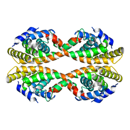
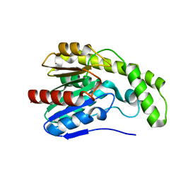
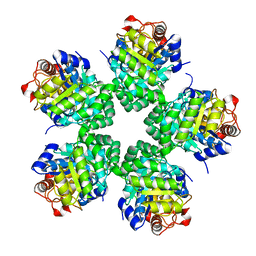
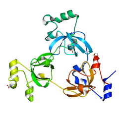
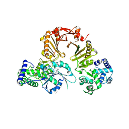
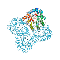
CRYSTAL STRUCTURE OF putative oxidoreductase from Streptococcus agalactiae 2603V/r
Descriptor: Oxidoreductase, Gfo/Idh/MocA family
Authors:Malashkevich, V.N, Toro, R, Meyer, A.J, Sauder, J.M, Burley, S.K, Almo, S.C, New York SGX Research Center for Structural Genomics (NYSGXRC)
Deposit date:2008-10-13
Release date:2008-10-21
Last modified:2023-12-27
Method:X-RAY DIFFRACTION (2 Å)
Cite:CRYSTAL STRUCTURE OF putative Gfo/Idh/MocA family oxidoreductase from Streptococcus agalactiae 2603V/r
To be Published
3DQ3
DownloadVisualize
BU of 3dq3 by Molmil
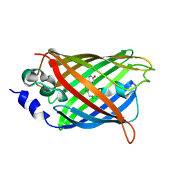
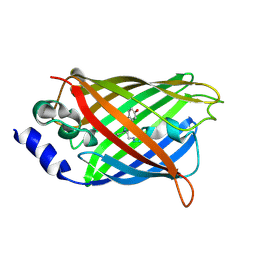
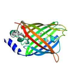
Structure of the Yellow Fluorescent Protein Citrine Frozen at 2500 Atmospheres: Structure 22 in a Series of 26 High Pressure Structures
Descriptor: Green fluorescent protein
Authors:Barstow, B, Kim, C.U.
Deposit date:2008-07-09
Release date:2008-09-23
Last modified:2024-10-09
Method:X-RAY DIFFRACTION (1.7 Å)
Cite:Alteration of citrine structure by hydrostatic pressure explains the accompanying spectral shift.
Proc.Natl.Acad.Sci.Usa, 105, 2008
3DQD
DownloadVisualize
BU of 3dqd by Molmil
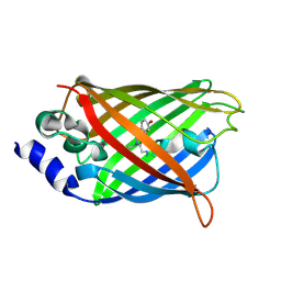
Structure of the Yellow Fluorescent Protein Citrine Frozen at 1250 Atmospheres Number 2: Structure 12 in a Series of 26 High Pressure Structures
Descriptor: Green fluorescent protein
Authors:Barstow, B, Kim, C.U.
Deposit date:2008-07-09
Release date:2008-09-23
Last modified:2023-11-15
Method:X-RAY DIFFRACTION (1.4 Å)
Cite:Alteration of citrine structure by hydrostatic pressure explains the accompanying spectral shift.
Proc.Natl.Acad.Sci.Usa, 105, 2008
3DVQ
DownloadVisualize
BU of 3dvq by Molmil
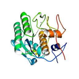
Proteinase K by LB nanotemplate method before high X-Ray dose on ESRF ID14-2 beamline
Descriptor: CALCIUM ION, Proteinase K
Authors:Pechkova, E, Tripathi, S.K, Nicolini, C.
Deposit date:2008-07-20
Release date:2009-07-07
Last modified:2023-11-01
Method:X-RAY DIFFRACTION (1.02 Å)
Cite:Radiation damage in protein structural characterization by Synchrotron Radiation: State of the art and Nanotechnology-based perspective
To be Published
3DW1
DownloadVisualize
BU of 3dw1 by Molmil
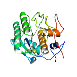
Proteinase K by LB nanotemplate method after the third step high X-Ray dose on ESRF ID14-2 beamline
Descriptor: CALCIUM ION, Proteinase K
Authors:Pechkova, E, Tripathi, S.K, Nicolini, C.
Deposit date:2008-07-21
Release date:2009-07-07
Last modified:2024-10-09
Method:X-RAY DIFFRACTION (1.03 Å)
Cite:Radiation damage in protein structural characterization by Synchrotron Radiation: State of the art and Nanotechnology-based perspective
To be Published
3EYB
DownloadVisualize
BU of 3eyb by Molmil
Structural and functional insights into the ligand binding domain of a non-duplicated RXR from the invertebrate chordate amphioxus
Descriptor: Nuclear hormone receptor RXR
Authors:Tocchini-Valentini, G.D, Rochel, N, Moras, D, Structural Proteomics in Europe (SPINE)
Deposit date:2008-10-20
Release date:2008-11-04
Last modified:2023-09-06
Method:X-RAY DIFFRACTION (2.79 Å)
Cite:Structural and functional insights into the ligand-binding domain of a nonduplicated retinoid X nuclear receptor from the invertebrate chordate amphioxus
J.Biol.Chem., 284, 2009
3EXO
DownloadVisualize
BU of 3exo by Molmil
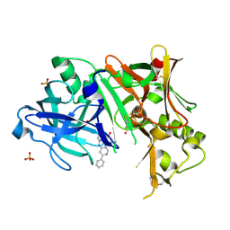
Crystal structure of BACE1 bound to inhibitor
Descriptor: Beta-secretase 1, GLYCEROL, N-{2-methyl-5-[(6-phenylpyrimidin-4-yl)amino]phenyl}methanesulfonamide, ...
Authors:Allison, T.J.
Deposit date:2008-10-16
Release date:2009-02-03
Last modified:2024-10-09
Method:X-RAY DIFFRACTION (2.1 Å)
Cite:Identification of a small molecule beta-secretase inhibitor that binds without catalytic aspartate engagement.
Bioorg.Med.Chem.Lett., 19, 2009
3DXR
DownloadVisualize
BU of 3dxr by Molmil
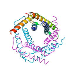
Crystal structure of the yeast inter-membrane space chaperone assembly TIM9.10
Descriptor: Mitochondrial import inner membrane translocase subunit TIM10, Mitochondrial import inner membrane translocase subunit TIM9
Authors:Webb, C.T, Gulbis, J.M.
Deposit date:2008-07-24
Release date:2008-12-23
Last modified:2023-08-30
Method:X-RAY DIFFRACTION (2.5 Å)
Cite:Structural and functional requirements for activity of the Tim9-Tim10 complex in mitochondrial protein import
Mol Biol Cell, 20, 2009
3EY4
DownloadVisualize
BU of 3ey4 by Molmil
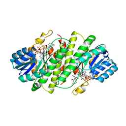
Further studies with the 2-amino-1,3-thiazol-4(5H)-one class of 11-hydroxysteroid dehydrogenase type 1 (11-HSD1) inhibitors: Reducing pregnane X receptor (PXR) activity and exploring activity in a monkey pharmacodynamic model
Descriptor: (5S)-2-{[(1S)-1-(4-fluorophenyl)ethyl]amino}-5-(1-hydroxy-1-methylethyl)-5-methyl-1,3-thiazol-4(5H)-one, 11-beta-Hydroxysteroid Dehydrogenase 1, NADPH DIHYDRO-NICOTINAMIDE-ADENINE-DINUCLEOTIDE PHOSPHATE
Authors:Zhang, J.D, Jordan, S.R, Li, V.
Deposit date:2008-10-17
Release date:2008-11-11
Last modified:2023-12-27
Method:X-RAY DIFFRACTION (3 Å)
Cite:Further studies with the 2-amino-1,3-thiazol-4(5H)-one class of 11-hydroxysteroid dehydrogenase type 1 (11-HSD1) inhibitors: Reducing pregnane X receptor (PXR) activity and exploring activity in a monkey pharmacodynamic model
To be Published, 2008
3DYI
DownloadVisualize
BU of 3dyi by Molmil
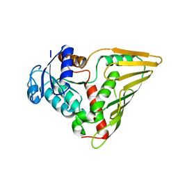
Snapshots of esterase D from lactobacillus rhamnosus: Insights into a rotation driven catalytic mechanism
Descriptor: CHLORIDE ION, Esterase D
Authors:Bennett, M.D, Delabre, M.-L, Holland, R, Norris, G.E.
Deposit date:2008-07-28
Release date:2009-07-28
Last modified:2023-11-01
Method:X-RAY DIFFRACTION (1.72 Å)
Cite:Snapshots of esterase D from lactobacillus rhamnosus: Insights into a rotation driven catalytic mechanism
To be Published
3EZ4
DownloadVisualize
BU of 3ez4 by Molmil
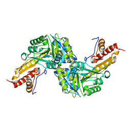
Crystal structure of 3-methyl-2-oxobutanoate hydroxymethyltransferase from Burkholderia pseudomallei
Descriptor: 2-AMINO-2-HYDROXYMETHYL-PROPANE-1,3-DIOL, 3-methyl-2-oxobutanoate hydroxymethyltransferase
Authors:Seattle Structural Genomics Center for Infectious Disease (SSGCID)
Deposit date:2008-10-22
Release date:2008-11-04
Last modified:2023-09-06
Method:X-RAY DIFFRACTION (2.1 Å)
Cite:Crystal structure of 3-methyl-2-oxobutanoate hydroxymethyltransferase from Burkholderia pseudomallei
To be Published
3F0A
DownloadVisualize
BU of 3f0a by Molmil
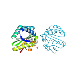
Structure of a putative n-acetyltransferase (ta0374) in complex with acetyl-coa from thermoplasma acidophilum
Descriptor: ACETYL COENZYME *A, CHLORIDE ION, N-ACETYLTRANSFERASE, ...
Authors:Filippova, E.V, Minasov, G, Shuvalova, L, Kiryukhina, O, Clancy, S, Joachimiak, A, Anderson, W.F, Midwest Center for Structural Genomics (MCSG)
Deposit date:2008-10-24
Release date:2008-11-11
Last modified:2023-11-15
Method:X-RAY DIFFRACTION (2.5 Å)
Cite:Crystal structure of the novel PaiA N-acetyltransferase from Thermoplasma acidophilum involved in the negative control of sporulation and degradative enzyme production.
Proteins, 79, 2011
3DQN
DownloadVisualize
BU of 3dqn by Molmil
Structure of the Yellow Fluorescent Protein Citrine Frozen at 500 Atmospheres: Structure 3 in a Series of 26 High Pressure Structures
Descriptor: Green fluorescent protein
Authors:Barstow, B, Kim, C.U.
Deposit date:2008-07-09
Release date:2008-09-23
Last modified:2024-10-16
Method:X-RAY DIFFRACTION (1.44 Å)
Cite:Alteration of citrine structure by hydrostatic pressure explains the accompanying spectral shift.
Proc.Natl.Acad.Sci.Usa, 105, 2008
2RHF
DownloadVisualize
BU of 2rhf by Molmil
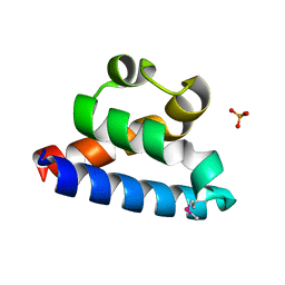
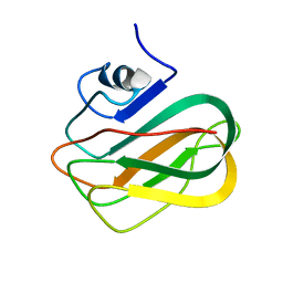
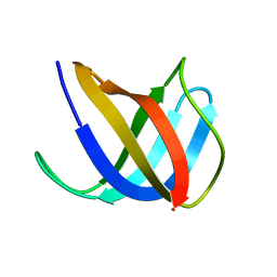
D. radiodurans RecQ HRDC domain 3
Descriptor: DNA helicase RecQ, PHOSPHATE ION
Authors:Keck, J.L, Killoran, M.P.
Deposit date:2007-10-09
Release date:2008-04-22
Last modified:2011-07-13
Method:X-RAY DIFFRACTION (1.1 Å)
Cite:Structure and function of the regulatory C-terminal HRDC domain from Deinococcus radiodurans RecQ.
Nucleic Acids Res., 36, 2008
2RHZ
DownloadVisualize
BU of 2rhz by Molmil
Crystal structure of the 3-MBT repeats from human L3MBTL1 with D355N point mutation
Descriptor: DI(HYDROXYETHYL)ETHER, Lethal(3)malignant brain tumor-like protein
Authors:Li, H, Patel, D.J.
Deposit date:2007-10-09
Release date:2007-12-11
Last modified:2023-11-15
Method:X-RAY DIFFRACTION (2.2 Å)
Cite:Structural Basis for Lower Lysine Methylation State-Specific Readout by MBT Repeats of L3MBTL1 and an Engineered PHD Finger.
Mol.Cell, 28, 2007
3DYD
DownloadVisualize
BU of 3dyd by Molmil
Human Tyrosine Aminotransferase
Descriptor: PYRIDOXAL-5'-PHOSPHATE, Tyrosine aminotransferase
Authors:Karlberg, T, Moche, M, Andersson, J, Arrowsmith, C.H, Berglund, H, Collins, R, Dahlgren, L.G, Edwards, A.M, Flodin, S, Flores, A, Graslund, S, Hammarstrom, M, Johansson, A, Johansson, I, Kotenyova, T, Lehtio, L, Nilsson, M.E, Nordlund, P, Nyman, T, Olesen, K, Persson, C, Sagemark, J, Thorsell, A.G, Tresaugues, L, Van Den Berg, S, Weigelt, J, Welin, M, Wikstrom, M, Wisniewska, M, Schuler, H, Structural Genomics Consortium (SGC)
Deposit date:2008-07-27
Release date:2008-08-19
Last modified:2023-08-30
Method:X-RAY DIFFRACTION (2.3 Å)
Cite:Human Tyrosine Aminotransferase
To be Published
3E0W
DownloadVisualize
BU of 3e0w by Molmil
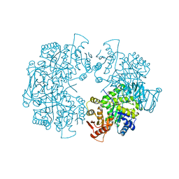
Crystal structure of pyruvate kinase from Leishmania mexicana
Descriptor: Pyruvate kinase
Authors:Tulloch, L.B, Gillmore, L.A, Walkinshaw, M.D.
Deposit date:2008-08-01
Release date:2008-08-19
Last modified:2023-08-30
Method:X-RAY DIFFRACTION (3.1 Å)
Cite:Sulphate removal induces a major conformational change in Leishmania mexicana pyruvate kinase in the crystalline state.
J.Mol.Biol., 383, 2008
2RVA
DownloadVisualize
BU of 2rva by Molmil
Solution structure of chitosan-binding module 2 derived from chitosanase/glucanase from Paenibacillus sp. IK-5
Descriptor: Glucanase
Authors:Shinya, S, Nishimura, S, Fukamizo, T.
Deposit date:2015-05-13
Release date:2016-04-06
Last modified:2024-05-15
Method:SOLUTION NMR
Cite:Mechanism of chitosan recognition by CBM32 carbohydrate-binding modules from a Paenibacillus sp. IK-5 chitosanase/glucanase.
Biochem.J., 473, 2016
3DPU
DownloadVisualize
BU of 3dpu by Molmil
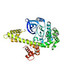
RocCOR domain tandem of Rab family protein (Roco)
Descriptor: Rab family protein
Authors:Gotthardt, K, Weyand, M, Kortholt, A, Van Haastert, P.J.M, Wittinghofer, A.
Deposit date:2008-07-09
Release date:2008-08-12
Last modified:2023-11-01
Method:X-RAY DIFFRACTION (2.9 Å)
Cite:Structure of the Roc-COR domain tandem of C. tepidum, a prokaryotic homologue of the human LRRK2 Parkinson kinase
Embo J., 27, 2008
3DQH
DownloadVisualize
BU of 3dqh by Molmil
Structure of the Yellow Fluorescent Protein Citrine Frozen at 1000 Atmospheres Number 5: Structure 9 in a Series of 26 High Pressure Structures
Descriptor: Green fluorescent protein
Authors:Barstow, B, Kim, C.U.
Deposit date:2008-07-09
Release date:2008-09-23
Last modified:2023-11-15
Method:X-RAY DIFFRACTION (1.45 Å)
Cite:Alteration of citrine structure by hydrostatic pressure explains the accompanying spectral shift.
Proc.Natl.Acad.Sci.Usa, 105, 2008
3DRJ
DownloadVisualize
BU of 3drj by Molmil
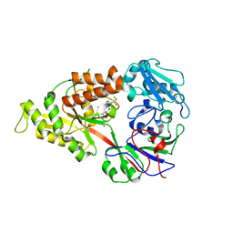
Crystal structure of Lactococcal OppA co-crystallized with pTH-related peptide in an open conformation
Descriptor: Oligopeptide-binding protein oppA, pTH-related peptide
Authors:Berntsson, R.P.-A, Doeven, M.K, Duurkens, R.H, Sengupta, D, Marrink, S.-J, Thunnissen, A.-M, Poolman, B, Slotboom, D.-J.
Deposit date:2008-07-11
Release date:2009-03-31
Last modified:2023-11-01
Method:X-RAY DIFFRACTION (1.5 Å)
Cite:The structural basis for peptide selection by the transport receptor OppA
Embo J., 28, 2009
3DSP
DownloadVisualize
BU of 3dsp by Molmil
Crystal structure of apo copper resistance protein CopK
Descriptor: Putative uncharacterized protein copK
Authors:Ash, M.-R, Maher, M.J.
Deposit date:2008-07-13
Release date:2009-03-10
Last modified:2023-11-01
Method:X-RAY DIFFRACTION (2.2 Å)
Cite:Unprecedented binding cooperativity between Cu(I) and Cu(II) in the copper resistance protein CopK from Cupriavidus metallidurans CH34: implications from structural studies by NMR spectroscopy and X-ray crystallography
J.Am.Chem.Soc., 131, 2009
3E02
DownloadVisualize
BU of 3e02 by Molmil
Crystal structure of a duf849 family protein (bxe_c0271) from burkholderia xenovorans lb400 at 1.90 A resolution
Descriptor: 1,2-ETHANEDIOL, ZINC ION, uncharacterized protein DUF849
Authors:Joint Center for Structural Genomics (JCSG)
Deposit date:2008-07-30
Release date:2008-08-12
Last modified:2024-10-30
Method:X-RAY DIFFRACTION (1.9 Å)
Cite:Crystal structure of protein of unknown function (DUF849) (YP_555544.1) from BURKHOLDERIA XENOVORANS LB400 at 1.90 A resolution
To be published
3I7B
DownloadVisualize
BU of 3i7b by Molmil
Calcium-Dependent Protein Kinase 1 from Toxoplasma gondii (TgCDPK1) in complex with bumped kinase inhibitor NM-PP1
Descriptor: 1,2-ETHANEDIOL, 1-(1-methylethyl)-3-(naphthalen-1-ylmethyl)-1H-pyrazolo[3,4-d]pyrimidin-4-amine, Calmodulin-domain protein kinase 1, ...
Authors:Larson, E.T, Merritt, E.A, Medical Structural Genomics of Pathogenic Protozoa (MSGPP)
Deposit date:2009-07-08
Release date:2010-02-09
Last modified:2023-09-06
Method:X-RAY DIFFRACTION (1.988 Å)
Cite:Toxoplasma gondii calcium-dependent protein kinase 1 is a target for selective kinase inhibitors.
Nat.Struct.Mol.Biol., 17, 2010
2VB3
DownloadVisualize
BU of 2vb3 by Molmil
Crystal structure of Ag(I)CusF
Descriptor: CATION EFFLUX SYSTEM PROTEIN CUSF, SILVER ION
Authors:Xue, Y, Davis, A.V, Balakrishnan, G, Stasser, J.P, Staehlin, B.M, Focia, P, Spiro, T.G, Penner-Hahn, J.E, O'Halloran, T.V.
Deposit date:2007-09-06
Release date:2007-12-18
Last modified:2023-12-13
Method:X-RAY DIFFRACTION (2.33 Å)
Cite:Cu(I) Recognition Via Cation-Pi and Methionine Interactions in Cusf.
Nat.Chem.Biol., 4, 2008

227111

건을2024-11-06부터공개중

PDB statisticsPDBj update infoContact PDBjnumon