Loading
PDBj
MenuPDBj@FacebookPDBj@TwitterPDBj@YouTubewwPDB FoundationwwPDB
RCSB PDBPDBeBMRBAdv. SearchSearch help

7FR7
DownloadVisualize
BU of 7fr7 by Molmil
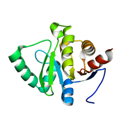
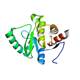
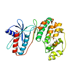
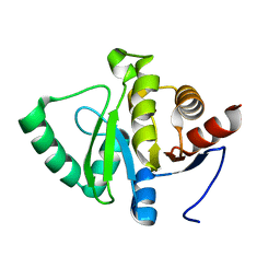
PanDDA analysis group deposition -- Crystal structure of SARS-CoV-2 NSP3 macrodomain in complex with Z431872694
Descriptor: 6-(azepane-1-carbonyl)pyrido[2,3-d]pyrimidine-2,4(1H,3H)-dione, Non-structural protein 3
Authors:Correy, G.J, Fraser, J.S.
Deposit date:2022-10-20
Release date:2022-11-09
Last modified:2024-05-22
Method:X-RAY DIFFRACTION (1 Å)
Cite:Ligand screen against SARS-CoV-2 NSP3 macrodomain
To be published
7FR8
DownloadVisualize
BU of 7fr8 by Molmil
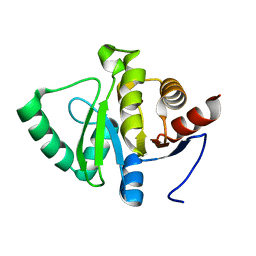
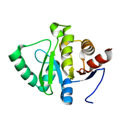
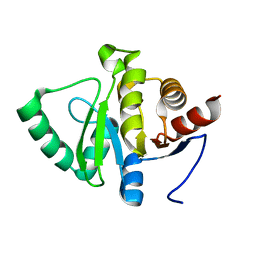
PanDDA analysis group deposition -- Crystal structure of SARS-CoV-2 NSP3 macrodomain in complex with Z2890408258 - (R) isomer
Descriptor: (2-hydroxy-3H-imidazo[4,5-b]pyridin-7-yl)[(2R)-2-methylmorpholin-4-yl]methanone, Non-structural protein 3
Authors:Correy, G.J, Fraser, J.S.
Deposit date:2022-10-20
Release date:2022-11-09
Last modified:2024-05-22
Method:X-RAY DIFFRACTION (1 Å)
Cite:Ligand screen against SARS-CoV-2 NSP3 macrodomain
To be published
7FR4
DownloadVisualize
BU of 7fr4 by Molmil
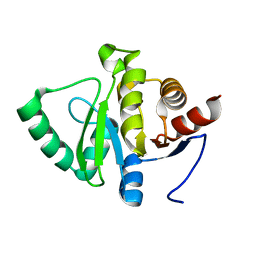
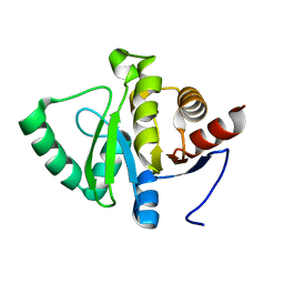
PanDDA analysis group deposition -- Crystal structure of SARS-CoV-2 NSP3 macrodomain in complex with PTERA_A26A - (S) isomer
Descriptor: 2-cyclohexyl-N-{(2S)-1-[(7H-purin-6-yl)amino]butan-2-yl}acetamide, DIMETHYL SULFOXIDE, Non-structural protein 3
Authors:Correy, G.J, Fraser, J.S.
Deposit date:2022-10-20
Release date:2022-11-09
Last modified:2024-05-22
Method:X-RAY DIFFRACTION (1 Å)
Cite:Ligand screen against SARS-CoV-2 NSP3 macrodomain
To be published
7FR1
DownloadVisualize
BU of 7fr1 by Molmil
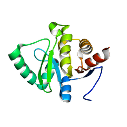
PanDDA analysis group deposition -- Crystal structure of SARS-CoV-2 NSP3 macrodomain in complex with Z1423250928
Descriptor: 1-methyl-N'-(7H-purin-6-yl)cyclopropane-1-carbohydrazide, Non-structural protein 3
Authors:Correy, G.J, Fraser, J.S.
Deposit date:2022-10-20
Release date:2022-11-09
Last modified:2024-05-22
Method:X-RAY DIFFRACTION (1.15 Å)
Cite:Ligand screen against SARS-CoV-2 NSP3 macrodomain
To be published
7FR3
DownloadVisualize
BU of 7fr3 by Molmil
PanDDA analysis group deposition -- Crystal structure of SARS-CoV-2 NSP3 macrodomain in complex with PTERA_A01A - (S) isomer
Descriptor: 3-cyclohexyl-N-{(2S)-1-[(9H-purin-6-yl)amino]butan-2-yl}propanamide, Non-structural protein 3
Authors:Correy, G.J, Fraser, J.S.
Deposit date:2022-10-20
Release date:2022-11-09
Last modified:2024-05-22
Method:X-RAY DIFFRACTION (1 Å)
Cite:Ligand screen against SARS-CoV-2 NSP3 macrodomain
To be published
7FRB
DownloadVisualize
BU of 7frb by Molmil
PanDDA analysis group deposition -- Crystal structure of SARS-CoV-2 NSP3 macrodomain in complex with Z5551426009 - (S) isomer
Descriptor: 1-methyl-N-{(2S)-3-methyl-2-[(9H-purin-6-yl)amino]butyl}cyclobutane-1-carboxamide, Non-structural protein 3
Authors:Correy, G.J, Fraser, J.S.
Deposit date:2022-10-20
Release date:2022-11-09
Last modified:2024-05-22
Method:X-RAY DIFFRACTION (1 Å)
Cite:Ligand screen against SARS-CoV-2 NSP3 macrodomain
To be published
7FRC
DownloadVisualize
BU of 7frc by Molmil
PanDDA analysis group deposition -- Crystal structure of SARS-CoV-2 NSP3 macrodomain in complex with PTERA_A05 - (R) isomer
Descriptor: 3-cyclohexyl-N-{(2R)-2-[(1H-pyrazolo[3,4-d]pyrimidin-4-yl)amino]butyl}propanamide, Non-structural protein 3
Authors:Correy, G.J, Fraser, J.S.
Deposit date:2022-10-20
Release date:2022-11-09
Last modified:2024-05-22
Method:X-RAY DIFFRACTION (1 Å)
Cite:Ligand screen against SARS-CoV-2 NSP3 macrodomain
To be published
7FRD
DownloadVisualize
BU of 7frd by Molmil
PanDDA analysis group deposition -- Crystal structure of SARS-CoV-2 NSP3 macrodomain in complex with PTERA_A25A - (S) isomer
Descriptor: 3-phenyl-N-{(2S)-1-[(7H-purin-6-yl)amino]butan-2-yl}propanamide, Non-structural protein 3
Authors:Correy, G.J, Fraser, J.S.
Deposit date:2022-10-20
Release date:2022-11-09
Last modified:2024-05-22
Method:X-RAY DIFFRACTION (1 Å)
Cite:Ligand screen against SARS-CoV-2 NSP3 macrodomain
To be published
7FR9
DownloadVisualize
BU of 7fr9 by Molmil
PanDDA analysis group deposition -- Crystal structure of SARS-CoV-2 NSP3 macrodomain in complex with Z1367095370
Descriptor: N-(1-methylcyclopropyl)-2,4-dioxo-1,2,3,4-tetrahydropyrido[2,3-d]pyrimidine-6-sulfonamide, Non-structural protein 3
Authors:Correy, G.J, Fraser, J.S.
Deposit date:2022-10-20
Release date:2022-11-09
Last modified:2024-05-22
Method:X-RAY DIFFRACTION (1 Å)
Cite:Ligand screen against SARS-CoV-2 NSP3 macrodomain
To be published
2A2Z
DownloadVisualize
BU of 2a2z by Molmil
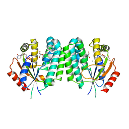
Crystal Structure of human deoxycytidine kinase in complex with deoxycytidine and uridine diphosphate
Descriptor: 2'-DEOXYCYTIDINE, CALCIUM ION, Deoxycytidine kinase, ...
Authors:Godsey, M.H, Ort, S, Sabini, E, Konrad, M, Lavie, A.
Deposit date:2005-06-23
Release date:2006-01-17
Last modified:2023-08-23
Method:X-RAY DIFFRACTION (3.02 Å)
Cite:Structural basis for the preference of UTP over ATP in human deoxycytidine kinase: illuminating the role of main-chain reorganization.
Biochemistry, 45, 2006
3OOC
DownloadVisualize
BU of 3ooc by Molmil
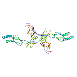
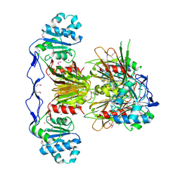
Crystal structure of the membrane fusion protein CusB from Escherichia coli
Descriptor: Cation efflux system protein cusB
Authors:Su, C.-C, Yang, F, Long, F, Reyon, D, Routh, M.D, Kuo, D.W, Mokhtari, A.K, Van Ornam, J.D, Rabe, K.L, Hoy, J.A, Lee, Y.J, Rajashankar, K.R, Yu, E.W.
Deposit date:2010-08-30
Release date:2010-12-29
Last modified:2024-02-21
Method:X-RAY DIFFRACTION (3.404 Å)
Cite:Crystal structure of the membrane fusion protein CusB from Escherichia coli.
J.Mol.Biol., 393, 2009
3OJP
DownloadVisualize
BU of 3ojp by Molmil
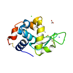
D52N Mutant of Hen Egg White Lysozyme (HEWL)
Descriptor: ACETATE ION, CHLORIDE ION, Lysozyme C, ...
Authors:O'Meara, F, Bradley, J, O'Rourke, P.E, Webb, H, Tynan-Connolly, B.M, Nielsen, J.E.
Deposit date:2010-08-23
Release date:2011-03-02
Last modified:2023-09-06
Method:X-RAY DIFFRACTION (1.81 Å)
Cite:D52N Mutant of Hen Egg White Lysozyme (HEWL)
TO BE PUBLISHED
3OIA
DownloadVisualize
BU of 3oia by Molmil
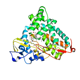
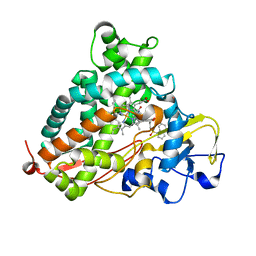
Crystal Structure of Cytochrome P450cam crystallized in the presence of a tethered substrate analog AdaC1-C8GluEtg-Bio
Descriptor: Camphor 5-monooxygenase, N-{15-oxo-19-[(3aS,4S,6aR)-2-oxohexahydro-1H-thieno[3,4-d]imidazol-4-yl]-4,7,10-trioxa-14-azanonadec-1-yl}-N'-(8-{[(3S,5S,7S)-tricyclo[3.3.1.1~3,7~]dec-1-ylcarbonyl]amino}octyl)pentanediamide, POTASSIUM ION, ...
Authors:Lee, Y.-T, Wilson, R.F, Glazer, E.C, Goodin, D.B.
Deposit date:2010-08-18
Release date:2010-11-17
Last modified:2023-09-06
Method:X-RAY DIFFRACTION (1.65 Å)
Cite:Crystal Structure of Cytochrome P450cam crystallized in the presence of a tethered substrate analog AdaC1-C8GluEtg-Bio
To be Published
3OYA
DownloadVisualize
BU of 3oya by Molmil
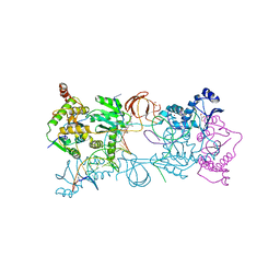
Crystal structure of the Prototype Foamy Virus (PFV) intasome in complex with magnesium and raltegravir at 2.65 resolution
Descriptor: AMMONIUM ION, DNA (5'-D(*AP*TP*TP*GP*TP*CP*AP*TP*GP*GP*AP*AP*TP*TP*TP*CP*GP*CP*A)-3'), DNA (5'-D(*TP*GP*CP*GP*AP*AP*AP*TP*TP*CP*CP*AP*TP*GP*AP*CP*A)-3'), ...
Authors:Hare, S, Cherepanov, P.
Deposit date:2010-09-23
Release date:2010-10-20
Last modified:2023-09-06
Method:X-RAY DIFFRACTION (2.65 Å)
Cite:Molecular mechanisms of retroviral integrase inhibition and the evolution of viral resistance.
Proc.Natl.Acad.Sci.USA, 107, 2010
3OZL
DownloadVisualize
BU of 3ozl by Molmil
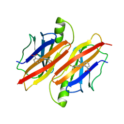
Crystal structure of human transthyretin variant A25T in complex with flufenamic acid.
Descriptor: 2-[[3-(TRIFLUOROMETHYL)PHENYL]AMINO] BENZOIC ACID, Transthyretin
Authors:Lima, L.M.T.R, Palmieri, L.C, Foguel, D, Palhano, F.L.
Deposit date:2010-09-25
Release date:2011-09-28
Last modified:2023-09-06
Method:X-RAY DIFFRACTION (1.9 Å)
Cite:Crystal structure of human transthyretin variant A25T in complex with flufenamic acid.
To be Published
3P6H
DownloadVisualize
BU of 3p6h by Molmil
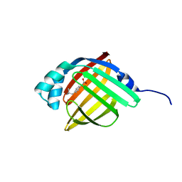
Human adipocyte lipid-binding protein FABP4 in complex with (S)-ibuprofen
Descriptor: Fatty acid-binding protein, adipocyte, IBUPROFEN
Authors:Gonzalez, J.M, Pozharski, E.
Deposit date:2010-10-11
Release date:2011-04-13
Last modified:2024-02-21
Method:X-RAY DIFFRACTION (1.15 Å)
Cite:Structural analysis of ibuprofen binding to human adipocyte fatty-acid binding protein (FABP4).
Acta Crystallogr F Struct Biol Commun, 71, 2015
3P6T
DownloadVisualize
BU of 3p6t by Molmil
Crystal Structure of Cytochrome P450cam crystallized in the presence of a tethered substrate analog AdaC2-C8-Dans
Descriptor: Camphor 5-monooxygenase, N-[8-({[5-(dimethylamino)naphthalen-1-yl]sulfonyl}amino)octyl]-2-[(3S,5S,7S)-tricyclo[3.3.1.1~3,7~]dec-1-yl]acetamide, POTASSIUM ION, ...
Authors:Lee, Y.-T, Wilson, R.F, Glazer, E.C, Goodin, D.B.
Deposit date:2010-10-11
Release date:2010-11-17
Last modified:2024-02-21
Method:X-RAY DIFFRACTION (1.9 Å)
Cite:Crystal Structure of Cytochrome P450cam crystallized in the presence of a tethered substrate analog AdaC2-C8-Dans
To be Published
3PEB
DownloadVisualize
BU of 3peb by Molmil
The Structure of a Creatine_N Superfamily domain of a dipeptidase from Streptococcus thermophilus.
Descriptor: 1,2-ETHANEDIOL, 2-[N-CYCLOHEXYLAMINO]ETHANE SULFONIC ACID, Dipeptidase, ...
Authors:Cuff, M.E, Mack, J.C, Clancy, S, Joachimiak, A, Midwest Center for Structural Genomics (MCSG)
Deposit date:2010-10-25
Release date:2010-11-03
Last modified:2017-11-08
Method:X-RAY DIFFRACTION (1.86 Å)
Cite:The Structure of a Creatine_N Superfamily domain of a dipeptidase from Streptococcus thermophilus.
TO BE PUBLISHED
3PCS
DownloadVisualize
BU of 3pcs by Molmil
Structure of EspG-PAK2 autoinhibitory Ialpha3 helix complex
Descriptor: EspG, Serine/threonine-protein kinase PAK 2
Authors:Tomchick, D.R, Alto, N.M, Selyunin, A.S.
Deposit date:2010-10-21
Release date:2011-01-05
Last modified:2023-09-06
Method:X-RAY DIFFRACTION (2.86 Å)
Cite:The assembly of a GTPase-kinase signalling complex by a bacterial catalytic scaffold.
Nature, 469, 2011
3PMI
DownloadVisualize
BU of 3pmi by Molmil
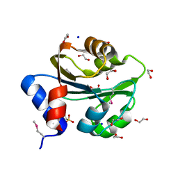
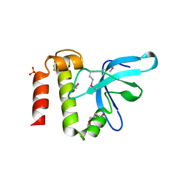
PWWP Domain of Human Mutated Melanoma-Associated Antigen 1
Descriptor: DI(HYDROXYETHYL)ETHER, PWWP domain-containing protein MUM1, SULFATE ION, ...
Authors:Lam, R, Zeng, H, Loppnau, P, Bountra, C, Weigelt, J, Arrowsmith, C.H, Edwards, A.M, Bochkarev, A, Min, J, Wu, H, Structural Genomics Consortium (SGC)
Deposit date:2010-11-17
Release date:2010-12-15
Last modified:2017-11-08
Method:X-RAY DIFFRACTION (2.82 Å)
Cite:Structural and histone binding ability characterizations of human PWWP domains.
Plos One, 6, 2011
3PMT
DownloadVisualize
BU of 3pmt by Molmil
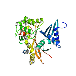
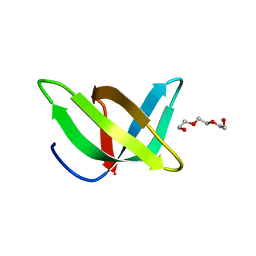
Crystal structure of the Tudor domain of human Tudor domain-containing protein 3
Descriptor: TETRAETHYLENE GLYCOL, Tudor domain-containing protein 3
Authors:Lam, R, Bian, C.B, Guo, Y.H, Xu, C, Kania, J, Bountra, C, Weigelt, J, Arrowsmith, C.H, Edwards, A.M, Bochkarev, A, Min, J, Structural Genomics Consortium (SGC)
Deposit date:2010-11-18
Release date:2010-12-01
Last modified:2024-05-22
Method:X-RAY DIFFRACTION (1.8 Å)
Cite:Crystal Structure of TDRD3 and Methyl-Arginine Binding Characterization of TDRD3, SMN and SPF30.
Plos One, 7, 2012
3PET
DownloadVisualize
BU of 3pet by Molmil
Crystal structure of a putative adhesin (BF0245) from Bacteroides fragilis NCTC 9343 at 2.07 A resolution
Descriptor: ACETATE ION, CHLORIDE ION, Putative adhesin, ...
Authors:Joint Center for Structural Genomics (JCSG)
Deposit date:2010-10-27
Release date:2011-02-09
Last modified:2023-02-01
Method:X-RAY DIFFRACTION (2.07 Å)
Cite:Crystal structure of a putative adhesin (BF0245) from Bacteroides fragilis NCTC 9343 at 2.07 A resolution
To be published
3PF6
DownloadVisualize
BU of 3pf6 by Molmil
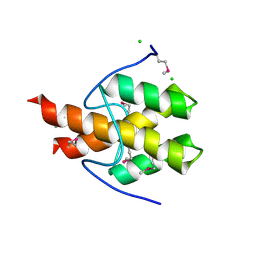
The structure of uncharacterized protein PP-LUZ7_gp033 from Pseudomonas phage LUZ7.
Descriptor: CHLORIDE ION, hypothetical protein PP-LUZ7_gp033
Authors:Cuff, M.E, Evdokimova, E, Liu, F, Edwards, A, Savchenko, A, Joachimiak, A, Midwest Center for Structural Genomics (MCSG)
Deposit date:2010-10-28
Release date:2010-11-10
Last modified:2017-11-08
Method:X-RAY DIFFRACTION (1.6 Å)
Cite:The structure of uncharacterized protein PP-LUZ7_gp033 from Pseudomonas phage LUZ7.
TO BE PUBLISHED
3PG1
DownloadVisualize
BU of 3pg1 by Molmil
MAP kinase LmaMPK10 from Leishmania major (1.95 angs resolution)
Descriptor: Mitogen-activated protein kinase, putative (Map kinase-like protein)
Authors:Horjales, S, Buschiazzo, A.
Deposit date:2010-10-29
Release date:2011-11-16
Last modified:2024-02-21
Method:X-RAY DIFFRACTION (1.95 Å)
Cite:The Crystal Structure of the MAP Kinase LmaMPK10 from Leishmania Major Reveals Parasite-Specific Features and Regulatory Mechanisms.
Structure, 20, 2012
3PFN
DownloadVisualize
BU of 3pfn by Molmil
Crystal Structure of human NAD kinase
Descriptor: NAD kinase, UNKNOWN ATOM OR ION
Authors:Wang, H, Tempel, W, Wernimont, A.K, Tong, Y, Guan, X, Shen, Y, Li, Y, Arrowsmith, C.H, Edwards, A.M, Bountra, C, Weigelt, J, Park, H, Structural Genomics Consortium (SGC)
Deposit date:2010-10-28
Release date:2010-11-10
Last modified:2017-11-08
Method:X-RAY DIFFRACTION (2.7 Å)
Cite:Crystal Structure of human NAD kinase
to be published

222415

건을2024-07-10부터공개중

PDB statisticsPDBj update infoContact PDBjnumon