1T3P
 
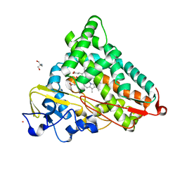
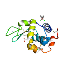 | | Half-sandwich arene ruthenium(II)-enzyme complex | | 分子名称: | ACETATE ION, CHLORIDE ION, Lysozyme C, ... | | 著者 | McNae, I.W, Fishburne, K, Habtemariam, A, Hunter, T.M, Melchart, M, Wang, F, Walkinshaw, M.D, Sadler, P.J. | | 登録日 | 2004-04-27 | | 公開日 | 2005-07-26 | | 最終更新日 | 2023-08-23 | | 実験手法 | X-RAY DIFFRACTION (1.6 Å) | | 主引用文献 | Half-sandwich arene ruthenium(II)-enzyme complex
CHEM.COMMUN.(CAMB.), 16, 2004
|
|
1T0O
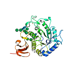 | | The structure of alpha-galactosidase from Trichoderma reesei complexed with beta-D-galactose | | 分子名称: | 2-acetamido-2-deoxy-beta-D-glucopyranose-(1-4)-2-acetamido-2-deoxy-beta-D-glucopyranose, alpha-D-mannopyranose-(1-3)-[alpha-D-mannopyranose-(1-6)]alpha-D-mannopyranose-(1-6)-[alpha-D-mannopyranose-(1-3)]beta-D-mannopyranose-(1-4)-2-acetamido-2-deoxy-beta-D-glucopyranose-(1-4)-2-acetamido-2-deoxy-beta-D-glucopyranose, alpha-D-mannopyranose-(1-3)-beta-D-mannopyranose-(1-4)-2-acetamido-2-deoxy-beta-D-glucopyranose-(1-4)-2-acetamido-2-deoxy-beta-D-glucopyranose, ... | | 著者 | Golubev, A.M, Nagem, R.A.P, Brandao Neto, J.R, Neustroev, K.N, Eneyskaya, E.V, Kulminskaya, A.A, Shabalin, K.A, Savel'ev, A.N, Polikarpov, I. | | 登録日 | 2004-04-12 | | 公開日 | 2004-10-12 | | 最終更新日 | 2023-08-23 | | 実験手法 | X-RAY DIFFRACTION (1.96 Å) | | 主引用文献 | Crystal structure of alpha-galactosidase from Trichoderma reesei and its complex with galactose: implications for catalytic mechanism
J.Mol.Biol., 339, 2004
|
|
1T4G
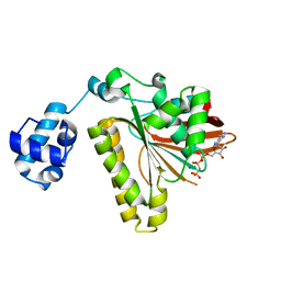 | | ATPase in complex with AMP-PNP | | 分子名称: | DNA repair and recombination protein radA, MAGNESIUM ION, PHOSPHOAMINOPHOSPHONIC ACID-ADENYLATE ESTER | | 著者 | Wu, Y, He, Y, Moya, I.A, Qian, X, Luo, Y. | | 登録日 | 2004-04-29 | | 公開日 | 2004-08-17 | | 最終更新日 | 2023-08-23 | | 実験手法 | X-RAY DIFFRACTION (2 Å) | | 主引用文献 | Crystal Structure of Archaeal Recombinase RadA; A Snapshot of Its Extended Conformation.
Mol.Cell, 15, 2004
|
|
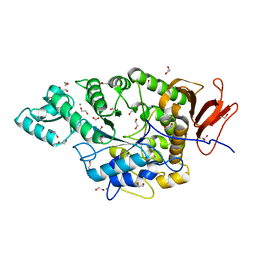
4NU9
 | | 2.30 Angstrom resolution crystal structure of betaine aldehyde dehydrogenase (betB) from Staphylococcus aureus with BME-free Cys289 | | 分子名称: | Betaine aldehyde dehydrogenase, SODIUM ION | | 著者 | Halavaty, A.S, Minasov, G, Dubrovska, I, Stam, J, Shuvalova, L, Kwon, K, Anderson, W.F, Center for Structural Genomics of Infectious Diseases (CSGID) | | 登録日 | 2013-12-03 | | 公開日 | 2013-12-11 | | 最終更新日 | 2023-09-20 | | 実験手法 | X-RAY DIFFRACTION (2.3 Å) | | 主引用文献 | Structural and functional analysis of betaine aldehyde dehydrogenase from Staphylococcus aureus.
Acta Crystallogr.,Sect.D, 71, 2015
|
|
7JJT
 | | Ruminococcus bromii amylase Amy5 (RBR_07800) | | 分子名称: | 1,2-ETHANEDIOL, ACETATE ION, Alpha-amylase, ... | | 著者 | Cerqueira, F, Koropatkin, N. | | 登録日 | 2020-07-27 | | 公開日 | 2021-06-23 | | 最終更新日 | 2023-10-18 | | 実験手法 | X-RAY DIFFRACTION (1.66 Å) | | 主引用文献 | The structures of the GH13_36 amylases from Eubacterium rectale and Ruminococcus bromii reveal subsite architectures that favor maltose production
Amylase, 4, 2020
|
|
1T6V
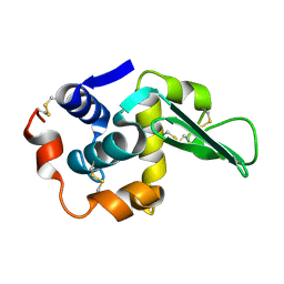
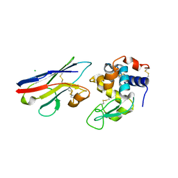 | | Crystal structure analysis of the nurse shark new antigen receptor (NAR) variable domain in complex with lysozyme | | 分子名称: | CHLORIDE ION, Lysozyme C, novel antigen receptor | | 著者 | Stanfield, R.L, Dooley, H, Flajnik, M.F, Wilson, I.A. | | 登録日 | 2004-05-07 | | 公開日 | 2004-08-24 | | 最終更新日 | 2023-08-23 | | 実験手法 | X-RAY DIFFRACTION (1.7 Å) | | 主引用文献 | Crystal structure of a shark single-domain antibody V region in complex with lysozyme.
Science, 305, 2004
|
|
4NYU
 | | Structure of Vibrio cholerae chitin de-N-acetylase in complex with acetate ion (ACT) in C 2 2 21 | | 分子名称: | ACETATE ION, CALCIUM ION, Deacetylase DA1, ... | | 著者 | Albesa-Jove, D, Andres, E, Biarnes, X, Planas, A, Guerin, M.E. | | 登録日 | 2013-12-11 | | 公開日 | 2014-08-13 | | 最終更新日 | 2023-11-08 | | 実験手法 | X-RAY DIFFRACTION (2.03 Å) | | 主引用文献 | Structural basis of chitin oligosaccharide deacetylation.
Angew.Chem.Int.Ed.Engl., 53, 2014
|
|
1T87
 | | Crystal Structure of the Ferrous CO-bound Cytochrome P450cam (C334A) | | 分子名称: | 2-AMINO-2-HYDROXYMETHYL-PROPANE-1,3-DIOL, CAMPHOR, CARBON MONOXIDE, ... | | 著者 | Nagano, S, Tosha, T, Ishimori, K, Morishima, I, Poulos, T.L. | | 登録日 | 2004-05-11 | | 公開日 | 2004-05-25 | | 最終更新日 | 2024-02-14 | | 実験手法 | X-RAY DIFFRACTION (1.8 Å) | | 主引用文献 | Crystal structure of the cytochrome p450cam mutant that exhibits the same spectral perturbations induced by putidaredoxin binding.
J.Biol.Chem., 279, 2004
|
|
1TA4
 | |
1T88
 | | Crystal Structure of the Ferrous Cytochrome P450cam (C334A) | | 分子名称: | 2-AMINO-2-HYDROXYMETHYL-PROPANE-1,3-DIOL, CAMPHOR, Cytochrome P450-cam, ... | | 著者 | Nagano, S, Tosha, T, Ishimori, K, Morishima, I, Poulos, T.L. | | 登録日 | 2004-05-11 | | 公開日 | 2004-05-25 | | 最終更新日 | 2024-02-14 | | 実験手法 | X-RAY DIFFRACTION (1.9 Å) | | 主引用文献 | Crystal structure of the cytochrome p450cam mutant that exhibits the same spectral perturbations induced by putidaredoxin binding.
J.Biol.Chem., 279, 2004
|
|
1TDY
 | |
1VQL
 | |
4NXL
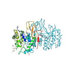
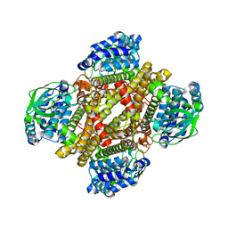 | | Dibenzothiophene monooxygenase (DszC) from Rhodococcus erythropolis | | 分子名称: | DszC | | 著者 | Zhang, L, Duan, X, Li, X, Rao, Z. | | 登録日 | 2013-12-09 | | 公開日 | 2014-07-23 | | 最終更新日 | 2024-02-28 | | 実験手法 | X-RAY DIFFRACTION (2.3 Å) | | 主引用文献 | Structural insights into the stabilization of active, tetrameric DszC by its C-terminus.
Proteins, 82, 2014
|
|
4O4P
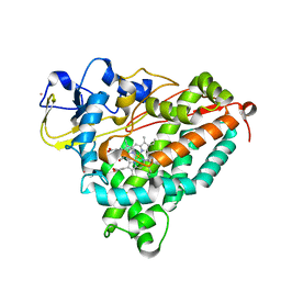
 | | Structure of P450 BM3 A82F F87V in complex with S-omeprazol | | 分子名称: | 6-methoxy-2-{[(4-methoxy-3,5-dimethylpyridin-2-yl)methyl]sulfanyl}-1H-benzimidazole, Bifunctional P-450/NADPH-P450 reductase, PROTOPORPHYRIN IX CONTAINING FE | | 著者 | Leys, D. | | 登録日 | 2013-12-19 | | 公開日 | 2014-01-29 | | 最終更新日 | 2023-09-20 | | 実験手法 | X-RAY DIFFRACTION (1.83 Å) | | 主引用文献 | Human P450-like oxidation of diverse proton pump inhibitor drugs by 'gatekeeper' mutants of flavocytochrome P450 BM3.
Biochem.J., 460, 2014
|
|
4NZB
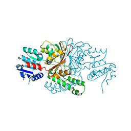
 | | NS9283 bound to Ls-AChBP | | 分子名称: | 2-acetamido-2-deoxy-beta-D-glucopyranose, 3-[3-(pyridin-3-yl)-1,2,4-oxadiazol-5-yl]benzonitrile, ACETATE ION, ... | | 著者 | Olsen, J.A, Kastrup, J.S, Gajhede, M. | | 登録日 | 2013-12-11 | | 公開日 | 2014-07-09 | | 最終更新日 | 2023-11-08 | | 実験手法 | X-RAY DIFFRACTION (2.68 Å) | | 主引用文献 | Structural and functional studies of the modulator NS9283 reveal agonist-like mechanism of action at alpha 4 beta 2 nicotinic acetylcholine receptors.
J.Biol.Chem., 289, 2014
|
|
4NZ4
 | | Structure of Vibrio cholerae chitin de-N-acetylase in complex with 2-(ACETYLAMINO)-2-DEOXY-A-D-GLUCOPYRANOSE (NDG) and zinc ion | | 分子名称: | 2-acetamido-2-deoxy-alpha-D-glucopyranose, ACETATE ION, CALCIUM ION, ... | | 著者 | Albesa-Jove, D, Andres, E, Biarnes, X, Planas, A, Guerin, M.E. | | 登録日 | 2013-12-11 | | 公開日 | 2014-08-13 | | 最終更新日 | 2023-11-08 | | 実験手法 | X-RAY DIFFRACTION (1.944 Å) | | 主引用文献 | Structural basis of chitin oligosaccharide deacetylation.
Angew.Chem.Int.Ed.Engl., 53, 2014
|
|
1VKM
 | |
7JJ1
 | |
5R8A
 | | PanDDA analysis group deposition INTERLEUKIN-1 BETA -- Fragment Z1492796719 in complex with INTERLEUKIN-1 BETA | | 分子名称: | Interleukin-1 beta, SULFATE ION, ~{N}-[(3~{R})-1,2,3,4-tetrahydroquinolin-3-yl]ethanamide | | 著者 | De Nicola, G.F, Nichols, C.E. | | 登録日 | 2020-03-03 | | 公開日 | 2020-04-22 | | 最終更新日 | 2024-03-06 | | 実験手法 | X-RAY DIFFRACTION (1.47 Å) | | 主引用文献 | Mining the PDB for Tractable Cases Where X-ray Crystallography Combined with Fragment Screens Can Be Used to Systematically Design Protein-Protein Inhibitors: Two Test Cases Illustrated by IL1 beta-IL1R and p38 alpha-TAB1 Complexes.
J.Med.Chem., 63, 2020
|
|
5R8O
 | |
5R97
 | | PanDDA analysis group deposition Form1 MAP kinase p38-alpha -- Fragment N13662a in complex with MAP kinase p38-alpha | | 分子名称: | 4-(piperidin-1-yl)-1,2,5-oxadiazol-3-amine, CHLORIDE ION, MAGNESIUM ION, ... | | 著者 | De Nicola, G.F, Nichols, C.E. | | 登録日 | 2020-03-04 | | 公開日 | 2020-07-22 | | 最終更新日 | 2024-03-06 | | 実験手法 | X-RAY DIFFRACTION (1.438 Å) | | 主引用文献 | Mining the PDB for Tractable Cases Where X-ray Crystallography Combined with Fragment Screens Can Be Used to Systematically Design Protein-Protein Inhibitors: Two Test Cases Illustrated by IL1 beta-IL1R and p38 alpha-TAB1 Complexes.
J.Med.Chem., 63, 2020
|
|
4NZZ
 | |
5R9L
 | |
5R8M
 | |
5RA3
 | |


