8OZZ
 
 | |
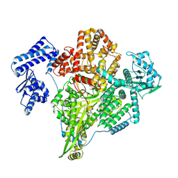
8OVG
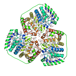
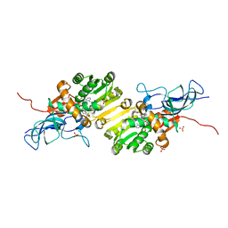
 | | Human Mitochondrial Lon Y186E Mutant ADP Bound | | 分子名称: | Lon protease homolog, mitochondrial | | 著者 | Kereiche, S, Bauer, J.A, Matyas, P, Novacek, J, Kutejova, E. | | 登録日 | 2023-04-26 | | 公開日 | 2024-05-08 | | 最終更新日 | 2024-05-15 | | 実験手法 | ELECTRON MICROSCOPY (8.47 Å) | | 主引用文献 | Polyphosphate and tyrosine phosphorylation in the N-terminal domain of the human mitochondrial Lon protease disrupts its functions.
Sci Rep, 14, 2024
|
|
8P8E
 | |
7PYN
 | | Structure of an LPMO (expressed in E.coli) at 2.31x10^5 Gy | | 分子名称: | Auxiliary activity 9, COPPER (II) ION, SULFATE ION | | 著者 | Tandrup, T, Muderspach, S.J, Banerjee, S, Ipsen, J.O, Rollan, C.H, Norholm, M.H.H, Johansen, K.S, Lo Leggio, L. | | 登録日 | 2021-10-10 | | 公開日 | 2022-08-24 | | 最終更新日 | 2024-01-31 | | 実験手法 | X-RAY DIFFRACTION (1.4 Å) | | 主引用文献 | Changes in active-site geometry on X-ray photoreduction of a lytic polysaccharide monooxygenase active-site copper and saccharide binding.
Iucrj, 9, 2022
|
|
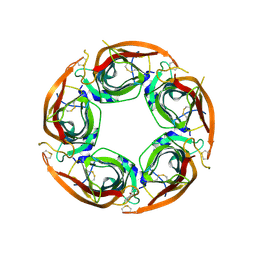
8P22
 | | X-ray structure of acetylcholine-binding protein (AChBP) in complex with IOTA376. | | 分子名称: | 2-[(2~{R})-1-ethylimidazolidin-2-yl]-6-pyridin-2-yl-pyridine, Acetylcholine-binding protein, GLYCEROL, ... | | 著者 | Cederfelt, D, Boronat, P, Dobritzsch, D, Hennig, S, Fitzgerald, E.A, de Esch, I.J.P, Danielson, U.H. | | 登録日 | 2023-05-14 | | 公開日 | 2024-05-08 | | 実験手法 | X-RAY DIFFRACTION (2.2 Å) | | 主引用文献 | Elucidating the regulation of ligand gated ion channels via biophysical studies of ligand-induced conformational dynamics of acetylcholine binding proteins
To Be Published
|
|
8OV9
 | |
8P14
 | |
8P1P
 | | USP28 in complex with AZ1 | | 分子名称: | 2-[[5-bromanyl-2-[[4-fluoranyl-3-(trifluoromethyl)phenyl]methoxy]phenyl]methylamino]ethanol, CHLORIDE ION, DIMETHYL SULFOXIDE, ... | | 著者 | Sauer, F, Karal-Nair, R, Kisker, C. | | 登録日 | 2023-05-12 | | 公開日 | 2024-05-22 | | 実験手法 | X-RAY DIFFRACTION (2.76 Å) | | 主引用文献 | USP28 in complex with AZ1
To Be Published
|
|
5L0Q
 | | Crystal structure of the complex between ADAM10 D+C domain and a conformation specific mAb 8C7. | | 分子名称: | 2-acetamido-2-deoxy-beta-D-glucopyranose, Disintegrin and metalloproteinase domain-containing protein 10, MAGNESIUM ION, ... | | 著者 | Xu, K, Saha, N, Nikolov, D.B. | | 登録日 | 2016-07-28 | | 公開日 | 2016-11-09 | | 最終更新日 | 2023-10-04 | | 実験手法 | X-RAY DIFFRACTION (2.759 Å) | | 主引用文献 | An activated form of ADAM10 is tumor selective and regulates cancer stem-like cells and tumor growth.
J.Exp.Med., 213, 2016
|
|
8P1K
 | |
8P2O
 | |
8P2P
 | |
8P11
 | | X-ray structure of acetylcholine-binding protein (AChBP) in complex with FL003044. | | 分子名称: | 4-(4-chlorophenyl)piperidin-4-ol, Acetylcholine-binding protein, CHLORIDE ION, ... | | 著者 | Cederfelt, D, Boronat, P, Dobritzsch, D, Hennig, S, Fitzgerald, E.A, de Esch, I.J.P, Danielson, U.H. | | 登録日 | 2023-05-11 | | 公開日 | 2024-05-08 | | 実験手法 | X-RAY DIFFRACTION (1.9 Å) | | 主引用文献 | Elucidating the regulation of ligand gated ion channels via biophysical studies of ligand-induced conformational dynamics of acetylcholine binding proteins
To Be Published
|
|
8OV5
 | |
7PYL
 | | Structure of an LPMO (expressed in E.coli) at 1.49x10^4 Gy | | 分子名称: | ACETATE ION, Auxiliary activity 9, COPPER (II) ION, ... | | 著者 | Tandrup, T, Muderspach, S.J, Banerjee, S, Ipsen, J.O, Rollan, C.H, Norholm, M.H.H, Johansen, K.S, Lo Leggio, L. | | 登録日 | 2021-10-10 | | 公開日 | 2022-08-24 | | 最終更新日 | 2024-01-31 | | 実験手法 | X-RAY DIFFRACTION (1.7 Å) | | 主引用文献 | Changes in active-site geometry on X-ray photoreduction of a lytic polysaccharide monooxygenase active-site copper and saccharide binding.
Iucrj, 9, 2022
|
|
8OXR
 | |
8OYQ
 | |
7PYM
 | | Structure of an LPMO (expressed in E.coli) at 5.61x10^4 Gy | | 分子名称: | Auxiliary activity 9, COPPER (II) ION, SULFATE ION | | 著者 | Tandrup, T, Muderspach, S.J, Banerjee, S, Ipsen, J.O, Rollan, C.H, Norholm, M.H.H, Johansen, K.S, Lo Leggio, L. | | 登録日 | 2021-10-10 | | 公開日 | 2022-08-24 | | 最終更新日 | 2024-01-31 | | 実験手法 | X-RAY DIFFRACTION (1.75 Å) | | 主引用文献 | Changes in active-site geometry on X-ray photoreduction of a lytic polysaccharide monooxygenase active-site copper and saccharide binding.
Iucrj, 9, 2022
|
|
8P7G
 | |
8OV8
 | | Crystal structure of Ene-reductase 1 from black poplar mushroom in complex to NADP | | 分子名称: | Ene-reductase 1, NADP NICOTINAMIDE-ADENINE-DINUCLEOTIDE PHOSPHATE, SODIUM ION, ... | | 著者 | Korf, L, Essen, L.-O, Karrer, D, Ruehl, M. | | 登録日 | 2023-04-25 | | 公開日 | 2024-05-15 | | 実験手法 | X-RAY DIFFRACTION (1.9 Å) | | 主引用文献 | Shifting the substrate scope of an ene/yne-reductase by loop engineering
To Be Published
|
|
8OYD
 | |
1ASJ
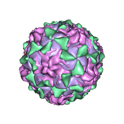 | | P1/MAHONEY POLIOVIRUS, AT CRYOGENIC TEMPERATURE | | 分子名称: | MYRISTIC ACID, P1/MAHONEY POLIOVIRUS, SPHINGOSINE | | 著者 | Wien, M.W, Curry, S, Filman, D.J, Hogle, J.M. | | 登録日 | 1997-08-11 | | 公開日 | 1997-12-03 | | 最終更新日 | 2023-08-09 | | 実験手法 | X-RAY DIFFRACTION (2.9 Å) | | 主引用文献 | Structural studies of poliovirus mutants that overcome receptor defects.
Nat.Struct.Biol., 4, 1997
|
|
8P9M
 | |
8OYZ
 | |
8OZ9
 | |


